Chapter 1: Introduction
Learning objectives
After studying this chapter, you should be able to:
Explain the purpose of this textbook and how it relates to the ASR Recommended References.
Name the three ASR certification credentials this textbook supports, and describe the exam format and topic weighting.
Explain why this textbook should not be your only study source.
Apply a structured study cycle that pairs each chapter with the Recommended References.
1.1 Purpose of This Textbook
This textbook is written for candidates preparing for the certification exams administered by the Academy of Surgical Research (ASR). It gives an overview of the information contained in the ASR's Recommended References, the reading list the Academy posts on its website for exam candidates, and explains how the topics in those references fit together.
It is important to understand what this book is and is not. The questions on each exam are drawn from the content of the Recommended References themselves. A topic covered in the references can appear on an exam even if this book never mentions it. For that reason, do not use this textbook as your only study source. Think of it as a guided tour of the material: it shows you the major landmarks, explains how they connect, and tells you where to look more closely, but the references are the territory itself.
1.2 The ASR Certifications
The Academy administers three certification exams, each aimed at a different professional role in surgical research:
SRT (Surgical Research Technician): for individuals who support surgical research, for example by preparing animals, instruments, and the operating room, and by assisting with peri-operative care.
SRS (Surgical Research Specialist): for individuals with broader, more advanced responsibility for surgical research procedures and their oversight.
SRA (Surgical Research Anesthetist): for individuals whose focus is anesthesia, analgesia, and patient monitoring in research animals.
Although the three exams emphasize different areas, they share a common foundation: the regulatory framework that governs animal surgery, aseptic technique, peri-operative care, anesthesia and analgesia, anatomy and physiology, instrumentation, surgical technique, and emergency management. The chapters of this book build that foundation in a logical order, beginning with regulations and asepsis, moving through anesthesia and analgesia, and ending with surgical and emergency procedures.
⚠ Check current guidance
Eligibility requirements, exam format, and the list of Recommended References are set by the ASR and can change. Before you begin studying, confirm the current requirements and the current Recommended References list for your exam on the ASR website. The role descriptions above are general summaries, not official ASR definitions.
1.3 The Certification Exams
The SRS and SRT exams draw on the same broad body of knowledge, weighted toward anatomy and physiology, analgesia, anesthesia, surgery, sutures, and instruments. The SRA exam also requires knowledge of asepsis, because the anesthetist works as part of the surgical team. Table 1.1 shows the approximate weighting of each topic and the chapters of this book that cover it, and Figure 1.1 shows the same weightings graphically.
Table 1.1. Approximate topic weighting on the SRS and SRT exams, and where each topic is covered.
| Topic | Approximate weight | Key content | Chapters |
|---|---|---|---|
| Anatomy and physiology (rodent and large animal) | 15–25% | Muscle, cardiovascular, and respiratory anatomy and physiology | 14, 12 |
| Analgesia (rodent and large animal) | 15–25% | Pain pathway, pain assessment, analgesic drugs and their modes of action | 13, 6, 11 |
| Surgical knowledge | 10–20% | Terminology, procedures, positioning, dissection, hemostasis, vascular access ports, telemetry implants | 16, 17, 4 |
| Suture knowledge | 10–20% | Suture materials and sizes (generic as well as brand names), needles, suture patterns | 16 |
| Instruments | 10–20% | Identification and proper use | 15 |
| Anesthesia (rodent and large animal) | 10–20% | Inhalant and injectable agents, equipment, vaporizers, breathing systems | 5–10, 12 |
| Peri-operative care | 5–15% | Fasting, monitoring, immediate post-operative recovery | 5, 12 |
| Dose calculations | 5–15% | Dose, volume, fluid rate, drip rate, and dilution calculations | Appendix A |
| Wound healing | 5–10% | Stages, factors affecting healing, wound classification | 16 |
| Regulatory review | 5–10% | AVMA, IACUC, AWA, USDA, AAALAC, controlled substances | 2 |
| Emergency techniques | 5–10% | Recognizing emergencies, drug selection, fluid therapy | 18, 5 |
| Aseptic procedures | 5–10% | Surgical suite management, record keeping, sterilization, surgical preparation | 3, 4 |
| Radiology and imaging | 2–5% | Radiology and imaging techniques | (not covered in detail; see the Recommended References) |
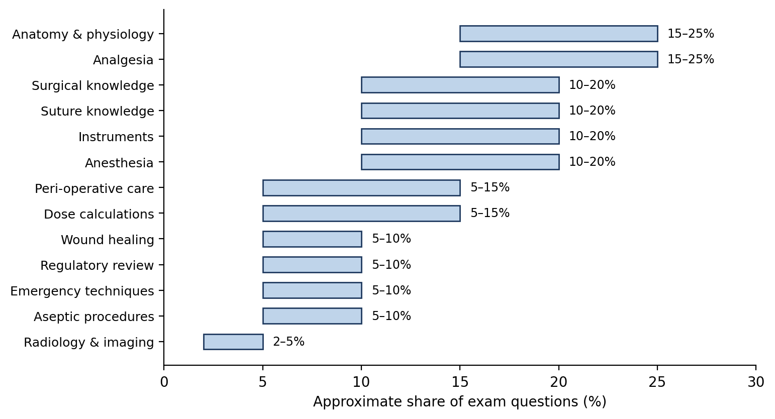
Figure 1.1. Approximate topic weighting on the SRS and SRT exams.
Exam Format
The exam has 200 questions: about 50 based on pictures (for example, instrument identification) and 150 written (didactic) questions.
All questions are multiple choice, matching, or true/false. Each is worth 0.5 points, so answer every question; there is no benefit to leaving one blank.
A passing score requires at least 136 of 200 correct (68%).
Dose calculation questions are standard word problems that provide all the information needed. Simple ones take a single step; harder ones require unit conversion or several steps (Appendix A).
Key Recommended References
Among the ASR Recommended References, the following are especially useful: Veterinary Anesthesia and Analgesia (Lumb and Jones), particularly for definitions; the Ethicon Wound Closure Manual; Clinical Textbook for Veterinary Technicians, for its chapters on surgery, anesthesia, analgesia, and sterilization; Pain Management in Animals; Laboratory Animal Anaesthesia; Small Animal Surgical Nursing; and Experimental and Surgical Technique in the Rat.
⚠ Check current guidance
Exam format, number of questions, passing score, and topic weightings are summarized from study materials and may change. Confirm current details, and the current Recommended References list, with the ASR before your exam.
1.4 How This Book Is Organized
Each chapter explains not only the rules and techniques of surgical research but also the reasoning behind them, because understanding why a practice exists makes it easier to remember and to apply in situations a rule does not anticipate. Each chapter also includes features designed to help you study:
Learning objectives at the start of each chapter tell you what you should be able to do once you have finished it.
Figures and tables summarize relationships, sequences, and comparisons that are hard to hold in your head from text alone.
"Why it matters" boxes connect a rule or technique to its purpose: animal welfare, personnel safety, or the quality of research data.
"Check current guidance" boxes flag information that may have been revised since publication. Always confirm these points against the current version of the source document.
Key terms, review questions, and an answer key at the end of each chapter let you test yourself.
Use the book actively. Write in the margins, add notes from your reading of the references, and use the review questions to find gaps you need to fill. Figure 1.2 shows a simple study cycle that works well with this book.
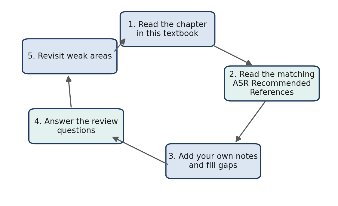
Figure 1.2. A suggested study cycle. Use each chapter as a starting point, then return to the Recommended References to deepen and confirm your understanding.
1.5 Keeping Current
Regulations, guidelines, and recommended drug protocols are revised over time, and new editions of key references are published periodically. When this book and a current official source disagree, the current official source takes precedence. Readers who find an error or inconsistency are encouraged to report it so that future printings can be corrected.
Why it matters
Surgical research professionals are expected to work from current standards. Building the habit of checking the source document, and noting its edition and date, is part of professional practice, not only exam preparation.
Key Terms
Academy of Surgical Research (ASR): The professional organization that administers the SRT, SRS, and SRA certification exams and publishes guidelines for surgical research with animals.
Recommended References: The reading list posted by the ASR on its website; the source from which certification exam questions are drawn.
SRT: Surgical Research Technician certification.
SRS: Surgical Research Specialist certification.
SRA: Surgical Research Anesthetist certification.
Review Questions
1. Where do ASR certification exam questions ultimately come from?
A. This textbook
B. The ASR Recommended References
C. Questions submitted by past candidates
D. The Animal Welfare Act only
2. Which statement best describes how this textbook should be used?
A. As a complete replacement for the Recommended References
B. As the only resource needed for the SRA exam
C. As a structured companion to reading the Recommended References
D. Only after passing the exam
3. Which three ASR certification exams does this textbook support?
A. SRS, SRT, and SRA
B. LATG, LAT, and ALAT
C. DVM, DACVAA, and DACLAM
D. CPIA, CMAR, and RLATG
4. What is the purpose of the "Check current guidance" boxes in this book?
A. To mark material that will not be on the exam
B. To flag information that may have been revised since publication
C. To list optional reading
D. To summarize each section
5. If this textbook and a current official source disagree, which should you follow?
A. This textbook
B. Whichever is easier to remember
C. The current official source
D. The older of the two
6. How many correct answers out of 200 are needed to pass the certification exam?
A. 100
B. 120
C. 136
D. 180
7. Which topics carry the greatest weight on the SRS and SRT exams?
A. Radiology and imaging
B. Anatomy and physiology, and analgesia
C. Regulatory review
D. Wound healing
Answer Key
1. B. Exam questions are drawn from the content of the ASR Recommended References. This textbook is a companion to them, not a substitute.
2. C. Topics in the references can appear on an exam even if this book does not cover them, so it should be used alongside the references.
3. A. The book supports the SRT, SRS, and SRA exams administered by the Academy. The other options are credentials from other organizations.
4. B. Regulations, guidelines, and protocols change over time. These boxes mark points to confirm against the current source document.
5. C. Current official sources always take precedence. Working from current standards is part of professional practice.
6. C. A passing score requires at least 136 of 200 questions correct (68%).
7. B. Anatomy and physiology and analgesia are each about 15–25% of the exam; radiology and imaging is only about 2–5%.
Chapter 2: Regulatory & Accreditation Overview
Learning objectives
After studying this chapter, you should be able to:
Distinguish between laws and regulations, federal policy, professional guidelines, and voluntary accreditation, and name the organization behind each major document.
Summarize the role of the USDA, NRC, OLAW, FDA, EPA, AAALAC International, AVMA, ACVA, and ASR in research animal surgery.
Define major and minor surgical procedures and explain when multiple major survival surgery can be justified.
Describe regulatory expectations for pain relief, euthanasia, peri-operative care, aseptic technique, surgical facilities, and records.
Explain why surgical facilities are divided into clean, mixed, and contaminated areas.
Surgery on research animals takes place within a framework of overlapping laws, policies, guidelines, and accreditation standards. A single surgical procedure might be governed at the same time by federal law, by the conditions attached to a research grant, by the standards of a voluntary accrediting body, and by the regulations that apply to studies submitted to the FDA. Each document has a different author, a different scope, and a different level of authority, and they do not always use the same words for the same idea.
Understanding this framework matters for two reasons. The first is animal welfare: these documents set the minimum standards that protect animals from unnecessary pain, distress, and infection. The second is data quality: an animal in pain, recovering from an infection, or stressed by poor care is a different physiological subject from a healthy one, and that difference can confound results. Good regulatory compliance and good science point in the same direction.
This chapter is divided into two parts. Section 2.1 introduces the agencies and organizations and the documents each one produces. Section 2.2 then compares what those documents say about specific aspects of surgery. On the certification exams, it is common to be asked not only what a rule is, but which document states it.
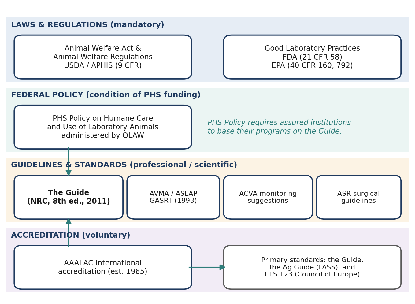
Figure 2.1. The oversight landscape for research animal surgery in the United States, grouped by type of authority. Arrows show key relationships between documents.
2.1 Agencies
2.1.1 United States Department of Agriculture (USDA)
The Animal Welfare Act (AWA) is the federal law that regulates the treatment of certain animals used in research, exhibition, and commerce. It was originally passed by Congress in 1966 and has been amended several times since. The AWA is administered by the USDA's Animal and Plant Health Inspection Service (APHIS).
A law passed by Congress usually sets out broad requirements; the agency responsible for it then writes regulations that explain in detail how those requirements are to be met. The regulations that implement the AWA are known as the Animal Welfare Regulations and are published in Title 9 of the Code of Federal Regulations (9 CFR). This chapter is based on the November 2013 version. The provisions that apply to research facilities are found in Subpart C (Research Facilities) of Part 2 of these regulations.
Because the AWA is federal law, its requirements are mandatory, and USDA inspectors verify compliance at registered research facilities. However, the AWA does not cover every animal used in research. Its regulations exclude rats of the genus Rattus and mice of the genus Mus bred for use in research, as well as birds bred for research, and they do not cover cold-blooded animals. This is one reason the rules for rodent surgery differ from document to document, as you will see in Section 2.2: a requirement that comes only from the AWA may not apply to a purpose-bred mouse, while a requirement from the PHS Policy or the Guide will.
⚠ Check current guidance
The Animal Welfare Regulations are amended periodically, and the scope of covered species has been the subject of rulemaking (for example, regarding birds). Confirm the current text of 9 CFR Parts 1–3 rather than relying on the November 2013 version described here.
2.1.2 National Research Council (NRC)
The Guide for the Care and Use of Laboratory Animals, usually called simply "the Guide," is published by the National Research Council. The edition cited throughout this book is the 8th edition (NRC 2011).
The Guide is deliberately less specific than the Animal Welfare Act. It is written in general terms so that its recommendations can be applied across the very wide range of institutions and settings that produce or use animals for research, teaching, and testing. Rather than prescribing exactly how something must be done (an engineering standard), the Guide often describes the outcome that must be achieved (a performance standard) and leaves the method to professional judgment. That flexibility is why Institutional Animal Care and Use Committees (IACUCs) have such a central role under the Guide: they interpret and implement its recommendations, oversee the institution's animal care and use program, and evaluate how well it is working.
Although the Guide is not itself a law, it carries real authority. The PHS Policy (Section 2.1.3) requires institutions to base their animal care and use programs on the Guide, and AAALAC International uses it as a primary standard for accreditation (Section 2.1.6).
Appendix B of the Guide reprints the U.S. Government Principles for the Utilization and Care of Vertebrate Animals Used in Testing, Research, and Training. The Guide also affirms that the use of laboratory animals is guided by the principles of the 3 Rs, a concept first described by William Russell and Rex Burch in 1959:
Replacement: replacing animals with non-animal models where acceptable alternatives exist.
Reduction: reducing the number of animals to the fewest needed to obtain statistically significant data.
Refinement: refining animal care and use to minimize pain and distress and enhance animal well-being.
Good surgical practice is one of the most direct applications of refinement. Skilled technique, effective anesthesia and analgesia, and attentive post-operative care all reduce pain and distress. They also contribute to reduction, because animals that recover well produce more consistent data and fewer are lost to complications.
With regard to surgery, the Guide gives specific information on the following topics, each of which is covered in later chapters of this book:
Training
Pre-surgical planning
Surgical facilities
Surgical procedures
Aseptic technique
Intraoperative monitoring
Postoperative care
⚠ Check current guidance
The 8th edition of the Guide (2011) is the edition cited in this book. A revised edition has been in development; check whether a newer edition has been published and adopted by OLAW and AAALAC before your exam.
2.1.3 Office of Laboratory Animal Welfare (OLAW)
The Office of Laboratory Animal Welfare (OLAW), part of the National Institutes of Health, implements the Public Health Service (PHS) Policy on Humane Care and Use of Laboratory Animals. This chapter refers to the version reprinted in 2015. The PHS Policy is mandated by the Health Research Extension Act of 1985, and it incorporates the U.S. Government Principles for the Utilization and Care of Vertebrate Animals Used in Testing, Research, and Training.
The PHS Policy is not a law in the same sense as the AWA. Instead, compliance is a condition of funding: institutions that receive PHS support, such as NIH grants, must comply with it. An institution does this by filing an Animal Welfare Assurance with OLAW, which describes its program and commits it to following the Policy. OLAW also provides guidance, instruction, and educational materials to the institutions and individuals who must comply.
One important difference from the AWA is scope. The PHS Policy applies to all live vertebrate animals used in PHS-supported activities, including the purpose-bred rats, mice, and birds that the AWA excludes. For a researcher working with rodents on an NIH grant, the PHS Policy and the Guide are therefore usually the governing documents.
⚠ Check current guidance
Confirm the current edition of the PHS Policy and any recent OLAW guidance notices, which are published regularly and can change expectations for topics covered in this chapter.
2.1.4 Food and Drug Administration (FDA)
The FDA's Good Laboratory Practice (GLP) regulations, found at 21 CFR Part 58, apply to nonclinical laboratory studies, including animal studies, that support applications for products the FDA regulates, such as drugs and medical devices. GLPs are primarily concerned with the integrity and reliability of study data, so that regulators can trust the results submitted to them.
To achieve this, GLPs include specific mandates for:
Personnel training, so that everyone conducting the study is qualified and their qualifications are documented.
Record keeping, so that every step of the study can be reconstructed.
Quality assurance, through an independent quality assurance unit that monitors the study.
Standard operating procedures (SOPs), which must be developed and followed for all scientific and husbandry procedures performed in the facility, from cleaning procedures to the surgical preparation of animals.
The guiding principle of GLP work is often summarized as "If it isn't recorded, it didn't happen." An undocumented anesthetic dose, a skipped scrub step, or an unrecorded post-operative observation cannot be proven to have occurred, and that can call the validity of an entire study into question.
⚠ Check current guidance
Check the current text of 21 CFR Part 58, which has been the subject of proposed revisions.
2.1.5 Environmental Protection Agency (EPA)
The EPA has GLP regulations similar to the FDA's (40 CFR Part 160 for studies under the federal pesticide law, and 40 CFR Part 792 for studies under the Toxic Substances Control Act). They apply to research studies in which a surgical procedure, a device, or the administration of a compound is a study component, and the EPA also regulates waste management. In practice, working under EPA or FDA GLPs greatly increases the amount of SOP compliance and record keeping required compared with non-GLP research.
2.1.6 AAALAC International
The Association for the Assessment and Accreditation of Laboratory Animal Care (AAALAC International) was formed in 1965 and offers an accreditation program for qualified institutions. Accreditation is voluntary: it is not a requirement for operating a research facility. Institutions seek it because it demonstrates, through independent peer review, that their animal care and use program meets a high standard. Once granted, accreditation must be maintained. Standards are verified by periodic inspections (site visits), and accreditation is lost if they are not met.
AAALAC uses three primary standards to evaluate programs:
The Guide for the Care and Use of Laboratory Animals, 8th edition (NRC 2011);
The Guide for the Care and Use of Agricultural Animals in Research and Teaching (the "Ag Guide"), FASS 2010; and
The European Convention for the Protection of Vertebrate Animals Used for Experimental and Other Scientific Purposes, Council of Europe (ETS 123).
⚠ Check current guidance
The Ag Guide has been revised since the 2010 edition cited here. AAALAC also publishes "Reference Resources" that it considers in its evaluations. Check AAALAC's website for the current editions of its primary standards.
2.1.7 American Veterinary Medical Association (AVMA)
The AVMA began developing Guidelines for Animal Surgery in Research and Teaching (GASRT), but the project stalled over one contentious issue: whether non-veterinarians should perform surgery. The AVMA board approved the original report up to the section on personnel and sent the remainder to the American Society of Laboratory Animal Practitioners (ASLAP). ASLAP reviewed the guidelines and published them in the September 1993 issue of the American Journal of Veterinary Research.
The resulting guidelines emphasize the importance of veterinary participation and oversight in animal research, while allowing surgical procedures to be performed by qualified non-veterinary surgeons. This balance is directly relevant to ASR-certified professionals, many of whom are non-veterinarians who perform or assist with surgery under veterinary oversight.
⚠ Check current guidance
The AVMA also publishes the AVMA Guidelines for the Euthanasia of Animals, which are widely referenced (and required by the PHS Policy) for euthanasia methods. Make sure you are using the latest edition when studying euthanasia (see Section 2.2.5).
2.1.8 American College of Veterinary Anesthesiologists (ACVA)
In 1994, the ACVA released "Suggestions for Monitoring Anesthetized Patients." These were originally published in the Journal of the American Veterinary Medical Association (JAVMA, 1995; 206(7): 936–937) and were updated in 2009. They cover:
Circulation
Oxygenation
Ventilation
Temperature
Neuromuscular blockade
Record keeping
The recovery period
Personnel
Sedation
The suggestions were originally written for clinical veterinarians treating client-owned animals, but they are often adopted for research animal anesthesia as well. The ACVA also released a position paper on the treatment of pain in animals, modified on March 17, 2006. These documents form a foundation for Chapter 12 (Patient Monitoring During Anesthesia) and Chapter 13 (Pain Assessment & Management).
⚠ Check current guidance
The ACVA has since been renamed the American College of Veterinary Anesthesia and Analgesia (ACVAA), and its monitoring and pain guidance may have been revised. Check the ACVAA website for the current versions.
2.1.9 Academy of Surgical Research (ASR)
The ASR has published peer-reviewed guidelines in its Journal of Investigative Surgery (JIS):
Guidelines for Training in Surgical Research with Animals. JIS, 1989; 2(2): 263–268.
Guidelines for Training in Surgical Research with Animals. JIS, 2009; 22: 218–225.
Guidelines for Rodent Survival Surgery. JIS, 2009; 22: 445–451.
These guidelines are closely aligned with the Guide's standards for surgical training and practice.
⚠ Check current guidance
The ASR may have published additional or updated guidelines since these were issued. Check the ASR website and the current Recommended References list.
Table 2.1. Summary of the major agencies, organizations, and documents.
| Organization | Key document | Type of authority | Notes |
|---|---|---|---|
| USDA / APHIS | Animal Welfare Act; Animal Welfare Regulations (9 CFR) | Federal law and regulations | Passed 1966. Research facilities: Subpart C. Excludes purpose-bred Rattus and Mus, and birds bred for research. |
| NRC | Guide for the Care and Use of Laboratory Animals (8th ed., 2011) | Guideline (performance standards) | Basis for PHS Policy compliance and AAALAC accreditation. Endorses the 3 Rs. |
| OLAW (NIH) | PHS Policy (reprinted 2015) | Federal policy; condition of PHS funding | Mandated by the Health Research Extension Act of 1985. Covers all live vertebrates. |
| FDA | Good Laboratory Practices (21 CFR 58) | Federal regulation | Training, records, QA, SOPs. "If it isn't recorded, it didn't happen." |
| EPA | Good Laboratory Practices (40 CFR 160, 792) | Federal regulation | Similar to FDA GLPs; also covers waste management. |
| AAALAC International | Accreditation program | Voluntary accreditation | Formed 1965. Standards: the Guide, the Ag Guide, ETS 123. |
| AVMA / ASLAP | Guidelines for Animal Surgery in Research and Teaching (1993) | Professional guideline | Veterinary oversight; permits qualified non-veterinary surgeons. |
| ACVA | Suggestions for Monitoring Anesthetized Patients (1994; updated 2009) | Professional guideline | Clinical in origin; widely adopted in research. |
| ASR | Training and rodent survival surgery guidelines (JIS) | Professional guideline | Published 1989 and 2009. |
2.2 Specific Regulations Relating to Surgery
Each of the documents described above addresses surgery, but with different levels of detail. As a general pattern, the AWA states firm minimum requirements, the Guide describes a broader and more detailed standard of practice, the PHS Policy tends to echo the AWA and the Guide, and AAALAC evaluates programs against the Guide. The sections below compare their positions topic by topic.
2.2.1 Major Procedures
Both the AWA and the Guide define a major surgical procedure as any surgical intervention that penetrates and exposes a body cavity, or that produces substantial impairment of physical or physiologic functions. The Guide gives examples such as laparotomies, thoracotomies, craniotomies, joint replacements, and limb amputations.
Notice that the definition has two independent parts. A procedure is major if it meets either one. A limb amputation does not open a body cavity, but it is still major because it substantially impairs physical function. This classification matters because it determines the level of asepsis, the facilities required, and whether rules on multiple surgeries apply. Figure 2.2 shows the decision process.
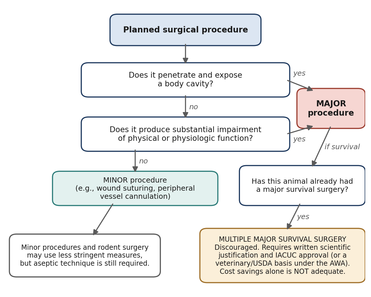
Figure 2.2. Classifying a surgical procedure as major or minor, and identifying when the rules on multiple major survival surgery apply.
2.2.2 Multiple Major Survival Surgery
A survival surgery is one from which the animal is allowed to recover from anesthesia. Performing more than one major survival surgery on the same animal is a significant welfare concern: each procedure adds pain, physiological stress, recovery time, and the risk of complications. For this reason every major document restricts it, although each words its conditions slightly differently.
The AWA allows multiple major survival surgery only if:
It is justified for scientific reasons by the principal investigator, in writing; or
It is required as a routine veterinary procedure or to protect the health or well-being of the animal, as determined by the attending veterinarian; or
A USDA administrator determines that it fulfills appropriate special circumstances.
The Guide discourages multiple major surgeries but permits them when they are scientifically justified and approved by the IACUC, if:
The surgeries are related components of a research project; or
They will conserve animal resources; or
They are needed for clinical reasons.
The Guide is explicit that cost savings alone is NOT an adequate reason. It also requires that the IACUC pay particular attention to the animal's well-being through continuing evaluation of outcomes.
The PHS Policy is similar in tone. AAALAC strongly discourages multiple major survival surgery but permits it if it is scientifically justified and approved by the IACUC, and if the surgeries are related components of a research project and deemed essential.
Table 2.2. Positions on multiple major survival surgery.
| Document | Position | Acceptable bases |
|---|---|---|
| AWA | Allowed only under specific conditions | Written scientific justification by the PI; veterinary care or animal well-being (attending veterinarian); special circumstances (USDA administrator) |
| The Guide | Discouraged; permitted with justification and IACUC approval | Related components of a project; conserving animal resources; clinical need. Cost savings alone is NOT adequate. Continuing evaluation of outcomes. |
| PHS Policy | Similar in tone to the AWA and the Guide | As above |
| AAALAC | Strongly discouraged; permitted with justification and IACUC approval | Related components of a research project and deemed essential |
Why it matters
The "conserve animal resources" justification connects to the 3 Rs. Using one animal for two related procedures may reduce the total number of animals used, but only if the welfare cost to that individual animal is acceptable. The IACUC's job is to weigh those two considerations.
2.2.3 Minor Procedures
The Guide describes a minor procedure as one that does not expose a body cavity and causes little or no physical impairment. Its examples include wound suturing; peripheral vessel cannulation; routine farm animal procedures such as castration, dehorning, and repair of prolapses; and most procedures routinely done on an "outpatient" basis in veterinary clinical practice. The AWA does not define minor procedures.
"Minor" does not mean "unimportant." Minor procedures still require aseptic technique, appropriate anesthesia and analgesia, and post-procedure monitoring. The classification only changes how stringent some of those measures need to be (see Section 2.2.7).
2.2.4 Pain & Distress
Because animals cannot report pain in words, regulators use a human benchmark. The AWA states that any procedure that could reasonably be expected to cause more than slight or momentary pain or distress in a human is to be considered painful. This is defined as pain in excess of that caused by minor procedures such as injections. For such procedures, the AWA:
Requires the IACUC to ensure that pain and distress will be avoided or minimized;
Requires consultation with, or guidance from, a veterinarian concerning pain-relieving drugs; and
Requires that any procedure that would cause more than slight or momentary pain be performed with appropriate analgesics, anesthetics, or tranquilizers.
The PHS's U.S. Government Principles for the Utilization and Care of Vertebrate Animals Used in Testing, Research, and Training echo the AWA on the use of analgesics.
The Guide is less specific than the AWA on this point, but it makes several important statements:
It references Recognition and Alleviation of Pain and Distress in Laboratory Animals (1992), published by the Institute for Laboratory Animal Research (ILAR).
It states that different species express pain and suffering in different ways, so personnel must be familiar with both species-specific and individual expressions of pain.
It specifies that sedatives, anxiolytics, and neuromuscular blocking agents are not analgesics and should not be used as such.
Why it matters
The warning about neuromuscular blocking agents is especially important. These drugs paralyze skeletal muscle, including the muscles an animal would use to show pain, but they provide no pain relief and no unconsciousness. An animal that is paralyzed but not adequately anesthetized can feel pain while appearing perfectly still. Sedatives and anxiolytics can similarly make an animal look calm without addressing its pain.
⚠ Check current guidance
The 1992 ILAR report has been followed by separate National Research Council reports on recognition and alleviation of distress (2008) and of pain (2009). Check which of these appear in the current ASR Recommended References.
2.2.5 Euthanasia
The AWA and the PHS Policy both require that animals that would otherwise suffer chronic or severe pain or distress that cannot be relieved be humanely euthanized at the end of the procedure or, if appropriate, during the procedure. Defined humane endpoints, and a plan to act on them, are part of responsible surgical planning.
⚠ Check current guidance
The PHS Policy requires euthanasia methods consistent with the AVMA Guidelines for the Euthanasia of Animals unless a deviation is justified and approved by the IACUC. Those guidelines are revised periodically; use the current edition.
2.2.6 Pre- & Post-Operative Care
The AWA states that pre- and post-operative care will be provided in accordance with established veterinary medical and nursing procedures. The Guide goes further, recommending a "team concept" because a well-coordinated team often increases the likelihood of a successful surgical outcome. It also recommends continuing and thorough assessment of surgical outcomes, so that problems are identified and procedures improved. Modifying standard techniques is acceptable, as long as the modification does not compromise the animals' well-being.
According to the Guide, pre-surgical planning should include input from all members of the surgical team. Table 2.3 summarizes the elements that planning should cover.
Table 2.3. Elements of pre-surgical planning recommended by the Guide.
| Planning element | Questions the team should answer |
|---|---|
| Responsible personnel | Who will perform surgery, anesthesia, monitoring, and post-operative care? Are they trained? |
| Roles and needs | What will each person do, and what does each need in order to do it? |
| Equipment and supplies | Which instruments, drugs, monitors, and consumables are required, and are they available and sterile? |
| Facilities | Which rooms will be used for preparation, surgery, and recovery? |
| Pre-operative health assessment | How will the animal's fitness for anesthesia and surgery be evaluated? |
| Post-operative care | Who will provide care and analgesia, how often will the animal be checked, and what are the endpoints? Responsibility is shared between the investigator and the veterinarian. |
While recovering from anesthesia, an animal should be kept in a clean, dry area and observed often by trained personnel. During recovery, attention should be paid to body temperature, cardiovascular and respiratory function, and signs of pain and distress. After recovery, animals should be observed for food and water intake and elimination, signs of post-operative pain, signs of infection, and the appearance of the incision(s). The Guide also expects good care and timely removal of staples, sutures, clips, and bandages. Chapter 5 covers peri-operative care in detail.
2.2.7 Aseptic Technique
Asepsis means preventing contamination of the surgical site by microorganisms. Even a small surgical infection causes pain, delays healing, can lead to death, and introduces an inflammatory response that may alter the very measurements a study is trying to make. Table 2.4 compares what the AWA and the Guide require.
Table 2.4. Aseptic technique requirements under the AWA and the Guide.
| Animal Welfare Act | The Guide | |
|---|---|---|
| Applies to | All survival surgery | All major survival surgery |
| Required elements | Surgical gloves Masks Sterile instruments |
Clipping of hair at, and disinfection of, the surgical site Preparation of the surgeon Decontaminated surgical attire Surgical scrub Sterile surgical gloves Sterile instruments, supplies, and implants |
| Surgical technique | — | General asepsis, gentle tissue handling, minimal dissection of tissue, appropriate use of instruments, effective hemostasis, and correct use of suture materials and patterns |
| Minor procedures and rodents | — | May use less stringent measures, but aseptic procedures and instruments are still required |
The Guide's list of surgical technique principles (gentle tissue handling, minimal dissection, hemostasis, and correct suture use) is closely related to Halsted's principles of surgery, which are covered in Chapter 16. They belong in a discussion of asepsis because damaged, devitalized, or bloody tissue is far more vulnerable to infection than healthy, well-perfused tissue. Chapters 3 and 4 cover sterilization and aseptic surgery in detail.
2.2.8 Facilities
The AWA states that major surgical procedures on non-rodent species must be conducted only in facilities intended for that purpose and maintained under aseptic conditions. Minor procedures and rodent surgery do not require a dedicated facility, but they must still be performed using aseptic procedures.
The Guide states that:
Unless an exception is specifically justified as an essential component of the research protocol and approved by the IACUC, aseptic surgery should be conducted in dedicated facilities or spaces.
Generally, agricultural animals maintained for biomedical research should undergo surgery with techniques and in facilities compatible with these guidelines.
For most survival surgery on rodents, aquatic species, and birds, an animal procedure laboratory is recommended, provided it is dedicated to surgery and related activities when used for this purpose and is managed to minimize contamination from other activities.
The Guide describes a surgical facility in terms of designated areas that become progressively cleaner as you approach the operating room (Figure 2.3):
Clean: the operating room (OR), scrub room, and sterile supply rooms.
Mixed (clean/dirty): hallways between ORs, prep rooms, the recovery room, storage rooms, and similar spaces.
Contaminated (dirty): prep rooms, dressing rooms, offices, and animal housing rooms.
Prep rooms can fall into either the mixed or the contaminated category, depending on the design of the facility and how the room is used. These areas are commonly separated by physical barriers, but separation can also be achieved by distance, or by timing activities so that appropriate cleaning and disinfection occur between them. Traffic and the number of personnel should be kept to a minimum, rooms should be designed for ease of cleaning and disinfection and appropriate ventilation, and only required equipment and supplies should be kept in the operating room, with storage minimized.
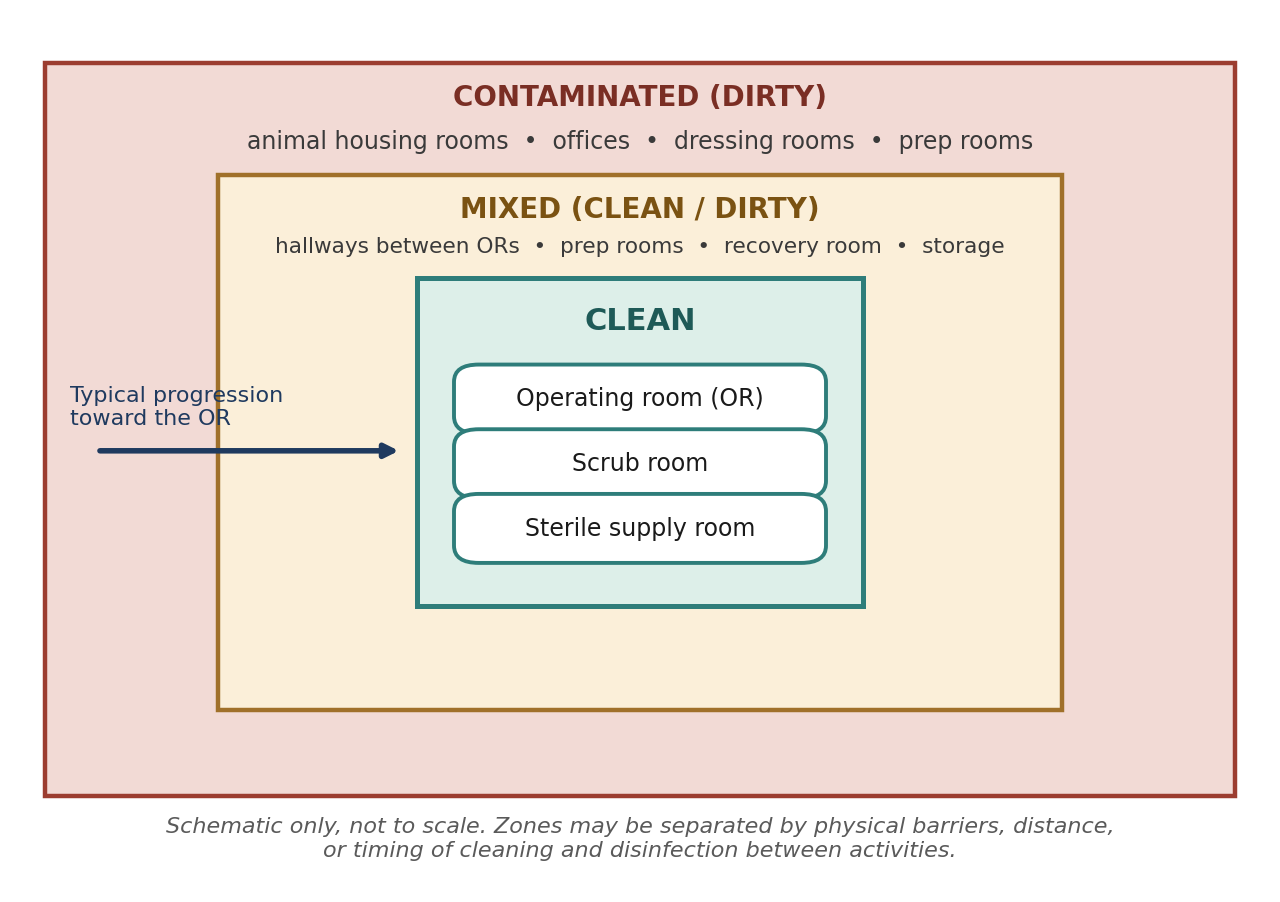
Figure 2.3. Schematic of designated areas in a surgical facility, from contaminated areas at the periphery to clean areas at the core.
Why it matters
Every person, cart, and box that enters the operating room brings microorganisms with it, and every surface is a place where dust and contamination can settle. Zoning, minimal traffic, and minimal storage all reduce the microbial load near the surgical site. Rooms designed for easy cleaning make it practical to keep that load low day after day.
2.2.9 Surgical Records
Records are required by several documents, for different reasons:
The AWA includes record-keeping requirements that support completion of the facility's annual report and the functions of the IACUC.
OLAW also has annual reporting and record-keeping requirements.
The Guide discusses record keeping with regard to colony management, quality control, controlled substances, and medical records. It recommends maintaining a history of surgical procedures and post-operative care, especially for dogs, cats, nonhuman primates, and farm animals. Good records are useful for the continuing assessment of surgical outcomes.
GLP regulations require that, for surgical protocols performed under GLP, documentation must exist showing that all parts of the protocol were followed in compliance.
All of these lead to the same practical rule, already introduced under GLPs: if it is not documented, it did not happen. Accurate anesthesia records, surgical notes, and post-operative observation logs protect the animal, by allowing problems to be recognized and trends noticed; protect the institution, by demonstrating compliance; and protect the science, by making the study reproducible.
Chapter Summary
Research animal surgery in the United States is governed by a layered framework. The Animal Welfare Act (USDA/APHIS) sets mandatory minimum requirements but excludes purpose-bred rats, mice, and birds. The PHS Policy (OLAW) applies to all live vertebrates in PHS-funded work and requires programs based on the Guide (NRC), which uses performance standards interpreted by the IACUC. GLP regulations (FDA and EPA) focus on data integrity through training, SOPs, quality assurance, and records. AAALAC International provides voluntary accreditation against the Guide and related standards. Professional guidance comes from the AVMA/ASLAP, the ACVA, and the ASR.
For surgery specifically, these documents define major and minor procedures, restrict multiple major survival surgery, require pain relief and humane euthanasia, set expectations for peri-operative care and asepsis, describe appropriate facilities, and require thorough records. Knowing which document says what is a frequent subject of exam questions.
Key Terms
3 Rs: Replacement, reduction, and refinement; the guiding principles for the humane use of animals in research.
AAALAC International: Association for the Assessment and Accreditation of Laboratory Animal Care; offers voluntary accreditation, formed in 1965.
Animal Welfare Act (AWA): Federal law passed in 1966, administered by USDA APHIS, regulating the treatment of covered animals in research.
Animal Welfare Regulations: The regulations implementing the AWA, published in Title 9 of the Code of Federal Regulations.
Aseptic technique: Procedures that prevent contamination of the surgical site by microorganisms.
Good Laboratory Practices (GLPs): FDA and EPA regulations governing the conduct of studies submitted to those agencies, emphasizing training, SOPs, quality assurance, and records.
The Guide: The NRC's Guide for the Care and Use of Laboratory Animals (8th ed., 2011).
IACUC: Institutional Animal Care and Use Committee; interprets, implements, oversees, and evaluates an institution's animal care and use program.
Major surgical procedure: A procedure that penetrates and exposes a body cavity or produces substantial impairment of physical or physiologic function.
Minor surgical procedure: A procedure that does not expose a body cavity and causes little or no physical impairment (Guide definition; not defined by the AWA).
Multiple major survival surgery: More than one major surgery from which the same animal is allowed to recover; restricted by all major oversight documents.
OLAW: Office of Laboratory Animal Welfare; implements the PHS Policy.
PHS Policy: Public Health Service Policy on Humane Care and Use of Laboratory Animals; mandated by the Health Research Extension Act of 1985.
Standard operating procedure (SOP): A written, approved procedure describing exactly how a task is performed.
Survival surgery: Surgery from which the animal is allowed to recover from anesthesia.
Review Questions
1. The Animal Welfare Act and Animal Welfare Regulations are administered by which agency?
A. OLAW
B. USDA Animal and Plant Health Inspection Service (APHIS)
C. FDA
D. AAALAC International
2. Which of the following best describes AAALAC International accreditation?
A. It is required by federal law for all research facilities
B. It is voluntary, and standards must be verified by periodic inspections or accreditation is lost
C. It replaces the need for an IACUC
D. It applies only to FDA-regulated studies
3. According to the AWA and the Guide, which procedure is a MAJOR surgical procedure?
A. Peripheral vessel cannulation
B. Wound suturing
C. Laparotomy
D. Subcutaneous injection
4. Under the Guide, which is NOT an acceptable justification for multiple major survival surgery?
A. The surgeries are related components of a research project
B. It will conserve animal resources
C. It is needed for clinical reasons
D. It will reduce the cost of the study
5. Which statement about minor procedures is correct?
A. The AWA defines minor procedures in detail
B. The Guide describes them as procedures that do not expose a body cavity and cause little or no physical impairment
C. Minor procedures never require aseptic technique
D. Castration is always a major procedure
6. Under the AWA, a procedure is considered painful if it would reasonably be expected to cause, in a human:
A. Any sensation at all
B. More than slight or momentary pain or distress
C. Pain lasting more than 24 hours
D. Pain requiring hospitalization
7. The Guide specifies that which class of drugs must NOT be used as analgesics?
A. Opioids
B. Local anesthetics
C. Neuromuscular blocking agents
D. Non-steroidal anti-inflammatory drugs
8. Under the AWA, which items are required for all survival surgery?
A. Surgical gloves, masks, and sterile instruments
B. Gowns, caps, and shoe covers
C. A dedicated operating room and recovery suite
D. A board-certified veterinary surgeon
9. In a surgical suite, which area is classified as CLEAN?
A. Animal housing room
B. Office
C. Scrub room
D. Dressing room
10. The phrase "If it isn't recorded, it didn't happen" is most closely associated with:
A. The 3 Rs
B. Good Laboratory Practices (GLPs)
C. AAALAC site visits
D. The AVMA surgery guidelines
Answer Key
1. B. The AWA is administered by USDA APHIS, and its regulations are published in Title 9 of the Code of Federal Regulations.
2. B. AAALAC accreditation is not a requirement for facility operation. It is voluntary, and accredited institutions must maintain standards verified by periodic inspections.
3. C. A major procedure penetrates and exposes a body cavity or produces substantial impairment of physical or physiologic function. A laparotomy opens the abdominal cavity. The other options are minor procedures or not surgery at all.
4. D. The Guide permits multiple major survival surgery when scientifically justified and IACUC-approved for the first three reasons. Cost savings alone is not an adequate reason.
5. B. The Guide provides the definition and examples, including routine farm animal procedures such as castration. The AWA does not define minor procedures, and aseptic procedures are still required.
6. B. The AWA standard is more than slight or momentary pain or distress in a human, meaning in excess of that caused by minor procedures such as injections.
7. C. Sedatives, anxiolytics, and neuromuscular blocking agents are not analgesics. Neuromuscular blockers paralyze without relieving pain, so they can hide pain rather than treat it.
8. A. The AWA requires aseptic procedures including surgical gloves, masks, and sterile instruments for all survival surgery. The Guide adds further requirements for major survival surgery.
9. C. Clean areas are the operating room, scrub room, and sterile supply rooms. Housing rooms, offices, and dressing rooms are contaminated (dirty) areas.
10. B. GLP regulations mandate documentation showing that every part of a protocol was followed. Undocumented work cannot be shown to have been done.
Chapter 3: Sterilants, Disinfectants & Antiseptics
Learning objectives
After studying this chapter, you should be able to:
Define sanitization, disinfection, antisepsis, asepsis, and sterilization, and explain how they differ.
Compare high-, intermediate-, and low-level disinfectants and their appropriate uses in a surgical facility.
Compare iodophors, chlorhexidine, and alcohol as tissue antiseptics.
Describe the principles, advantages, and limitations of filtration, radiation, thermal, liquid chemical, and gas sterilization.
Prepare, label, and store surgical packs so that sterility is achieved and maintained.
Every surgical infection begins with a microorganism reaching the surgical site. Some arrive on the instruments, some from the surrounding environment, some from the surgeon's hands, and some from the animal's own skin. This chapter describes the chemical and physical tools used to remove or destroy those microorganisms. Chapter 4 then explains how these tools are combined into aseptic surgical practice.
The most important idea in this chapter is that the terms sanitize, disinfect, antiseptic, and sterilize are not interchangeable. Each describes a different degree of microbial kill, on a different kind of surface, using a different kind of agent. Choosing the wrong one, such as using a disinfectant where a sterilant is required, is one of the most common and most serious errors in surgical preparation.
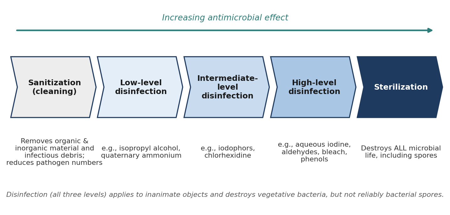
Figure 3.1. The continuum of microbial control, from sanitization to sterilization, with examples of agents at each level of disinfection.
3.1 Sanitization & Disinfection
The surgical environment includes the rooms themselves and everything in them: structures, equipment, lights, tables, and other surfaces. All of it should be sanitized and disinfected according to established daily, weekly, and monthly cleaning protocols. Daily protocols typically address surfaces that are used or contaminated during each surgical day, while weekly and monthly protocols address areas that collect contamination more slowly, such as walls, ceilings, light fixtures, and storage areas. Written schedules make sure no area is overlooked and provide a record that cleaning was done.
3.1.1 Definitions
Sanitization is the removal of organic and inorganic material and infectious debris in order to reduce the number of pathogens. It is fundamentally a cleaning step: scrubbing, washing, and rinsing physically remove blood, tissue, hair, dust, and the microorganisms carried with them.
Disinfection is the inactivation of most pathogenic organisms on inanimate objects and surfaces, except for some highly resistant forms. In practice, disinfection implies the destruction of the vegetative (actively growing) forms of bacteria, but not bacterial spores. A disinfectant may be antimicrobial (killing microorganisms), bacteriostatic (preventing their growth), or both.
Sanitization should come first. Organic material shields microorganisms from contact with a disinfectant, and many disinfectants are partly inactivated by it, so a disinfectant applied to a dirty surface will be far less effective than the same product applied to a clean one.
Microorganisms differ greatly in how well they resist chemical agents. Figure 3.2 shows the generally accepted order of resistance. It explains why a product that easily kills vegetative bacteria and enveloped viruses may have little or no effect on bacterial spores, which can only be reliably destroyed by sterilization.
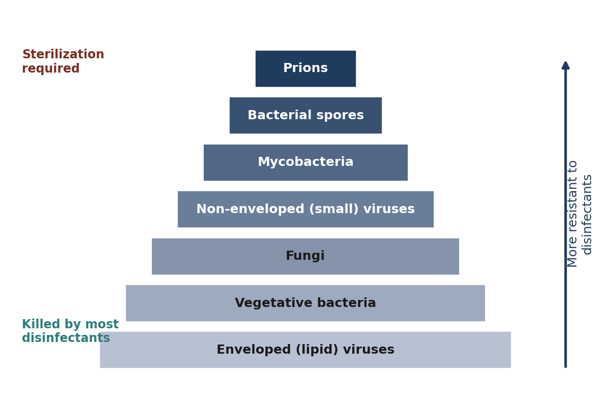
Figure 3.2. General hierarchy of microbial resistance to chemical disinfectants. Organisms near the top require more potent agents or sterilization.
3.1.2 Methods
Disinfection applies to inanimate objects and uses chemicals. Disinfectants are rated as high-, intermediate-, or low-level based on their efficacy against microorganisms. For cleaning surfaces in a surgical area, intermediate-level and some low-level disinfectants are appropriate.
High-level disinfectants are used for disinfecting instruments between procedures, for delicate endoscopic equipment that cannot be steam sterilized, and for cleaning critical surfaces. They include:
Aqueous iodine, which contains high levels of free iodine. It is cytotoxic and stains surfaces, and requires 30 minutes of exposure for disinfection.
Aldehydes, which require 30–45 minutes of exposure for disinfection.
Sodium hypochlorite (bleach), which is toxic and corrosive. Disinfection requires 3,000 ppm for 45–60 minutes.
Phenol compounds (carbolic acid), which require 30 minutes for disinfection but are no longer commonly used because of possible toxicity.
Intermediate-level disinfectants are used for cleaning surfaces. Both of the main examples are also used as tissue antiseptics (Section 3.2):
Iodophors (povidone-iodine) are iodine complexed with surfactants or polymers, which release free iodine slowly. Diluting them lowers cytotoxicity and increases bactericidal activity, because dilution increases the amount of free iodine available. Iodophors are rapidly deactivated in the presence of organic matter, have residual activity of 4–6 hours, and may be mildly irritating.
Chlorhexidine has a rapid onset and long residual activity (8–12 hours), is not deactivated by organic matter, and is non-irritating. It is considered superior to iodophors because of its residual action, which persists even after the solution has dried.
Low-level disinfectants are used for disinfecting instruments between rodent procedures and for cleaning critical surfaces:
Isopropyl alcohol is bactericidal but ineffective against most spores and fungi. It has minimal residual effect, is inhibited by organic debris, and is cytotoxic. It is also a degreaser. Surface decontamination requires 30 minutes of exposure. Alcohol is also used as part of the aseptic surgical preparation of the patient's skin.
Quaternary ammonium compounds ("quats," such as Zephiran) are bactericidal and work by dissolving the outer coatings of some pathogens. Resistant strains of some bacteria, including Staphylococcus aureus and Pseudomonas, are common, and quats are ineffective against spores and some viruses. They may even support the growth of some types of bacteria. According to the CDC, quats are not an appropriate sterilant. They have low toxicity in stable solutions and require 10–30 minutes for surface decontamination.
Table 3.1. Disinfectants by level, with uses and required exposure times.
| Level | Agent | Exposure time | Key properties |
|---|---|---|---|
| High | Aqueous iodine | 30 min | High free iodine; cytotoxic; stains surfaces |
| High | Aldehydes | 30–45 min | — |
| High | Sodium hypochlorite (bleach) | 3,000 ppm for 45–60 min | Toxic; corrosive |
| High | Phenol compounds | 30 min | No longer commonly used (possible toxicity) |
| Intermediate | Iodophors (povidone-iodine) | — | Slow-release iodine; deactivated by organic matter; residual 4–6 h; mildly irritating |
| Intermediate | Chlorhexidine | — | Rapid onset; residual 8–12 h; not deactivated by organic matter; non-irritating |
| Low | Isopropyl alcohol | 30 min | Bactericidal; not effective on most spores/fungi; inhibited by organic debris; degreaser |
| Low | Quaternary ammonium | 10–30 min | Resistant strains common; not sporicidal; not a sterilant (CDC) |
Why it matters
Contact time is part of the method, not an optional extra. A disinfectant that is wiped on and immediately wiped off has not had time to act. If a product requires 10 minutes of wet contact, the surface must stay wet for 10 minutes.
⚠ Check current guidance
Exposure times and concentrations vary between commercial formulations. Always follow the manufacturer's label, which is the legally approved directions for use of a registered disinfectant.
3.2 Tissue Antiseptics
An antiseptic is a substance that inhibits or destroys microorganisms on or in living tissue. Like a disinfectant, it may be antimicrobial, bacteriostatic, or both. Antiseptics are used for preparing the surgical site on the patient and for the surgeon's scrub of the hands and forearms before gowning and gloving. Many intermediate-level disinfectants are also acceptable antiseptics, because they balance effective microbial kill with tolerable effects on tissue.
Two related terms complete the vocabulary. Asepsis is a state of freedom from disease-causing contaminants. Aseptic technique is the set of steps required to prevent contamination of the surgical site with infectious agents. Antiseptics are one tool of aseptic technique; Chapter 4 describes the rest.
Skin cannot be sterilized without destroying it. Microorganisms live not only on the surface but also in hair follicles and sweat glands, where antiseptics cannot reach them. The goal of skin antisepsis is therefore to reduce the microbial population as much as possible at the start of surgery and, ideally, to keep it low for the duration of the procedure. This is why residual activity is so valuable.
The common antiseptics are:
Iodophors are available as tinctures, solutions, and detergents (scrubs). Like chlorhexidine, they work by contact time. They are a principal surgical antiseptic.
Chlorhexidine is considered more effective than the iodophors because of its residual activity. It is available as tinctures, solutions, and detergents. Its antiseptic activity depends on contact time, so it requires sufficient duration of skin contact to be effective. It can be irritating to mucosal surfaces and may induce allergic responses.
Alcohol is useful for low-level antisepsis. It evaporates rapidly and leaves no residue, has a minimal residual effect, and is inhibited by organic debris. Standard human and veterinary surgical texts consider it not sufficient as the primary antiseptic. It is useful in conjunction with iodophors or chlorhexidine, because it breaks up surface oils and surface tension.
Table 3.2. Comparison of common tissue antiseptics.
| Property | Iodophors | Chlorhexidine | Alcohol |
|---|---|---|---|
| Forms available | Tinctures, solutions, detergents | Tinctures, solutions, detergents | Solution |
| Mode of action | Contact time | Contact time | Rapid; evaporates |
| Residual activity | 4–6 hours | 8–12 hours, even when dried | Minimal |
| Effect of organic matter | Rapidly deactivated | Not deactivated | Inhibited |
| Cautions | May be mildly irritating | Irritating to mucosa; possible allergic responses | Cytotoxic; not sufficient as primary antiseptic |
| Role | Principal antiseptic | Principal antiseptic; considered more effective | Adjunct; degreases skin and breaks surface tension |
Why it matters
Alternating a scrub of iodophor or chlorhexidine with alcohol is common in surgical skin preparation. The alcohol removes skin oils and helps the antiseptic contact the skin, but it is the iodophor or chlorhexidine that provides lasting antimicrobial activity. Chapter 4 describes the full preparation procedure.
3.3 Methods of Sterilization
Sterilization is the physical or chemical destruction of all microbial life, including transmissible agents. Unlike disinfection, it is an absolute standard: an item is either sterile or it is not. Everything that enters a body cavity, the bloodstream, or a surgical wound (instruments, implants, catheters, sutures, and fluids) must be sterile. Table 3.4 at the end of this section compares the methods.
3.3.1 Filtration
Filtration can be used for gases or liquids. It works by separating out particulate matter of known sizes using a membrane, so that microorganisms larger than the membrane's pores are physically held back. It is commonly used for pharmaceutical liquids. Filtration is useful for heat-sensitive media, offers high throughput, and provides absolute sterilization of the filtered material. Its limitation is that it cannot differentiate between particles of similar size: anything smaller than the pores passes through, and anything larger is retained, regardless of what it is.
3.3.2 Radiation
Radiation sterilization usually uses gamma radiation from Cobalt 60. It penetrates relatively impervious materials such as metal and plastic, so items can be sterilized in their final sealed packaging, and it leaves no chemical residue. However, radiation can start a structural change in materials, especially some polymers, and that change may continue to develop over months, making some plastics brittle or discolored. Because the process is expensive, it is usually used by manufacturers of single-use sterile supplies rather than by research facilities.
3.3.3 Thermal
Moist heat in the form of saturated steam under pressure (autoclave steam sterilization) is the most widely used and most dependable sterilization process. Thermal methods rely on high temperature to denature bacterial proteins, and sterilization is a function of time and temperature: the higher the temperature, the shorter the time required.
There are two types of thermal sterilization:
Dry heat, such as a hot bead sterilizer or an oven, is used for moisture-sensitive materials such as oils, powders, and petroleum products. Hot bead sterilizers are useful for sterilizing surgical instruments during multiple rodent surgeries. Only the part of the instrument buried in the heated beads is sterilized, and the instrument must be allowed to cool before it touches tissue.
[ PHOTO PLACEHOLDER ] Suggested source: Wikimedia Commons (commons.wikimedia.org), search "glass bead sterilizer." Check the image's license and give the attribution it requires. |
|---|
Figure 3.3. A hot bead (glass bead) sterilizer used for re-sterilizing instrument tips between rodent surgeries.
Wet heat, which includes boiling water and the autoclave, kills bacteria through the coagulation of critical proteins. Moist heat transfers energy far more efficiently than dry heat, which is why it is faster at a given temperature.
Boiling water is a poor sterilant at ambient pressure because its temperature is relatively low. Its effect may be enhanced by adding sodium hydroxide or sodium carbonate. Boiling is destructive to instruments, especially glassware and rubber.
The autoclave uses steam under pressure, which allows temperatures higher than ordinary steam can provide.
Successful autoclaving depends on steam reaching every surface of every item. Materials and wrappings must not be damaged by heat, moisture, and pressure, but must be permeable to steam. The surgical pack must be completely saturated with steam for effective sterilization, for example 121 °C for 13–15 minutes (5–10 minutes of kill time plus 3–8 minutes as a safety margin). Larger or denser packs require more time for adequate steam saturation.
The time it takes the autoclave to reach temperature and saturate the pack is called the heat-up time; pre-vacuum autoclaves achieve it in about 12 minutes by actively removing air before steam is admitted. Pre-vacuum autoclaves can be used for emergency "flash" autoclaving, and because they also reach higher temperatures they require shorter exposure times (131 °C for 3 minutes). After a cycle, loads should be vented for about 10 minutes to prevent condensation, since wet packs can draw microorganisms through the wrap (see Section 3.4). Figure 3.4 shows the phases of a cycle.
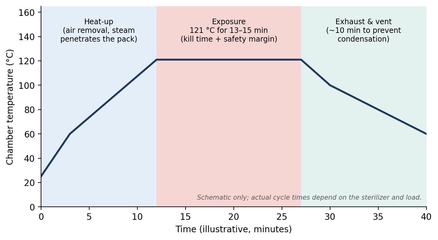
Figure 3.4. The phases of a steam autoclave cycle: heat-up, exposure at sterilizing temperature, and exhaust/venting.
Why it matters
Air is the enemy of steam sterilization. Pockets of trapped air insulate items from steam, so a pack can sit in a chamber at 121 °C while its center never reaches that temperature. Dense packs, tightly closed instruments, and overloaded chambers all trap air, which is why pack preparation (Section 3.4) is as important as the cycle settings.
⚠ Check current guidance
Autoclave cycle parameters, including flash (immediate-use) sterilization times and temperatures, depend on the sterilizer, the load, and the packaging. Use the sterilizer manufacturer's validated cycles and current professional standards (for example, AAMI guidance) rather than a single set of numbers.
3.3.4 Liquid Chemical
When using chemicals for sterilization, it is critical to distinguish sterilants from disinfectants. Sterilants are designed to kill all microorganisms; disinfectants are not. Disinfectants are NOT adequate for sterilizing instruments, implants, or catheters.
Chemical sterilants are regulated by the FDA and the EPA as medical devices. Approved chemical sterilants say on the bottle that they are sterilants. Products labeled only as disinfectants should not be used for instrument or device sterilization. Reading the label is therefore a basic safety step.
Cold sterilants work by contact, so every surface must be in contact with the solution. Flush the sterilant through catheters and other lumens, open all latches and hinges, and make sure no air bubbles are trapped. Cold sterilants are also tissue irritants, so sterilized items must be completely rinsed with sterile water or saline to remove all chemicals before they contact tissue. The common cold sterilants are compared in Table 3.3.
Aldehydes (glutaraldehyde) require more than 12 hours of full immersion to sterilize resistant spores. Glutaraldehyde is noncorrosive, but activated solutions have a shelf life of less than 2 weeks. It is toxic to the skin, eyes, and respiratory tract. Glutaraldehyde solutions come in different strengths, and only those classified as sterilants should be used for instrument or device sterilization.
Formalin is a 37% aqueous solution of formaldehyde. It requires more than 24 hours of full immersion to sterilize resistant spores and is toxic to the skin, eyes, and respiratory tract.
Hydrogen peroxide as a 6% aqueous solution, with more than 30 minutes of exposure, may kill some resistant spores in addition to less hardy microorganisms. It is potentially explosive at high concentrations, corrosive, and irritating to the skin and eyes.
Peracetic acid (35%) was registered by the EPA as an antimicrobial in 1985 for indoor use on hard surfaces.
Table 3.3. Cold (liquid chemical) sterilants.
| Agent | Immersion time for resistant spores | Notes and hazards |
|---|---|---|
| Glutaraldehyde | > 12 hours | Noncorrosive; activated solution shelf life < 2 weeks; toxic to skin, eyes, and respiratory tract; use only products labeled as sterilants |
| Formalin (37% formaldehyde) | > 24 hours | Toxic to skin, eyes, and respiratory tract |
| Hydrogen peroxide (6%) | > 30 minutes (may kill some resistant spores) | Potentially explosive at high concentration; corrosive; skin and eye irritant |
| Peracetic acid (35%) | — | EPA-registered antimicrobial (1985) for indoor hard surfaces |
⚠ Check current guidance
Personnel exposure to glutaraldehyde and formaldehyde is regulated, and many facilities now restrict their use. Check your institution's occupational health requirements and the current product labels for exposure times and shelf life.
3.3.5 Gas Sterilization (Chemical)
Gas sterilization requires an enclosed chamber, similar to an autoclave. It is valuable for heat- and moisture-sensitive items that cannot be autoclaved.
Ethylene oxide (ETO) is a colorless gas that destroys microbial metabolic pathways by alkylation. It is flammable, explosive, toxic, and irritating to skin and mucous membranes. Older manual systems require 12 hours of exposure, and exposure time is inversely proportional to pressure. Newer automated systems (such as those made by 3M) use heat and moisture to decrease both the exposure and the aeration times. Aeration is needed after ETO sterilization because the gas is absorbed into plastics and rubber and must be allowed to dissipate before the item is safe to handle or implant.
Hydrogen peroxide gas is safer than ETO for the user, requires little ventilation, and has shorter exposure times. However, it cannot be used with absorbent materials such as paper or cloth (cellulose-based materials). It can also condense into water and soak the packaging, which prevents gas contact and allows microorganisms to "wick" through the wet packaging after sterilization. STERIS stopped supporting its use in a research environment around 2012.
⚠ Check current guidance
Gas sterilization systems, their manufacturer support, and their regulatory status change over time, and ETO in particular is subject to evolving environmental and workplace exposure rules. Check the current status of any system your facility uses.
Table 3.4. Comparison of sterilization methods.
| Method | Best used for | Advantages | Limitations |
|---|---|---|---|
| Filtration | Pharmaceutical liquids; gases | Suits heat-sensitive media; high throughput; absolute sterilization | Cannot distinguish similarly sized particles |
| Radiation (Cobalt 60) | Manufactured single-use supplies | Penetrates metal and plastic; no residue | Can alter polymers over months; costly |
| Dry heat | Oils, powders, petroleum products; instrument tips between rodent surgeries | Suitable for moisture-sensitive materials | Only the heated portion is sterilized; must cool before use |
| Boiling water | Not recommended | Simple | Poor sterilant; low temperature; damages glass and rubber |
| Steam autoclave | Most instruments, packs, and heat-stable items | Most widely used and most dependable | Requires heat-, moisture-, and pressure-tolerant materials; steam must penetrate |
| Liquid chemical | Heat-sensitive items | No heat required | Long immersion; full contact required; toxic; must rinse with sterile fluid |
| Ethylene oxide gas | Heat- and moisture-sensitive items | Penetrates packaging | Flammable, explosive, toxic; long exposure and aeration |
| Hydrogen peroxide gas | Heat-sensitive items in non-cellulose packaging | Safer for users; short cycle; little ventilation | No paper or cloth; condensation can cause wicking |
3.4 Sterilization of Surgical Supplies
Sterilization can only be as good as the preparation that precedes it. All instruments must be thoroughly cleaned before sterilization. Organic debris dramatically extends the time required. For example, glutaraldehyde sterilization time increases from 12 hours to 48 hours in the presence of organic debris. Debris also physically shields the microorganisms beneath it.
For most forms of sterilization, instruments should be dry. This is less important for steam autoclaving, and some residual moisture is actually important for ETO sterilization, which depends on humidity to work effectively.
Packs should be assembled so that the sterilizing agent can reach every surface and so that sterility can be verified (Figure 3.5):
Instruments should be in the open position, so that steam or gas can reach hinges and clasps.
Items should be placed so that they cannot accidentally puncture the wraps or sleeves, and protectors should be placed over sharp tips.
Multiple layers of sterilization indicators should be used to verify proper sterilization. Sterilization sleeves have indicators printed on them. Packs should have indicators inside the inner wrap and under or inside any containers or large objects, which are the places steam or gas reaches last.
Packs and sleeves should be dated on the day sterilization is performed.
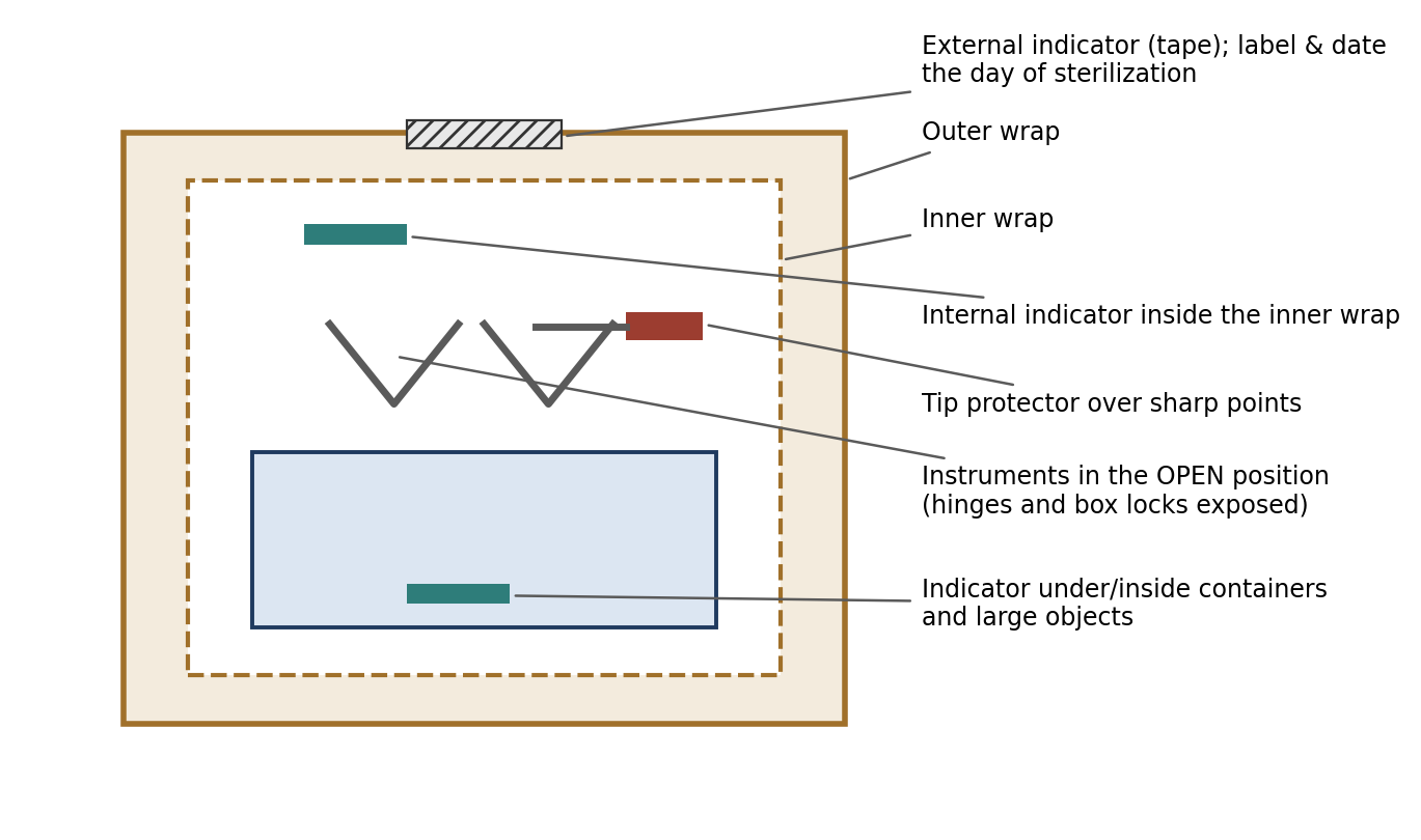
Figure 3.5. Preparing a double-wrapped instrument pack: open instruments, protected tips, and indicators placed where the sterilant reaches last.
[ PHOTO PLACEHOLDER ] Suggested source: Wikimedia Commons (commons.wikimedia.org), search "autoclave tape" or "sterilization indicator." Check the image's license and give the attribution it requires. |
|---|
Figure 3.6. Chemical sterilization indicators (autoclave tape or indicator strips) before and after a steam cycle, showing the color change.
Why it matters
An external indicator shows only that a pack was exposed to the sterilization process, not that conditions inside the pack were adequate. That is why indicators are also placed at the pack's most protected points. If the indicator in the center of the pack has changed, the rest of the pack was exposed too.
Once sterilized, packs must be stored so that they stay sterile:
Paper/plastic-wrapped items and packs are considered sterile for 12 months when stored in closed cabinets.
Cloth-wrapped items are considered sterile for six months when stored in closed cabinets.
Packs and sleeves that get wet or are damaged (for example, punctured) should be considered contaminated and non-sterile.
If there is any doubt about sterility, consider the item contaminated and non-sterile.
Table 3.5. Storage life of sterilized packs in closed cabinets.
| Wrap type | Considered sterile for | Becomes non-sterile if |
|---|---|---|
| Paper/plastic | 12 months | Wet, damaged, punctured, or of doubtful sterility |
| Cloth | Six months | Wet, damaged, punctured, or of doubtful sterility |
⚠ Check current guidance
Many facilities now use "event-related" sterility, in which a pack remains sterile until an event (such as moisture, damage, or a broken seal) compromises it, rather than a fixed expiration time. Follow your institution's SOP and current professional standards for pack shelf life.
Chapter Summary
Sanitization removes debris and reduces pathogen numbers; disinfection inactivates most pathogens on inanimate objects but not reliably spores; antisepsis reduces microorganisms on living tissue; and sterilization destroys all microbial life. Disinfectants are graded high, intermediate, or low, and each requires a specific contact time. Chlorhexidine and iodophors are the principal surgical antiseptics, with chlorhexidine favored for its residual activity and resistance to organic matter; alcohol is an adjunct, not a primary antiseptic.
Steam autoclaving is the most widely used and dependable sterilization method; filtration, radiation, dry heat, liquid chemical sterilants, and gas sterilization each serve specific needs. Only products labeled as sterilants may be used to sterilize instruments and devices. Instruments must be cleaned, packed open with protected tips, monitored with indicators placed in the pack's most protected locations, dated, and stored so that they stay dry and intact. When in doubt, an item is not sterile.
Key Terms
Antiseptic: A substance that inhibits or destroys microorganisms on or in living tissue.
Asepsis: A state of freedom from disease-causing contaminants.
Aseptic technique: The steps required to prevent contamination of the surgical site with infectious agents.
Autoclave: A chamber that sterilizes using saturated steam under pressure.
Bacteriostatic: Preventing the growth of bacteria without necessarily killing them.
Cold sterilant: A liquid chemical sterilant used at room temperature by full immersion.
Disinfection: Inactivation of most pathogenic organisms on inanimate objects, including vegetative bacteria but not spores.
Heat-up time: The time for an autoclave to reach temperature and saturate the pack with steam.
Iodophor: Iodine complexed with a surfactant or polymer that releases free iodine slowly (e.g., povidone-iodine).
Residual activity: Continued antimicrobial action of an agent after it has been applied.
Sanitization: Removal of organic and inorganic material and infectious debris to reduce pathogen numbers.
Sterilant: A product designed to kill all microorganisms; must be labeled as a sterilant.
Sterilization: The physical or chemical destruction of all microbial life, including transmissible agents.
Sterilization indicator: A device that changes appearance when exposed to sterilizing conditions, used to verify processing.
Vegetative bacteria: Bacteria in their actively growing (non-spore) form.
Wicking: Movement of microorganisms through wet packaging, compromising sterility.
Review Questions
1. Which process destroys ALL microbial life, including bacterial spores?
A. Sanitization
B. High-level disinfection
C. Sterilization
D. Antisepsis
2. An antiseptic differs from a disinfectant in that an antiseptic is:
A. Always more potent
B. Used on or in living tissue
C. Used only on instruments
D. Able to kill prions
3. Which antiseptic is considered superior to iodophors because of its residual activity and because it is not deactivated by organic matter?
A. Isopropyl alcohol
B. Chlorhexidine
C. Quaternary ammonium
D. Aqueous iodine
4. Why is alcohol NOT sufficient as the primary surgical skin antiseptic?
A. It stains the skin
B. It has minimal residual effect and is inhibited by organic debris
C. It is too slow to act
D. It is a sterilant
5. What is the most widely used and most dependable method of sterilization?
A. Ethylene oxide gas
B. Gamma radiation
C. Saturated steam under pressure (autoclave)
D. Boiling water
6. Which statement about boiling water is correct?
A. It is an effective sterilant at ambient pressure
B. It is a poor sterilant because its temperature is relatively low
C. It is gentle on glassware and rubber
D. It is the method of choice for implants
7. After an instrument is sterilized with a cold (liquid chemical) sterilant, it must be:
A. Air-dried without rinsing
B. Rinsed thoroughly with sterile water or saline before tissue contact
C. Rinsed with tap water
D. Wiped with alcohol only
8. A bottle labeled "disinfectant" should be used for:
A. Sterilizing catheters
B. Sterilizing implants
C. Surface disinfection, but not instrument or device sterilization
D. Any purpose, since disinfectants and sterilants are equivalent
9. Which material should NOT be processed with hydrogen peroxide gas sterilization?
A. Stainless steel instruments
B. Cloth or paper (cellulose-based) wraps
C. Plastic trays
D. Non-absorbent synthetic wraps
10. Under the storage guidance in this chapter, a cloth-wrapped pack stored in a closed cabinet is considered sterile for:
A. 30 days
B. Six months
C. 12 months
D. Indefinitely
Answer Key
1. C. Sterilization is the physical or chemical destruction of all microbial life. Disinfection, even at the high level, does not reliably destroy spores.
2. B. An antiseptic inhibits or destroys microorganisms on or in living tissue. Disinfectants are used on inanimate objects and surfaces.
3. B. Chlorhexidine has a rapid onset, residual activity of 8–12 hours (even when dried), and is not deactivated by organic matter, whereas iodophors are rapidly deactivated by organic matter.
4. B. Alcohol evaporates rapidly, has minimal residual effect, and is inhibited by organic debris. It is useful alongside an iodophor or chlorhexidine, not as the primary antiseptic.
5. C. Moist heat in the form of saturated steam under pressure is the most widely used and most dependable sterilization process.
6. B. At ambient pressure, boiling water has a relatively low temperature and is a poor sterilant. It is also destructive to instruments, especially glassware and rubber.
7. B. Cold sterilants are tissue irritants, so all chemical residue must be removed with sterile water or saline before the item contacts tissue.
8. C. Only products labeled as sterilants are designed to kill all microorganisms. Disinfectants are not adequate for instrument, implant, or catheter sterilization.
9. B. Hydrogen peroxide gas cannot be used with absorbent, cellulose-based materials such as paper or cloth.
10. B. Cloth-wrapped items are considered sterile for six months in closed cabinets; paper/plastic-wrapped items for 12 months. Any pack that becomes wet or damaged is non-sterile.
Chapter 4: Principles of Aseptic Surgery
Learning objectives
After studying this chapter, you should be able to:
Explain the goal of surgical asepsis and how bacteria cause surgical wound infection.
Describe the steps of aseptic patient preparation, from clipping through final skin antisepsis.
Identify surgical positions and the methods used to drape the patient and maintain a sterile field.
Perform, in the correct sequence, the surgeon scrub, aseptic gowning, and closed and open gloving.
Recognize breaks in aseptic technique, respond appropriately, and follow standard operating room conduct.
Surgical asepsis is not a single procedure but a series of best practices, each of which uses aseptic technique to prevent or minimize contamination of the surgical site with infectious agents. Because skin, air, and people can never be made completely sterile, the realistic goal is to lower the concentration of microorganisms below the level required for infection. Each practice in this chapter removes or blocks one source of contamination; together, they keep the total microbial load at the surgical site low enough that the animal's own defenses can handle what remains.
Asepsis begins before anyone enters the operating room. General attire for OR entry includes a cap, a mask, clean scrubs, booties (shoe covers), and gloves. Section 4.6 describes surgical attire in more detail.
4.1 Antimicrobial Therapy & Wound Infection
Understanding how surgical infections develop explains why each aseptic step matters. The major bacteria involved in surgical wound infections are listed in Table 4.1. Many of them are normal inhabitants of skin, the environment, or the gastrointestinal tract, which is why the patient's skin, the surgeon's hands, and the surrounding environment are the main sources of contamination.
Table 4.1. Major bacteria involved in surgical wound infections.
| Organism | Notes |
|---|---|
| Staphylococcus aureus | Common skin organism; resistant strains are common (see Chapter 3, quaternary ammonium compounds). |
| Coagulase-negative staphylococci | Common skin organisms. |
| Streptococcus pneumoniae | Gram-positive coccus. |
| Enterococcus spp. | Intestinal organisms. |
| Escherichia coli | Intestinal organism. |
| Pseudomonas multivorans | Now classified as Burkholderia multivorans. |
In general, bacteria do not damage tissue cells directly. Instead, their waste products, such as endotoxins and cytotoxins, damage and kill cells. The body provides bacteria with everything they need to multiply: food, shelter, humidity, and warmth. Bacteria generally remain localized until they gain entry to the bloodstream, at which point a local infection becomes a systemic one.
The body fights extracellular bacteria and other organisms through an antibody-mediated immune response. Antibodies recognize foreign material and call in phagocytes, which engulf and consume it, many of them dying in the process. This immune response produces the familiar signs of infection: increased temperature, the production of pus (largely dead phagocytes, bacteria, and tissue debris), and an influx of serous fluid.
Surgical infections prolong wound healing because of damage from bacterial toxins, physical debris in the wound, and changes in local circulation. An infected wound may break down, heal slowly, or require further treatment, and the inflammation and fever it causes may affect research data.
Antimicrobial Agents
Antibacterial therapy is species- and target-tissue-specific: a drug that is effective and safe in one species or tissue may not be in another. Antibacterial treatment can be prophylactic, a preventative strategy used before contamination becomes infection, or therapeutic, a healing strategy used to treat an established infection.
Antimicrobial agents are described by their effect:
Bactericidal: a substance that kills bacteria.
Bacteriostatic: a biological or chemical agent that stops bacteria from reproducing, while not necessarily killing them. The immune system must then clear the bacteria.
Other antimicrobial agents include antivirals, antifungals, and antiprotozoals.
Why it matters
Antibiotics are not a substitute for asepsis. Good aseptic technique prevents contamination in the first place; antibiotics, used after consultation with a veterinarian, are a backup when contamination is known or suspected (Section 4.6.6). Routine reliance on antibiotics can also introduce a variable into the study and encourage resistant bacteria.
4.2 Aseptic Patient Preparation
The patient's own skin and hair are a major source of contamination. Patient preparation removes as much of that contamination as possible in stages, moving from coarse cleaning in the preparation area to final antisepsis in the operating room (Figure 4.1).
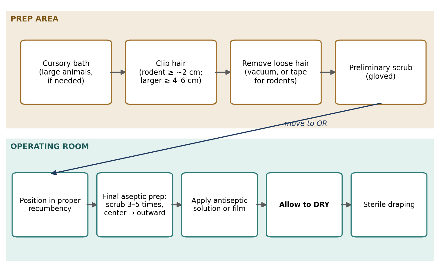
Figure 4.1. The sequence of aseptic patient preparation, from the preparation area to the operating room.
4.2.1 Cursory Animal Bathing
Cursory cleansing is typically done for larger animals, such as livestock, if needed. Some animals, especially livestock, carry large amounts of contaminants (feces, dirt, and bedding material) on their fur or skin, and these can be removed before surgery by cursory bathing or washing. Be aware that getting the animal wet may reduce its body temperature, especially if it is already anesthetized, because anesthesia impairs the body's ability to regulate temperature.
4.2.2 Preliminary Surgical Site Preparation
Removing hair. Hair should be clipped close to the skin around any potential incision sites to create a working surgical field with wide margins. The size of the clipped area depends on the procedure and is species-specific, but as a minimum:
In rodents, approximately 2 cm from the incision site.
In larger animals, 4–6 cm from the incision site.
Wide margins allow the incision to be extended if necessary and keep hair from falling into the surgical field. Shaving with a blade is not generally recommended, because the resulting skin trauma may lead to increased bacterial counts. For the same reason, clipping hair the day before surgery is associated with higher bacterial counts and should be avoided: the tiny skin injuries caused by clipping give bacteria time to multiply.
All loose hair should be removed from the animal and the preparation site with a vacuum. In rodents, sticky tape pressed onto the clipped hair may be used instead.
Preliminary scrub. Gloves should be worn for the following steps to minimize contamination from the technician's hands. A preliminary scrub reduces the contamination load before the formal aseptic preparation begins.
Aseptic surgical preparation of the skin. A typical scrub consists of applying chlorhexidine or povidone-iodine scrub with soaked gauze sponges, followed by a wipe with gauze sponges soaked in 70% isopropyl alcohol or sterile water. Key points of technique are:
With oily or dirty skin, an initial wipe with 70% isopropyl alcohol may be performed to remove oils and reduce surface tension.
The evaporation of alcohol can cause a significant loss of body heat, particularly in rodents and neonates, which have a large surface area relative to their body mass. This can be reduced by warming the containers of prep solution in a 100–105 °F water bath. Alcohol-soaked sponges should be damp but not dripping wet, and warm sterile water or saline may be used instead of alcohol.
The scrub should be performed 3–5 times.
Each scrub begins over the incision site and follows a concentric circle away from it (Figure 4.2). Never return to the incision site or a previously cleaned area with the same gauze sponge once you reach the outermost area.
When preparing a limb, it may be useful to tape the foot to an IV stand and suspend it for prepping.
When the final scrub has dried, apply povidone-iodine or chlorhexidine solution to the surgical site.
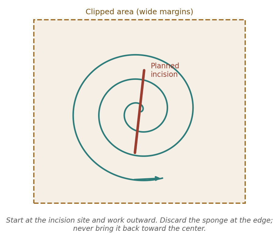
Figure 4.2. The concentric scrub pattern. Each pass moves contaminants away from the planned incision.
Normally, a final surgical site preparation is performed after the animal has been moved to the operating room and placed in proper recumbency, because moving and positioning the animal can contaminate a site that was prepared earlier. A povidone-iodine or chlorhexidine film antiseptic system may then also be applied to the skin, for example 3M DuraPrep (0.7% iodine povacrylex and 74% isopropyl alcohol) or ChloraPrep (2% chlorhexidine gluconate in 70% isopropyl alcohol).
Finally, allow the antiseptic solution to dry before sterile draping. Antiseptics need contact time to work, wet skin can wick contamination through drapes, and adhesive drapes will not stick to wet skin.
Why it matters
The concentric pattern and the "never go back" rule exist because a sponge picks up microorganisms as it moves. Once it has touched the less clean periphery, bringing it back to the center would deposit those organisms right where the incision will be made.
⚠ Check current guidance
Recommendations on the number of scrub cycles, the use of alcohol versus saline between scrubs, and the choice of antiseptic continue to be studied. Check current veterinary surgical references and your institution's SOP.
4.3 Surgical Mindset
Aseptic technique depends on honesty and vigilance. Contamination is usually invisible, so the only safe approach is to act on suspicion rather than wait for proof. The guiding rule is simple: if in doubt, assume contamination and correct it.
If there is a question whether a step was performed incorrectly, assume that it was and start over.
If a prepared surgical site may have become contaminated, redo the entire prep.
If a drape in the operating field may have been contaminated by contact with non-sterile material, replace it or, if that is impractical, cover the area with a new sterile drape.
If an instrument may be contaminated, remove it from further use and request a sterile replacement.
If a sterile glove may have been perforated or contaminated, discard it and aseptically put on a new sterile glove or gloves.
Developing this mindset means being willing to speak up about your own errors and those you see others make. The cost of restarting a step is a few minutes; the cost of an infection can be the animal's welfare, its value to the study, and sometimes its life.
4.4 Surgical Positioning
The surgeon should direct the positioning of the animal on the table. Surgeons often have specific positioning requirements that allow them to see the surgical site better, and these vary from surgeon to surgeon.
Positions are named for the part of the body in contact with the table (Figure 4.3):
Right lateral recumbency: lying on its right side (left lateral recumbency is the reverse).
Dorsal recumbency: lying on its back.
Sternal or ventral recumbency: lying on its sternum.
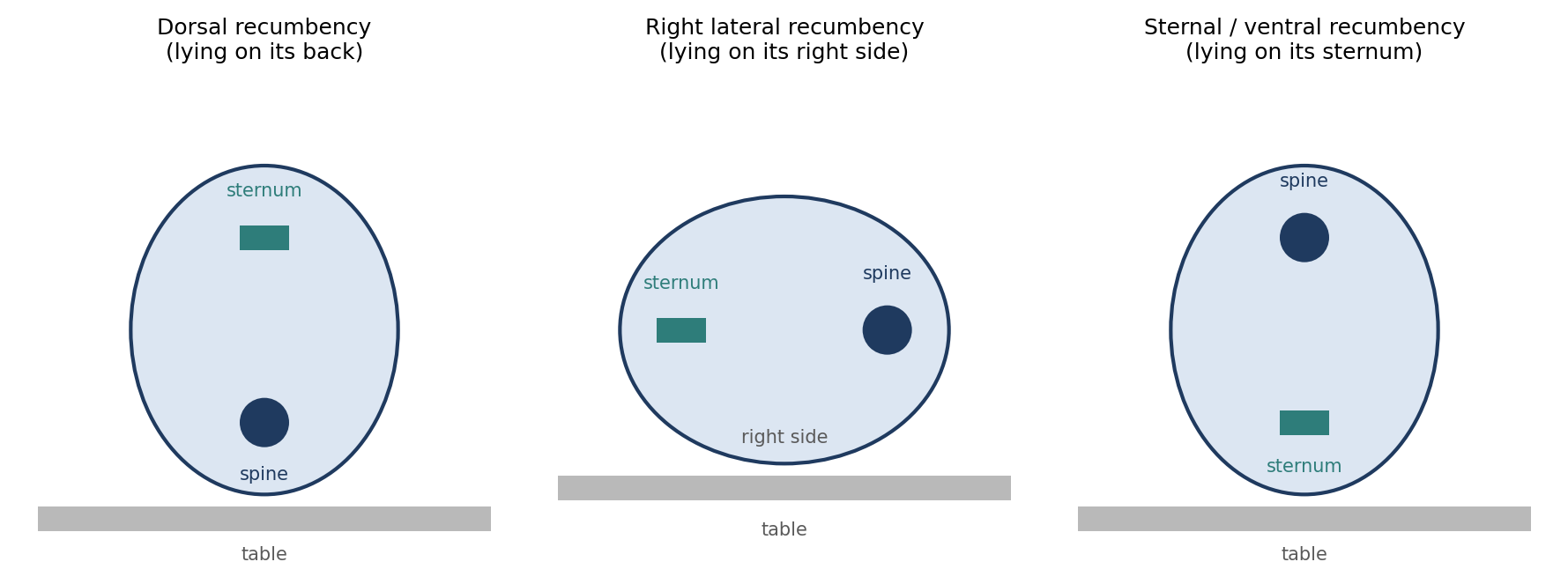
Figure 4.3. Common surgical positions, shown as cross-sections of the trunk. Each is named for the surface in contact with the table.
The animal may be held in place with sandbags, troughs, rolled towels, "beanbags," limb ties, or incisor clips (for rodents). Take care that limb ties do not occlude normal blood flow; an overtightened tie can cause swelling, nerve damage, or tissue injury during a long procedure.
Limb Preparation and Positioning
Before prepping, wrap the foot in VetWrap or a similar bandage material.
When the animal is brought into the OR, suspend the limb from an IV pole and then prepare it.
Cut the suspending tape close to the limb, place a sterile stockinette on the foot, and unroll it toward the body.
Pass the wrapped limb through a hole in the sterile drape if needed.
Wrap the limb in an adhesive incise drape such as Steri-Drape.
Stereotaxic Positioning
For procedures where skull stability is important, the animal may be placed in a stereotaxic frame. The nose clamp and the ear or jaw bars must not be so tight that they occlude the nares or cause trauma to the eardrums, jaw, or skin.
Maintaining sterility with a stereotaxic frame is difficult. The horizontal slides must remain accessible for moving the manipulator arms, yet they are also handled while the animal is being positioned, before the sterile field has been created. Wrapping the frame in stockinette before sterilizing it helps: holes are cut to allow insertion of the ear bars, and once the animal is positioned, the stockinette is cut off and the animal is draped.
[ PHOTO PLACEHOLDER ] Suggested source: Wikimedia Commons (commons.wikimedia.org), search "stereotaxic frame" or "stereotactic apparatus rat." Check the image's license and give the attribution it requires. |
|---|
Figure 4.4. A rodent stereotaxic frame, showing the ear bars, nose clamp, and manipulator arm.
4.5 Aseptic Draping & Sterile Field Maintenance
Once the animal is properly positioned and aseptically prepared, a sterile field is created by placing sterile drapes to isolate the sterile "working" field. In general, the sterile field should be large enough to prevent accidental contamination of the incision site(s), the operating team, and the sterile instruments and equipment.
Sterile drapes can make it difficult to physically monitor respiratory and cardiovascular function during surgery, because they cover the chest, mucous membranes, and limbs. This is why electronic monitoring should be used as a safety net whenever possible (Chapter 12).
In some cases, especially with larger animals, a Mayo stand or overhead table positioned above the animal can isolate the head and neck. Draped, it gives surgeons a sterile surface for instrument trays while leaving the anesthetist room to see and reach the head and neck, without interfering with surgical asepsis.
Table 4.2. Draping recommendations for rodents and non-rodents.
| Rodent | Non-rodent | |
|---|---|---|
| Minimum | A sterile fenestrated drape whose opening reveals only the surgical site | A sterile fenestrated drape sufficient to cover the animal and surgical field |
| Coverage | Commonly covers the entire animal | Cloth or paper drapes covering all areas except those immediately adjacent to the incision site(s) |
| Options | Clear plastic adhesive surgical drapes; a table drape placed before positioning to enlarge the field | Plastic adhesive incise drape (e.g., 3M Steri-Drape) over the fenestration; sterile bandaging patches (e.g., Tegaderm), which are more conveniently sized |
| Securing | — | Towel clamps or sterile surgical staples |
| Equipment | — | Drape or sleeve non-sterilizable equipment in the field (drill cables, fluoroscopic C-arms, imaging equipment); sterilize light handles or provide sterile covers |
An adhesive incise drape extends the sterile field right to the edge of the incision, covering the small strip of prepared (but not sterile) skin between the drape opening and the wound. Sterile light handles allow the surgeon to adjust the operating lights without breaking asepsis.
4.6 Surgical Personnel Attire & Preparation
4.6.1 Attire & Operating Personnel Preparation
People are a major source of contamination in the operating room. Skin constantly sheds microscopic flakes carrying bacteria, hair sheds, and breath carries droplets. Surgical attire contains these sources.
Street clothes, especially shoes, are a major source of contaminants. All personnel working in the OR should replace street clothing with clean scrubs, and scrub tops should be tucked into the pants to reduce dispersion of skin debris.
During hair clipping and prep, a clean lab coat or Tyvek coverall can be worn to protect the scrubs. This is especially important when working with rabbits. Alternatively, a lint roller can be used to remove fur from scrubs before entering the OR.
Street shoes should be replaced with sanitized OR-specific shoes or covered with shoe covers.
Safety glasses are recommended, and they are mandatory for nonhuman primate surgery because of the risk of zoonotic disease transmission through splashes to the eyes.
Surgical caps or hair nets should be worn to prevent contamination from shedding hair. Hair longer than shoulder length must be secured with pins or elastics so that it stays under the cap. Facial hair should also be covered.
Surgical masks should be worn to filter expired air. Masks are short-term filters only and should be replaced between surgical procedures, because they lose effectiveness as they become moist.
Exam gloves help minimize skin contaminants during preparation.
4.6.2 Surgeon Scrub
The surgeon scrub uses an antiseptic soap, such as chlorhexidine or povidone-iodine, to reduce the microbial population on the hands and forearms. Before beginning:
Remove all jewelry, including wristwatches.
Clip fingernails short to prevent tearing gloves and to decrease the bacterial load under the nails.
Because antiseptic solutions work by contact over time, the scrub should take about 10 minutes (about 5 minutes per cycle). During the scrub:
Keep the hands higher than the elbows throughout, so that water drains from the hands toward the elbows and not the reverse.
Once the scrub has begun, the hands and forearms should touch only sterile surfaces.
If you have a hand or finger wound, scrub it appropriately, dry it aseptically, and cover it with an appropriate sterile bandage to isolate it.
If there is any break in technique, start over.
A typical scrub protocol is:
Wet the hands and forearms.
Apply antiseptic scrub to both hands and forearms.
Use a brush to scrub under the nails and a sponge to scrub the skin.
Scrub the hands first, then the forearms.
Perform one cycle at a time: scrub, rinse, and repeat for up to 10 minutes.
Two approaches are used to make sure every surface is scrubbed adequately (Table 4.3).
Table 4.3. Anatomical and timed surgeon scrub methods.
| Method | How it works |
|---|---|
| Anatomical (counted) scrub | Each skin surface (the four sides of each finger, the back of the hand, and so on) is scrubbed a set number of times, ten to fifteen strokes. |
| Timed scrub | Each surface is scrubbed for a set amount of time (about 5 minutes per cycle). Some research suggests that a final covering of antiseptic soap before towel drying enhances antimicrobial activity during surgery. |
Aseptic towel drying. Dry the hands with a sterile towel, using opposite corners for each hand, so that if one hand is unknowingly contaminated, contaminants are not transferred to the other. Keep the hands and forearms well away from the scrubs and any non-sterile surface. Remember that scrubbed hands are clean but not sterile, and should be considered to contaminate any sterile object they touch.
⚠ Check current guidance
Current human and veterinary surgical guidance often supports shorter scrub times than the traditional 10 minutes, as well as alcohol-based surgical hand rubs as an alternative to brush scrubbing. Check your institution's SOP and current references.
4.6.3 Aseptic Gowning
Because scrubbed hands are not sterile, a sterile gown must be handled only by its inside surfaces, so that the outside stays sterile.
Pick up the gown by the collar or the inside shoulder seams.
If needed, gently shake the gown so that it unfolds completely, keeping it away from non-sterile surfaces.
Slip the arms into the sleeves.
Do not allow the hands to protrude from the cuffs if closed gloving will follow.
For batch surgeries of rodents, consider wearing the same sterile gown for multiple surgeries, or wearing sterile Tyvek sleeves that are changed after each batch. This requires greater attention to possible contamination of instruments or devices from touching non-sterile areas, skin, or scrubs.
4.6.4 Aseptic Gloving
Closed gloving is the recommended method because the hands never leave the gown sleeves, which minimizes the chance of contamination:
With the hand still inside the sleeve, lay the glove on the cuff above the palm, thumb side down and fingers pointing toward the elbow.
Grasp the palm side of the glove through the cuff.
With the other hand, also through its cuff, grasp the top side of the glove.
Pull the cuff of the glove over the cuff of the gown.
Advance the fingers past the gown cuff and into the glove.
Pull the glove cuff as far down the sleeve as possible. Done properly, the gown cuff covers the back of the hand to the fingers under the glove.
Repeat steps 1–6 with the other hand.
[ PHOTO PLACEHOLDER ] Suggested source: Wikimedia Commons (commons.wikimedia.org), search "closed gloving" or "surgical gloving technique." Alternatively, photograph your own facility's staff. Check any image's license and give the attribution it requires. |
|---|
Figure 4.5. The closed gloving sequence: glove placed on the sleeve cuff, glove cuff pulled over the gown cuff, and fingers advanced into the glove.
Open gloving is useful when performing multiple rodent surgeries and may be used for changing gloves, but it carries a much greater risk of contamination because the bare hands are exposed:
Extend the hands from the sleeves. Contamination risk may be lessened by keeping the fingers of the assisting hand below the gown cuff.
With the assisting hand, grasp the inside of the first glove's cuff and pull the glove onto the hand. Do not unroll the glove cuff yet.
Slip the gloved fingers beneath the cuff of the second glove (touching only its outside), slide the other hand in, and pull the glove over the hand and sleeve.
Taking care not to touch contaminated surfaces, unroll the glove cuffs over the gown sleeves and adjust the gloves as needed.
The principle behind open gloving is skin to skin, glove to glove: bare skin touches only the inside of a glove, and a gloved hand touches only the outside of the other glove. When changing gloves during a procedure, it is preferable to have a sterile assistant glove the surgeon rather than open glove. After donning, wipe gloves free of powder, because glove powder in a wound can cause inflammation and adhesions.
Table 4.4. Comparison of closed and open gloving.
| Closed gloving | Open gloving | |
|---|---|---|
| Hands | Remain inside the gown sleeves | Extended from the sleeves |
| Contamination risk | Minimal; recommended method | Much greater |
| Typical uses | Standard method for gloving after gowning | Multiple rodent surgeries; changing gloves |
⚠ Check current guidance
Powdered surgical gloves were banned in the United States by the FDA, effective January 2017. If your facility uses only powder-free gloves, the powder-removal step no longer applies, but it may still appear in exam references.
4.6.5 Aseptic Techniques
Sterile items must be transferred from their packaging to the sterile field without contamination. The general rule is that non-sterile personnel handle only the outside of packaging, and sterile personnel handle only what is inside.
Sterile surgical packs. Non-sterile personnel open the outer wrap, taking care not to contaminate the inside of the outer wrap or the inner wrap, and they must not lean over the sterile inner wrap. Sterile personnel then open the inner wrap and either use it as part of the sterile field or remove the instruments from the pack.
Surgical sleeves or foil packs. Non-sterile personnel open these using an aseptic hand-off technique. Sleeves and foil packs should not be opened directly over the sterile field: either the sterile person removes the item from the sleeve, or the non-sterile person drops it onto the sterile instrument table.
Sterile fluids. When a bottle is opened, a little fluid should first be poured into a waste basket to flush contaminants from the bottle rim. The sterile person then passes the bowl under the stream until it is full. The fluid should keep pouring until the bowl is removed from the stream, which prevents fluid from running back along the non-sterile outside of the bottle and dripping into the sterile bowl.
Fluids can also be withdrawn from a bottle or bag with a septum using a needle and syringe:
Wipe the septum clean with a 70% isopropyl alcohol wipe.
A non-sterile person holds the bottle or bag at a downward angle.
The sterile person pushes the needle through the septum and aspirates the fluid.
The needle is now potentially contaminated and should be discarded.
4.6.6 Breaches in Aseptic Techniques
Even careful teams experience breaks in asepsis. What matters is recognizing them and responding correctly (Table 4.5).
Table 4.5. Responding to breaks in aseptic technique.
| Breach | Response |
|---|---|
| Step possibly performed incorrectly | Assume it was; start over |
| Prepared site possibly contaminated (before surgery) | Redo the entire prep |
| Drape contaminated | Replace it, or cover with a fresh sterile drape |
| Instrument possibly contaminated | Remove from use; request a sterile replacement |
| Glove or gown damaged, punctured, or contaminated | Replace immediately; evaluate any instruments handled for contamination |
| Incision site contaminated | Remove visible contaminants; lavage with large quantities of sterile isotonic fluid; subsequent irrigation with povidone-iodine or chlorhexidine solution may help |
| Any of the above, or a break discovered after surgery | Antibiotics may be used after consultation with a veterinarian; monitor closely for 2–3 weeks |
After any breach, pay careful attention to the animal for 2–3 weeks after surgery and watch for signs of infection: erythema (redness), swelling, signs of pain or discomfort, abnormal discharge, loss of function, inappetence, reduced urine or fecal output, and any abnormal behavior.
4.6.7 Rodent Surgical Techniques
It is sometimes assumed that rodents do not develop surgical infections, but mice and rats can and do develop infections. In fact, rats are the most common model for infection in antibiotic testing. Small incisions, quick surgeries, and a good immune system coupled with a fast metabolism may allow less stringent aseptic conditions in rodents than in larger species, but attention to aseptic technique is still required. Sterile gloves, drapes, and surgical preps are recommended, and sterile gowns or sleeves may be useful.
Why it matters
Even a subclinical infection, one that causes no obvious signs, can alter immune function, body weight, behavior, and many physiological measurements. In rodent studies, where many animals are compared, uncontrolled infections add variability that can obscure real results.
4.6.8 Operating Room (OR) Conduct
Air in the operating room carries skin flakes, lint, and droplets, and every movement stirs it. OR behavior is designed to keep airborne contamination low and to protect the sterile field from contact with anything non-sterile.
Keep the number of personnel in the OR to a minimum, and keep traffic in and out of the OR to a minimum.
Avoid unnecessary movement, which disturbs air currents and can increase airborne contaminants.
Never pass non-sterile objects, such as equipment or non-sterilely gloved hands, over sterile fields. Non-sterile personnel should avoid sterile areas.
Everyone should always pay attention to the sterile field, watching for and pointing out any compromise in sterility or aseptic technique.
Sterile personnel should pass each other back to back.
When not using them, surgeons must keep their hands above waist level. Clasping the hands reduces the chance of brushing them against a non-sterile object.
Consider everything below the level of the table non-sterile, including portions of the gown, equipment cables, suction hoses, and dropped instruments. Only the front of the gown from the waist to the shoulders is considered sterile.
Instruments that become contaminated, whether on purpose (for example, scissors used to cut drapes or instruments that touch the contaminated underside of a drape) or by accident (for example, dropped off the sterile field), should be immediately removed from the sterile field and the sterile instrument table.
4.6.9 Standard Operating Room Conduct
The following standards apply to all sterile members of the surgical team. Figure 4.6 shows the sterile zones of a gowned team member.
The OR team should always face the sterile field and never turn their backs to sterile surfaces.
Surgeons should sit only if the entire procedure will be performed seated, because a seated surgeon's lap is below table level.
Surgeons should not touch or lean over non-sterile surfaces, and should not lean against the OR table, walls, or other equipment.
Arms and hands should remain in front of the gown, below the shoulders and above the waist. Never fold the arms (which puts the hands in the armpits); hands may be clasped in front.
Surgeons should stay close to the surgical field and not move around.
Gown sleeves are considered sterile from 2 inches above the elbow to the cuff. The back of the gown is non-sterile.
Anything below the edges of the instrument or surgery table tops is non-sterile. Do not touch anything below waist level or below the level of these tables.
Sterile instruments should never fall below the edge of the sterile table. Lift instruments; do not drag them. If there is any doubt about an item's sterility, consider it contaminated.
Drop used sponges and disposables into kick buckets; do not place them on the table drapes or back on the sterile instrument table.
Keep all sterile surfaces dry. Moisture allows bacteria to wick through drapes and gowns from non-sterile surfaces below (Chapter 3).
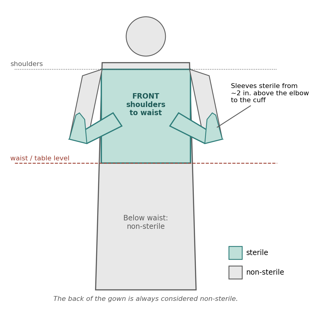
Figure 4.6. Sterile and non-sterile zones of a gowned surgical team member.
Chapter Summary
Surgical asepsis aims to keep microbial contamination below the level needed for infection. Bacteria such as Staphylococcus aureus and E. coli damage tissue through their toxins, and infection delays healing; antibiotics may be prophylactic or therapeutic but do not replace asepsis. Patient preparation proceeds from clipping (with wide margins, immediately before surgery) and hair removal, through a gloved preliminary scrub, to a final 3–5-cycle concentric scrub in the OR, application of antiseptic solution or film, and drying before draping.
The surgeon positions the animal, which is then draped to create a sterile field large enough to protect the incision, team, and instruments, with electronic monitoring compensating for reduced visibility. Personnel wear clean attire and perform a timed or anatomical scrub, then gown by touching only the inside of the gown and glove, preferably by the closed method. Throughout surgery, the team acts on any suspicion of contamination, keeps hands between waist and shoulders, treats everything below table level and the back of the gown as non-sterile, and keeps sterile surfaces dry.
Key Terms
Bactericidal: Kills bacteria.
Bacteriostatic: Stops bacteria from reproducing without necessarily killing them.
Closed gloving: Gloving in which the hands remain inside the gown sleeves; the recommended method.
Dorsal recumbency: Lying on the back.
Endotoxin: A toxic component of certain bacteria that damages host cells.
Erythema: Redness of the skin, a sign of inflammation or infection.
Fenestrated drape: A drape with an opening (fenestration) that exposes the surgical site.
Incise drape: A sterile adhesive plastic drape applied directly to the skin over the incision site.
Kick bucket: A wheeled waste container used to collect used sponges and disposables.
Lateral recumbency: Lying on the side (named for the side in contact with the table).
Open gloving: Gloving with the hands extended from the sleeves; greater risk of contamination.
Phagocyte: An immune cell that engulfs and consumes foreign material.
Prophylactic: Preventive; given before infection develops.
Sterile field: The area created by sterile drapes in which surgery is performed.
Sternal (ventral) recumbency: Lying on the sternum.
Stereotaxic frame: A device that holds the head rigidly, used for procedures requiring skull stability.
Surgical asepsis: The set of best practices that prevent or minimize contamination of the surgical site.
Therapeutic: Used to treat an established condition.
Review Questions
1. What is the ultimate goal of surgical asepsis?
A. To sterilize the patient's skin
B. To lower the concentration of microorganisms below the level required for infection
C. To eliminate the need for antibiotics
D. To kill all bacteria in the operating room
2. Why should hair NOT be clipped the day before surgery?
A. The hair grows back overnight
B. It is associated with higher bacterial counts due to skin trauma
C. It makes the animal cold
D. It violates the AWA
3. During the aseptic skin scrub, each pass of the gauze sponge should:
A. Start at the outer edge and move toward the incision
B. Start over the incision site and move outward in concentric circles
C. Move back and forth across the incision
D. Return to the center with the same sponge
4. An animal lying on its back is in which position?
A. Sternal recumbency
B. Ventral recumbency
C. Dorsal recumbency
D. Right lateral recumbency
5. Why should electronic monitoring be used whenever possible once an animal is draped?
A. Drapes make it difficult to physically monitor respiratory and cardiovascular function
B. It is required by AAALAC
C. It replaces the need for an anesthetist
D. Drapes increase heart rate
6. Once the surgeon scrub has begun, the surgeon should keep the hands:
A. Below the elbows so water drains off the fingertips
B. Higher than the elbows
C. At the sides
D. In the pockets of the scrub top
7. After the scrub and towel drying, the surgeon's hands are:
A. Sterile
B. Clean but not sterile
C. Contaminated and must be scrubbed again
D. Sterile only on the palms
8. Which gloving method is recommended because it minimizes the chance of contamination?
A. Open gloving
B. Closed gloving
C. Assisted open gloving with bare hands
D. Double open gloving
9. When pouring sterile fluid into a sterile bowl, the first portion should be:
A. Poured into the bowl to rinse it
B. Poured into a waste basket to flush the bottle rim
C. Discarded with the bottle cap
D. Used to wet the drapes
10. Which part of a sterile surgical gown is considered sterile?
A. The entire gown
B. The back and the sleeves
C. The front from the waist to the shoulders, and the sleeves from about 2 inches above the elbow to the cuff
D. Only the cuffs
Answer Key
1. B. Skin and the environment cannot be made sterile. Asepsis aims to keep the microbial load at the surgical site below the level needed to cause infection.
2. B. Clipping causes small skin injuries that bacteria colonize. The longer the interval before surgery, the higher the bacterial count, so clipping should be done shortly before surgery.
3. B. Working from the incision outward moves contaminants away from the cleanest area. A sponge that reaches the outer edge is never returned to the center.
4. C. Recumbency is named for the part of the body in contact with the table. Lying on the back means the dorsum is on the table: dorsal recumbency.
5. A. Sterile drapes cover the chest and mucous membranes, so electronic monitoring provides a safety net when the animal cannot easily be observed directly.
6. B. Keeping the hands above the elbows lets water drain from the cleanest area (hands) toward the less clean area (elbows), not the reverse.
7. B. Scrubbed hands are clean but not sterile and should be considered to contaminate any sterile object they touch. This is why gowns are handled only by their inside surfaces and closed gloving is preferred.
8. B. In closed gloving the hands never leave the gown sleeves, so bare skin never touches the outside of the gloves or gown.
9. B. Pouring a small amount into waste first flushes possible contaminants from the rim of the bottle before fluid flows into the sterile bowl.
10. C. Only the front of the gown between the waist and shoulders and the sleeves from about 2 inches above the elbow to the cuff are considered sterile. The back and everything below the waist are non-sterile.
Chapter 5: Peri-Operative Care
Learning objectives
After studying this chapter, you should be able to:
Describe the components and purpose of the pre-anesthetic evaluation.
Apply species-appropriate fasting guidelines and explain the risks of fasting in small mammals, birds, neonates, and ruminants.
Compare crystalloid and colloid fluids, and select appropriate fluids and rates for the surgical patient.
Compare airway devices and describe endotracheal intubation, including species-specific concerns and alternative methods.
Explain the importance of thermoregulation and describe post-operative recovery care for small and large animals.
Peri-operative care covers everything done for the patient around the time of surgery: before anesthesia, during the procedure, and through recovery. Surgery and anesthesia place heavy demands on the body. Anesthetic drugs depress breathing, circulation, and temperature regulation; surgery causes fluid and blood loss; and the recovering animal cannot yet protect its own airway or move normally. Good peri-operative care anticipates these problems and prevents them, so the animal arrives at surgery in the best possible condition and recovers safely.
5.1 Pre-Anesthetic Evaluation
A thorough history should be recorded and a complete physical exam performed before anesthesia. The purpose of this pre-anesthetic evaluation is to determine the patient's physical status: the presence or absence of disease, the severity of any pain, and the level of stress. More specifically, physical status describes the patient's medical condition and the overall efficiency and function of its organ systems.
The goal is to identify any deviations from normal that will affect anesthetic uptake, action, elimination, and safety. The nervous, cardiopulmonary, hepatic, and renal systems are the most important, because they control how anesthetic drugs are taken up, how the body responds to them, and how they are metabolized and excreted. The information gathered is used to decide which drugs to give and at approximately what doses.
A physical exam should include:
Attitude, physical condition, conformation, and temperament.
Palpation, percussion, and auscultation.
Vital parameters, such as heart rate (HR), respiratory rate (RR), temperature, capillary refill time (CRT), mucous membrane (MM) color, and pulse quality. These values also serve as baselines for intra-operative and post-operative monitoring, since a change from the animal's own normal is often more meaningful than a comparison with a textbook range.
Laboratory tests should be chosen based on the history and physical exam findings. Common tests include complete blood counts, blood chemistries, urinalysis, blood gases, clotting time and platelet counts, ECG, and radiography.
Table 5.1. Components of the pre-anesthetic physical exam and why each matters.
| Component | What it tells you |
|---|---|
| Attitude, condition, conformation, temperament | General health, body condition, and how the animal will tolerate handling and induction |
| Palpation, percussion, auscultation | Abnormalities of the heart, lungs, abdomen, and other organs |
| HR, RR, temperature | Cardiovascular, respiratory, and metabolic baselines |
| CRT, MM color, pulse quality | Perfusion and oxygenation baselines |
| Laboratory tests and imaging | Organ function, blood cell counts, clotting ability, and conditions not detectable on physical exam |
⚠ Check current guidance
Many institutions also assign a physical status classification (such as the ASA system) as part of the pre-anesthetic evaluation. Check whether your references or SOPs use one. Chapter 7 covers the effects of disease on anesthesia.
5.2 Fasting
Fasting before anesthesia reduces the volume of stomach contents, lowering the risk of vomiting or regurgitation and aspiration of stomach contents into the lungs. Generally, food is withheld for 12–18 hours in most species, with water available ad libitum.
Fasting is not advised in small mammals, birds, and neonates, which have high metabolic rates and limited energy reserves and may become hypoglycemic. Fasting is also not generally necessary in rabbits, rodents, and ruminants. Rabbits and rodents cannot vomit, so the main reason for fasting does not apply.
Ruminants are a special case. The rumen holds a large volume of fermenting contents that continue to produce gas, and the anesthetized ruminant cannot eructate (belch). Rumen tympany (bloat) can be prevented by passing a stomach tube. If fasting is required for a specific procedure:
Withhold food for 24–48 hours and water for 12–24 hours.
Use shorter times for small ruminants and longer times for large ruminants.
Do not fast neonates, because they are not yet "true ruminants."
Prolonged fasting combined with anesthesia can result in rumen stasis.
Table 5.2. Fasting guidelines by species group.
| Species group | Food | Water | Notes |
|---|---|---|---|
| Most species | Withhold 12–18 hours | Ad libitum | Reduces risk of vomiting and aspiration |
| Small mammals, birds, neonates | Fasting not advised | Ad libitum | Risk of hypoglycemia |
| Rabbits and rodents | Fasting not generally necessary | Ad libitum | Cannot vomit |
| Ruminants (if fasting required) | Withhold 24–48 hours | Withhold 12–24 hours | Shorter for small, longer for large ruminants; do not fast neonates; risk of rumen stasis |
⚠ Check current guidance
Current companion animal anesthesia guidelines often recommend shorter fasting periods for dogs and cats than the traditional 12–18 hours. Check current references and your institution's SOP for the species you work with.
5.3 Fluid Therapy
Surgical anesthesia and procedures can lead to dehydration. Fluids are lost through bleeding, evaporation from exposed tissues and the airway, and fluid shifts into injured tissue, while anesthetic drugs often lower blood pressure. Fluids are usually given intravenously (IV), but can also be given subcutaneously (SC), orally, or intra-osseously (IO). IO administration, into the marrow cavity of a bone, is used for severely dehydrated or traumatized patients with poor or inaccessible veins who need rapid absorption of fluid into the blood.
IV fluids during anesthesia help maintain adequate blood pressure, body temperature, and urine production, and they provide a ready route for drug administration in an emergency. Fluids should be warmed to about 37 °C to prevent hypothermia.
The common maintenance rate for lactated Ringer's solution (LRS) or isotonic saline is 5–10 mL/kg/hr. Use 10–15 mL/kg/hr for procedures that open a major body cavity or may cause excessive bleeding. Higher rates are recommended for ruminants, which lose fluid through heavy salivation.
⚠ Check current guidance
Recent fluid therapy guidelines for companion animals often recommend lower intra-operative rates than these traditional values, to avoid fluid overload. Check current references and your institution's SOP.
Fluids are grouped into crystalloids and colloids. The difference between them, shown in Figure 5.1, determines where the fluid goes once it enters the body.
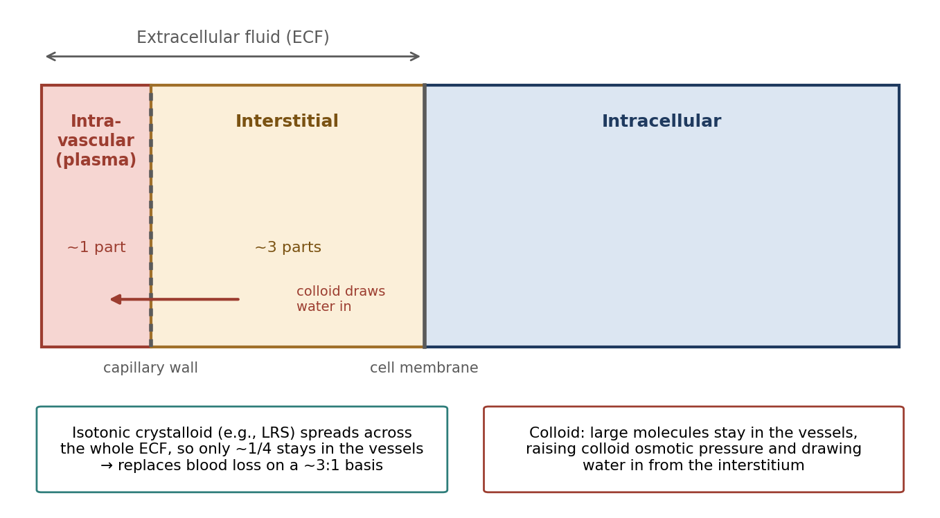
Figure 5.1. Body fluid compartments, and how crystalloid and colloid fluids distribute. Compartments are not drawn to scale.
5.3.1 Crystalloid Solutions
A crystalloid is any solution of crystalline solids dissolved in water. Water and small solutes such as sodium cross the capillary wall freely, so crystalloids do not stay in the bloodstream; they distribute throughout the extracellular fluid.
Lactated Ringer's solution closely approximates the electrolyte concentrations of extracellular fluid. It is isotonic, so it does not cause fluid to shift into or out of cells. Because it spreads throughout the extracellular fluid, it does not replace blood loss on a 1:1 basis; it replaces blood on about a 3:1 basis. It may be given rapidly in large volumes to re-expand the extracellular fluid volume.
Isotonic saline (0.9%) does not meet free water and electrolyte needs for maintenance, and excessive use may dilute extracellular electrolytes and buffers. It may be used to correct hyponatremia (low blood sodium) or metabolic alkalemia.
Hypotonic saline (0.45%) may be used as a hydrating solution, and for maintenance when supplemented with dextrose, potassium chloride, or both. It becomes isotonic when supplemented with 2.5% dextrose.
Hypertonic saline (3–5%) promotes rapid sodium replenishment in hyponatremia and is useful in managing shock, especially hemorrhagic shock, because it rapidly draws water from the tissues into the vessels. A dose of 4–6 mL/kg of 7.5% saline is used, and it should be given IV.
Dextrose solutions (2.5–50%) provide a source of free water for treating dehydration, because the dextrose is metabolized and leaves water behind. They are not effective as plasma expanders. Hypertonic dextrose solutions may be used as a caloric supplement.
Sodium bicarbonate is a hypertonic solution of sodium bicarbonate in sterile water for injection, used to treat metabolic acidosis. It causes sodium retention, so use it cautiously in patients with congestive heart failure or edema.
Table 5.3. Crystalloid solutions.
| Solution | Tonicity | Main uses | Cautions |
|---|---|---|---|
| Lactated Ringer's | Isotonic | Maintenance; re-expanding ECF volume | Replaces blood loss only ~3:1 |
| 0.9% saline | Isotonic | Hyponatremia; metabolic alkalemia | Does not meet maintenance needs; may dilute electrolytes and buffers |
| 0.45% saline | Hypotonic | Hydration; maintenance with dextrose and/or KCl | Becomes isotonic with 2.5% dextrose |
| Hypertonic saline (3–5%; 7.5% at 4–6 mL/kg) | Hypertonic | Hyponatremia; shock, especially hemorrhagic | Give IV |
| Dextrose (2.5–50%) | Varies | Free water for dehydration; calories (hypertonic) | Not a plasma expander |
| Sodium bicarbonate | Hypertonic | Metabolic acidosis | Sodium retention; caution in heart failure or edema |
⚠ Check current guidance
Hypertonic saline is described here as both a 3–5% solution and as a 7.5% solution dosed at 4–6 mL/kg. Concentrations and doses vary between products and references, so confirm the formulation you are using before calculating a dose.
5.3.2 Colloidal Solutions
Colloids are suspensions of large-molecular-weight particles, which tend to remain within the vascular compartment. They increase intravascular colloid osmotic pressure, which reduces further movement of water out of the vessels and may draw water from the interstitial space into the vessels. Colloids are used to expand vascular volume or to treat acute hypoproteinemia.
Plasma is used fresh to treat coagulopathies (clotting disorders); canine plasma may be stored at 2 °C for up to 30 days. Plasma stored at −70 °C can be used to treat vascular volume deficits or hypoproteinemia. It must be gradually thawed and warmed to 37 °C before infusion.
Whole blood should be used to treat severe anemia and blood loss greater than 10–15% of total blood volume. Peri-operatively, hematocrit should be maintained above 21–25% to ensure adequate oxygen delivery to peripheral tissues. If blood types are not matched, serious transfusion reactions (hemolysis or agglutination, depending on species) can occur.
Dextran solutions are low- to average-molecular-weight polysaccharides produced by bacterial enzymatic action on sucrose. They can be used for vascular volume expansion when blood products are not available, but they carry no oxygen, so care must be taken not to dilute the hematocrit to critical levels.
Hetastarch (hydroxyethyl starch) is used to expand plasma volume after losses from bleeding or severe injury. It is made by adding hydroxyethyl ether groups to the glucose units of amylopectin starch, and is supplied as a non-pyrogenic solution of 6% hetastarch in 0.9% saline. Its polymers range in molecular weight from 10,000 to 1,000,000. The smaller polymers are eliminated through the kidneys (40% within 24 hours), while the larger molecules are slowly broken down by serum amylase until they are small enough to be excreted. This gives hetastarch a more sustained ability to maintain vascular volume than dextran solutions.
Table 5.4. Colloid solutions.
| Colloid | Main uses | Key facts |
|---|---|---|
| Plasma | Fresh: coagulopathies. Frozen (−70 °C): volume deficits, hypoproteinemia | Canine plasma: up to 30 days at 2 °C. Thaw gradually and warm to 37 °C |
| Whole blood | Severe anemia; loss > 10–15% of blood volume | Keep hematocrit > 21–25%; match blood types |
| Dextran | Volume expansion when blood products are unavailable | No oxygen-carrying capacity; watch hematocrit |
| Hetastarch (6% in 0.9% saline) | Plasma volume expansion | Polymers 10,000–1,000,000; 40% renally cleared in 24 h; longer-lasting than dextran |
Why it matters
Crystalloids and colloids complement each other. Crystalloids replace losses from the whole extracellular space but leave the vessels quickly; colloids stay in the vessels and hold water there. Neither carries oxygen, which is why significant blood loss ultimately requires blood.
⚠ Check current guidance
Hydroxyethyl starch products have received FDA safety warnings regarding kidney injury, bleeding, and mortality in some human patient groups, and their use in veterinary patients is being reassessed. Blood product storage times also vary by product and species. Check current guidance before use.
5.4 Airway Management
Safe anesthesia requires that the patient's airway remain patent (open), with adequate ventilation and oxygenation. Anesthetic drugs relax the muscles that normally hold the airway open and depress the reflexes that protect it, such as swallowing and coughing. Supplemental oxygen may need to be provided during the pre-anesthetic, induction, maintenance, and recovery phases, by face mask, nasal cannula, flow-by, oxygen tent, or chamber.
5.4.1 Face Mask
The face mask is a low-tech method of delivering gas (oxygen, inhalant anesthetics, and so on) to the animal's respiratory system rather than to the general environment. However, it has significant limitations:
It does not provide an airtight seal, so inhalant anesthetic leaks into the room and exposes personnel.
It does not protect the airway, so aspiration and obstruction can occur.
It does not allow positive pressure ventilation.
5.4.2 Endotracheal Tube
An endotracheal tube (ETT) passes through the larynx into the trachea. It offers many advantages over a mask:
Maintains a patent airway, protecting against both aspiration and obstruction.
Protects the airway from foreign material during oral surgery and dental procedures.
Allows positive pressure ventilation.
Delivers oxygen, inhalant anesthetics, and test compounds effectively.
Provides an airtight seal if the cuff is properly inflated.
Provides easy access for suctioning the trachea or bronchi.
Decreases anatomical dead space if the tube is the correct size.
Endotracheal tubes may be made of polyvinyl chloride, silicone, plastic, or rubber. Red rubber is not recommended because it tends to crack, is difficult to clean, and is opaque. Other materials are less porous, more durable, and clear, which lets you see condensation inside the tube as the animal breathes.
The two basic designs are the Cole tube and the Murphy tube (Figure 5.2).
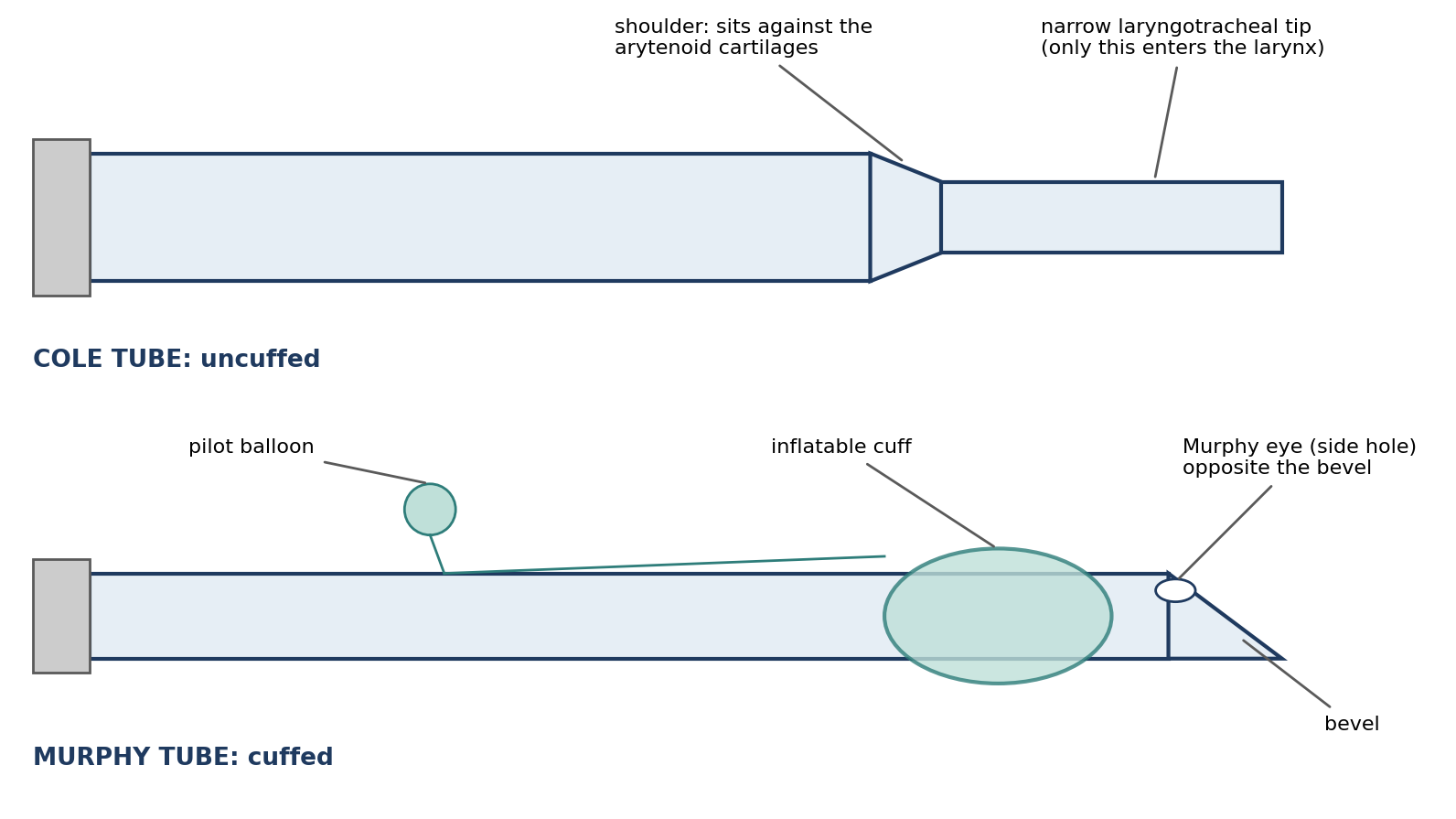
Figure 5.2. Cole and Murphy endotracheal tube designs.
The Cole tube is uncuffed. It has a "shoulder" that narrows to a thinner tip. The shoulder should sit against the arytenoid cartilages, forming a seal, and only the narrow tip should pass into the larynx and trachea. The shoulder should not enter or contact the larynx, because pressure on the laryngeal cartilages can cause damage and laryngeal dilation. Choosing the correct diameter for the laryngotracheal portion of the tube creates a seal, provided the tube stays properly positioned; movement in either direction can cause damage (too deep) or an improper seal (too shallow).
The Murphy tube is cuffed. It has an opening, called a Murphy eye or side hole, in the wall opposite the bevel, which allows gas to flow even if the end hole becomes occluded (for example, against the tracheal wall or by mucus). It provides a better seal than the Cole tube and does not require such precise placement.
Cuff Inflation
A Murphy tube's cuff should be inflated with the smallest amount of air that creates a seal at an airway pressure of 15–20 mmHg, depending on whether positive pressure ventilation is planned. The proper technique is:
Squeeze the reservoir bag to produce an airway pressure reading of 15–20 mmHg.
While holding that pressure, inject air into the cuff and listen for air leaking around the tube.
Stop injecting air as soon as the leak can no longer be heard.
Overinflating the cuff can rupture it or injure the airway. Excess pressure on the tracheal lining cuts off its blood supply, leading to ischemic injury, mucosal damage, pressure necrosis, and ultimately tracheal strictures.
⚠ Check current guidance
Many current references express cuff-seal airway pressure in centimeters of water (cm H₂O) rather than mmHg. Be sure you know which unit your anesthesia machine's manometer displays.
Other Endotracheal Tube Considerations
Some tubes are reinforced or armored with wire or plastic to prevent kinking when the head and neck are flexed, or when the breathing circuit must be placed off to the side. Depending on the brand, reinforced tubes may have smaller interior lumens, which increases airflow resistance, so they should not be used unnecessarily.
Intubation Procedure (Basic Technique)
The specific procedure varies between species, but the basic technique is:
Induce anesthesia. A sufficient depth must be reached to reduce the gag reflex.
Position the animal in dorsal, sternal, or lateral recumbency, depending on species and individual preference. Dorsal recumbency may be preferred for swine and primates.
Open the mouth. An assistant may help steady the animal or hold the mouth open.
Gently pull the tongue forward and out. Do not pull it out too far, especially in monkeys, or injury can result, and do not pull it hard over the lower incisors, which can cut its underside.
Optionally, apply topical lidocaine spray or liquid to the larynx to prevent laryngospasm. Additionally or alternatively, apply lidocaine (xylocaine) jelly to the tip of the tube.
Visualize the epiglottis and glottis. In most species a laryngoscope helps. Place the tip of the blade at the base of the epiglottis and use it to pull the tongue out, not push it down, which would hinder visualization. Do not put the blade directly on the epiglottis, which can cause trauma.
Measure the tube before inserting it (Figure 5.3). Hold the tube, or a second tube of the same size, against the animal with the tip at the approximate location of the thoracic inlet, and note the depth mark at the level of the incisors.
Introduce the tube into the trachea and advance it until the tip is at about the level of the thoracic inlet. If it is placed too deep, the tip can pass into one bronchus, interfering with ventilation. A stylet may help stiffen the tube in some species and with silicone tubes, but it must not extend past the tube's distal opening, or it may injure the trachea.
Secure the tube with tape, gauze, or rubber bands.
Inflate the cuff, if present, as described above.
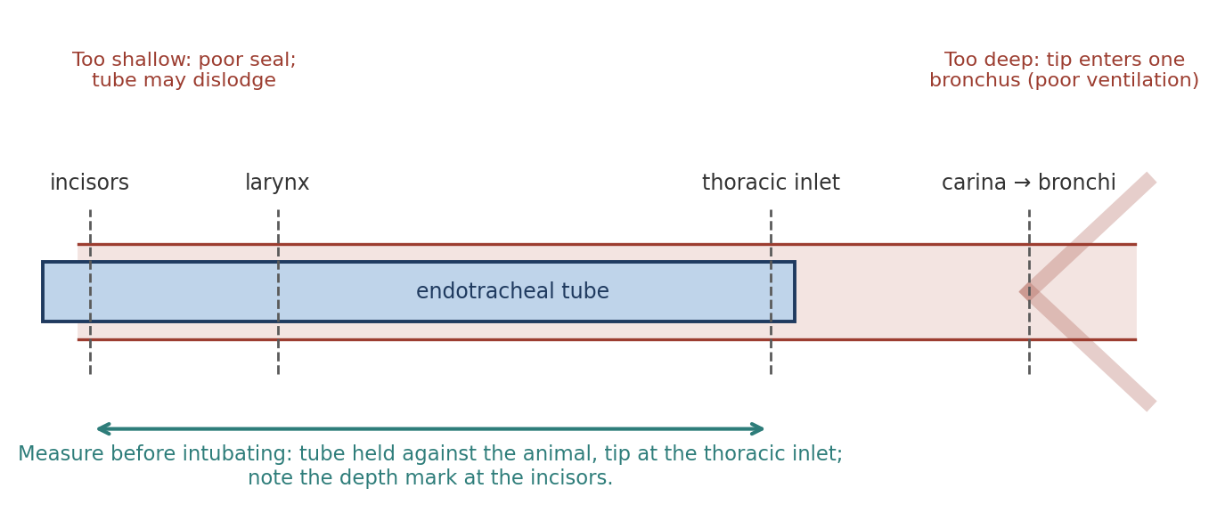
Figure 5.3. Measuring endotracheal tube depth. The tip should lie at about the thoracic inlet, above the carina.
Why it matters
Placing the tube too deep (endobronchial intubation) delivers oxygen and anesthetic to only one lung. The animal may become hypoxemic and its anesthetic depth may be hard to control, even though the tube appears to be working. Measuring before insertion is a simple way to prevent this.
Laryngoscopes
A laryngoscope is useful for visualizing the glottis and makes intubation easier, and it is preferred by most anesthetists. It provides illumination, a means of extending and depressing the tongue, and leverage for opening the mouth in nonhuman primates. The blade should not be used to manipulate the epiglottis directly, as this may cause trauma. In species with a long soft palate, such as dogs and swine, the epiglottis may be found behind the soft palate. In that case, use the tip of the endotracheal tube, not the laryngoscope blade, to gently sweep the epiglottis down.
Table 5.5. Laryngoscope blade styles.
| Style | Examples | Sizes |
|---|---|---|
| Straight | Miller, Wisconsin, Soper | Pediatric and adult |
| Curved | Macintosh, Bizarri-Guiffrida | Pediatric and adult |
[ PHOTO PLACEHOLDER ] Suggested source: Wikimedia Commons (commons.wikimedia.org), search "Miller blade" and "Macintosh blade." Check the image's license and give the attribution it requires. |
|---|
Figure 5.4. Straight (Miller) and curved (Macintosh) laryngoscope blades.
Species-Specific Concerns for Intubation
Airway anatomy varies widely between species, and each presents its own challenges (Table 5.6).
Table 5.6. Species-specific considerations for endotracheal intubation.
| Species | Challenges | Techniques and tips |
|---|---|---|
| Large animals (horses, cows, sheep, goats) | Usually not fasted, so the mouth may contain food; long distance from mouth to epiglottis makes visualization difficult | Flush debris from the mouth (aids visualization and prevents debris entering the bronchi); lubricate tube with water-soluble lubricant; specially made laryngoscopes |
| Cats | Pressure under the neck shifts the soft palate and obscures the view | Ensure proper anesthetic depth first. Benzocaine-containing topical sprays are contraindicated (risk of methemoglobinemia) |
| Rabbits | Very small glottis; difficult to see from the mouth | Extend the head back for blind intubation; watch for condensation or listen for breath sounds while advancing (if condensation stops, back up, redirect, and re-advance); guide cannula; LMA is a good alternative; endoscope-guided intubation is extremely successful |
| Rodents | Small size makes visualization difficult | Guide catheter or retrograde guide tube; intubation board or table; specially modified otoscope used as a laryngoscope |
| Swine | Highly vascular false pockets (pseudo-pharynx) outside the trachea bleed profusely if irritated; prone to laryngospasm; sharp angle from mouth to trachea | Gently twist the tube while advancing; lubricate with xylocaine gel (lubrication plus local analgesia against laryngospasm); dorsal recumbency straightens the path |
| Ruminants | Regurgitate rumen contents if anesthesia becomes too light | Intubate promptly; once intubated, lower the head below chest level and/or place a rumen tube with collection bag (which also prevents gas distension) |
Alternative Methods of Intubation
When direct visualization is difficult, several alternative methods can be used.
Guide-tube technique. Place a thin guide, such as a 5–8 French urinary catheter or an appropriately sized cardiovascular guide wire, into the trachea. Thread the endotracheal tube over it into the trachea, then remove the guide. Never force the guide, because the trachea is easily torn.
Retrograde intubation. After surgically preparing the skin over the trachea, insert a Venicath-style needle and catheter through the skin into the trachea just below (caudal to) the larynx, directed toward the head. Advance the catheter up through the larynx until it is visible in the mouth. Thread the endotracheal tube over the catheter into the trachea, remove the catheter, and finish advancing the tube. Secure the tube in place.
Laryngeal mask airway (LMA). An LMA is an alternative to both face masks and endotracheal tubes. It was developed for human pediatric patients weighing less than 5 kg; the LMA Unique™ Size 1 is the most commonly used in animals. When properly inserted, its mask lies flat against the laryngeal opening, forming a direct end-to-end junction between the upper airway and the tube that supplies gas to the bronchial tree (Figure 5.5). To insert it:
Insert the LMA with the cuff perpendicular to the teeth.
Once past the teeth, twist it 90 degrees so that the dorsal black line on the tube is at the midline, with the convex side against the hard palate.
Continue advancing until you meet unyielding resistance.
Overinflating the LMA cuff can cause lingual cyanosis (a blue tongue from compressed blood flow). The LMA is a good alternative for rabbits.
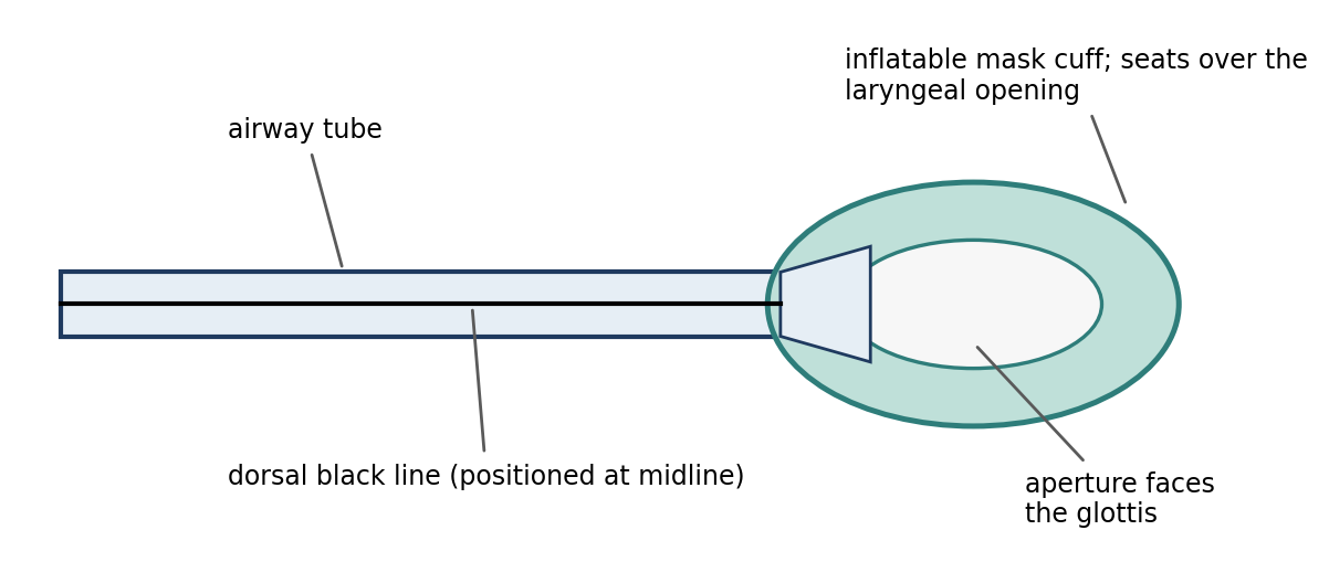
Figure 5.5. Basic structure of a laryngeal mask airway.
Endoscope-guided intubation. Laryngoscopy with a flexible or rigid endoscope is useful in patients with abnormal anatomy or pathology. The endoscope can be placed inside the endotracheal tube to guide it directly, or passed through the mouth beside the tube to watch it enter the glottis.
Table 5.7. Comparison of airway devices.
| Feature | Face mask | Endotracheal tube | Laryngeal mask airway |
|---|---|---|---|
| Airtight seal | No | Yes (with properly inflated cuff) | Seals at the laryngeal opening |
| Protects against aspiration | No | Yes | Partial |
| Positive pressure ventilation | No | Yes | Limited |
| Personnel exposure to inhalants | High | Low | Low to moderate |
| Skill required | Low | Moderate to high | Moderate |
⚠ Check current guidance
The LMA entries for aspiration protection, ventilation, and exposure in Table 5.7 are general comparisons rather than fixed standards; performance depends on fit and species. Veterinary-specific supraglottic airway devices are now available for several species, so check current references.
5.5 Thermoregulation
Maintaining body temperature is extremely important throughout the entire surgical period: pre-operative, intra-operative, and post-operative. Most anesthetic agents depress the thermoregulatory centers and metabolism, so the anesthetized animal both produces less heat and loses its normal responses to cold, such as shivering and vasoconstriction. Body heat is lost rapidly.
Small animals such as rodents have a higher surface-area-to-body-weight ratio than larger animals, so they lose heat faster and are more susceptible to hypothermia. Wet skin from surgical prep, cold surfaces, cold fluids, and an open body cavity all add to heat loss.
Body temperature is supported with heating blankets (water-circulating or warm-air blankets), heated tables, warm IV fluids, and warm intra-operative fluids such as lavage saline. Covering the extremities can also help maintain body temperature during surgery. Normothermic patients tolerate anesthesia better and recover faster.
Body temperature should be monitored continuously to avoid both hypothermia and hyperthermia, since heating devices can overheat or burn an animal that cannot move away from them. Hypothermia can interfere with normal platelet activity (and therefore clotting) and with heart rate.
Why it matters
Hypothermia is self-reinforcing: as temperature falls, drug metabolism slows, so anesthetics last longer and recovery is delayed, and the animal stays anesthetized and losing heat even longer. Preventing heat loss from the start is much easier than rewarming a cold patient.
5.6 Post-Operative Care & Recovery
Recovery is one of the highest-risk periods of anesthesia. The animal is no longer receiving the close attention of the surgical team, but it still cannot fully protect its airway, regulate its temperature, or move normally.
5.6.1 Small Animals
Place the animal in lateral recumbency.
For non-rodents, record vital signs every 15 minutes until the animal is conscious and extubated. Check rodents every 15–20 minutes until they are mobile.
If a rodent recovers in a cage, make sure it cannot bury its nose in a corner or in bedding, either of which can occlude the airway.
Continue thermoregulation with water-circulating heating pads, heat lamps, warm-air blankets, or an incubator until the animal can regulate its own temperature.
Turn the animal from left to right lateral recumbency and back every 15–30 minutes to prevent atelectasis (lung collapse) and fluid accumulation in the lower lung.
Remove the endotracheal tube when the swallowing reflex returns, since that shows the animal can protect its own airway.
If the animal is not intubated, pull the tongue forward so that it does not block the pharynx.
If the animal is returned to its home cage, remove water and food pans until it has recovered, to prevent drowning or choking.
If recovery is prolonged, IV fluids at a maintenance rate may be continued until the animal is conscious.
Oxygen therapy by mask or nasal oxygen is helpful, especially after a thoracotomy.
Give post-operative analgesics and sedatives as needed.
Do not place recovering animals with conscious animals, which may harm them. This is a particular risk with pigs, mice, and rats.
5.6.2 Large Animals (Livestock)
Take special care with horses and cows, which can severely injure themselves and personnel while struggling during recovery. Move the animal to a padded room or stall, if available.
Bandage and pad all limbs before anesthesia, and keep the padding in place until recovery is complete.
Remove food and water.
Have oxygen and suction readily available.
Record vital signs routinely until the return of the coughing, swallowing, and righting reflexes.
Keep ruminants in sternal recumbency with the head higher than the chest to prevent regurgitation and aspiration of rumen contents.
Table 5.8. Recovery care for small and large animals.
| Small animals | Large animals (livestock) | |
|---|---|---|
| Position | Lateral recumbency; turn every 15–30 min | Padded room or stall; ruminants sternal with head above chest |
| Monitoring | Vitals every 15 min until conscious and extubated (non-rodents); rodents every 15–20 min until mobile | Vitals routinely until coughing, swallowing, and righting reflexes return |
| Airway | Extubate when swallowing returns; tongue forward if not intubated; prevent nose burying (rodents) | Oxygen and suction readily available |
| Other | Continue heat support; remove food and water; analgesics; oxygen after thoracotomy; do not house with conscious animals | Pad and bandage limbs; remove food and water |
Chapter Summary
Peri-operative care starts with a pre-anesthetic evaluation: history, physical exam (which also establishes baselines), and laboratory tests, focused on the nervous, cardiopulmonary, hepatic, and renal systems. Fasting for 12–18 hours applies to most species, but not to small mammals, birds, and neonates (hypoglycemia risk), and is not generally needed in rabbits, rodents, and ruminants; ruminants that must be fasted follow longer, size-dependent schedules.
Warm fluids at 5–10 mL/kg/hr (10–15 for open body cavities or heavy bleeding) support blood pressure, temperature, and urine output. Crystalloids spread through the extracellular fluid and replace blood about 3:1; colloids stay in the vessels; whole blood is needed for severe anemia or losses over 10–15%, keeping hematocrit above 21–25%. A cuffed endotracheal tube best protects the airway; measure it to the thoracic inlet, inflate the cuff to seal at 15–20 mmHg, and know the species-specific challenges and alternatives such as guide tubes, retrograde intubation, and the LMA. Keep patients normothermic throughout, and monitor recovering animals closely in a safe position until they can protect their airway and regulate their temperature.
Key Terms
Atelectasis: Collapse of part of the lung, often in the dependent (lower) lung of a recumbent animal.
Colloid: A fluid containing large molecules that remain in the vascular compartment.
Colloid osmotic pressure: Osmotic pressure created by large molecules (such as proteins) that holds water in the vessels.
Cole tube: An uncuffed endotracheal tube with a shoulder that seats against the arytenoid cartilages.
Crystalloid: A solution of crystalline solids dissolved in water that distributes throughout the extracellular fluid.
Hematocrit: The percentage of blood volume made up of red blood cells.
Hypoproteinemia: Abnormally low protein levels in the blood.
Intra-osseous (IO): Administered into the marrow cavity of a bone.
Isotonic: Having the same solute concentration as body fluids, so no fluid shifts occur across cell membranes.
Laryngeal mask airway (LMA): A device whose mask seals over the laryngeal opening without entering the trachea.
Laryngospasm: Involuntary closure of the larynx, which obstructs the airway.
Murphy eye: A side hole near the tip of a Murphy tube that allows gas flow if the end hole is blocked.
Normothermia: Normal body temperature.
Patent: Open and unobstructed.
Physical status: The patient's medical condition and the function of its organ systems, assessed before anesthesia.
Retrograde intubation: Intubation over a catheter passed from the trachea up through the larynx into the mouth.
Rumen tympany: Bloat; distension of the rumen with gas.
Review Questions
1. Which organ systems are considered most important in the pre-anesthetic evaluation?
A. Integumentary, skeletal, and reproductive
B. Nervous, cardiopulmonary, hepatic, and renal
C. Endocrine and lymphatic
D. Digestive and urinary only
2. Which animals generally do NOT need to be fasted before anesthesia?
A. Dogs and cats
B. Rabbits, rodents, and ruminants
C. Nonhuman primates
D. Swine
3. What is the common maintenance rate for lactated Ringer's solution or isotonic saline during anesthesia?
A. 1–2 mL/kg/hr
B. 5–10 mL/kg/hr
C. 20–30 mL/kg/hr
D. 50 mL/kg/hr
4. Lactated Ringer's solution replaces blood loss on approximately what basis?
A. 1:1
B. 2:1
C. 3:1
D. 10:1
5. Colloid solutions expand blood volume primarily because they:
A. Carry oxygen
B. Contain large molecules that remain in the vascular compartment and raise colloid osmotic pressure
C. Are hypotonic
D. Contain dextrose
6. Peri-operatively, hematocrit should be maintained above:
A. 5–10%
B. 21–25%
C. 45–50%
D. 60%
7. Which is a disadvantage of a face mask compared with an endotracheal tube?
A. It allows positive pressure ventilation
B. It protects the airway from aspiration
C. It does not provide an airtight seal and exposes personnel to inhalant anesthetics
D. It decreases anatomical dead space
8. An endotracheal tube cuff should be inflated:
A. Until it feels firm to the touch
B. With the smallest amount of air that creates a seal at an airway pressure of 15–20 mmHg
C. With 20 mL of air in all species
D. Only after the animal has recovered
9. Why is intubation of swine potentially dangerous?
A. Swine have no epiglottis
B. Swine have highly vascular false pockets (pseudo-pharynx) that can bleed profusely, and they are prone to laryngospasm
C. Swine cannot be placed in dorsal recumbency
D. Swine require a Cole tube
10. How should a recovering ruminant be positioned?
A. Dorsal recumbency
B. Lateral recumbency with the head lowered
C. Sternal recumbency with the head higher than the chest
D. Standing immediately after extubation
Answer Key
1. B. The nervous, cardiopulmonary, hepatic, and renal systems most affect anesthetic uptake, action, elimination, and safety.
2. B. Fasting is not generally necessary in rabbits, rodents, and ruminants. Small mammals, birds, and neonates may become hypoglycemic if fasted.
3. B. The common maintenance rate is 5–10 mL/kg/hr, increased to 10–15 mL/kg/hr for procedures that open a major body cavity or risk excessive bleeding.
4. C. Because an isotonic crystalloid distributes throughout the extracellular fluid, only a fraction remains in the vessels, so about 3 mL is needed for each 1 mL of blood lost.
5. B. The large particles stay in the vessels, increasing colloid osmotic pressure, which limits water loss from the vessels and may draw water in from the interstitium.
6. B. A hematocrit above 21–25% is needed to ensure adequate oxygen delivery to peripheral tissues.
7. C. A face mask does not seal, so inhalant anesthetic leaks into the room. It also does not protect the airway or allow positive pressure ventilation.
8. B. Inject air while squeezing the reservoir bag to 15–20 mmHg and listening for a leak; stop as soon as no leak is heard. Overinflation can injure the trachea.
9. B. The pseudo-pharynx can bleed profusely if irritated, and swine are prone to laryngospasm. Lubrication with lidocaine (xylocaine) gel helps.
10. C. Sternal recumbency with the head higher than the chest helps prevent regurgitation and aspiration of rumen contents during recovery.
Chapter 6: Pre-Anesthetic Agents & Anesthetic Adjuncts
Learning objectives
After studying this chapter, you should be able to:
List the goals of pre-anesthetic medication and match each drug class to the goals it serves.
Compare the effects, uses, and species considerations of anticholinergics and tranquilizers.
Describe the effects and side effects of opioids, and distinguish full agonists, partial agonists, agonist-antagonists, and antagonists.
Describe alpha-2-adrenergic agonists, their reversal agents, and neuroleptanalgesic combinations.
Explain how depolarizing and non-depolarizing neuromuscular blocking agents work, how to monitor animals receiving them, and how they are reversed.
Pre-anesthetic agents are given before anesthesia (and adjuncts are given alongside it) to make the whole anesthetic experience safer and smoother for the animal. No single drug does everything, so premedication usually combines drugs from different classes. The goals of pre-anesthetic medication are to:
Aid in handling the patient by reducing stress and anxiety and inducing sedation.
Provide analgesia and muscle relaxation.
Decrease airway secretions and salivation.
Obtund (dull) autonomic reflex responses.
Decrease gastric fluid volume and acidity.
Suppress or prevent vomiting.
Decrease anesthetic requirements.
Promote smooth induction and recovery from anesthesia.
Table 6.1. Drug classes used for premedication and the main goals they serve.
| Drug class | Examples | Main goals served |
|---|---|---|
| Anticholinergics | Atropine, glycopyrrolate | Decrease secretions; prevent vagal bradycardia |
| Tranquilizers | Acepromazine, droperidol, diazepam, midazolam | Reduce anxiety; sedation; decrease anesthetic requirements; smoother recovery (no analgesia) |
| Opioids | Morphine, hydromorphone, fentanyl, buprenorphine, butorphanol | Analgesia; decrease anesthetic requirements |
| Alpha-2 agonists | Xylazine, detomidine, medetomidine, dexmedetomidine | Sedation, muscle relaxation, analgesia |
| Neuroleptanalgesics | Fentanyl/droperidol | Pronounced sedation and analgesia |
| Muscle relaxants / neuromuscular blockers | Guaifenesin; vecuronium, atracurium | Muscle relaxation (adjuncts to general anesthesia) |
⚠ Check current guidance
The drug information in this chapter reflects established references, but drug availability, approved species, and recommended combinations change over time, and many of these drugs are controlled substances. Always confirm doses and protocols with a veterinarian and current formularies before use.
6.1 Anticholinergics
Anticholinergics block certain receptors for the neurotransmitter acetylcholine, specifically the muscarinic receptors of the parasympathetic nervous system. The parasympathetic system, carried largely by the vagus nerve, slows the heart, increases secretions, and stimulates the gut. Blocking it produces the opposite effects. Anticholinergics:
Reduce oral and respiratory secretions.
Prevent vagal inhibition of the heart (bradycardia) and gastrointestinal stimulation (they inhibit intestinal peristalsis).
Reduce vagus nerve responses, such as vomiting and laryngospasm.
Relieve cholinergic-mediated bronchoconstriction and promote bronchodilation.
Dilate the pupils and reduce tear secretion.
Are the treatment of choice for opioid-, xylazine-, and vagal-reflex-induced bradycardia.
Can cause sinus tachycardia, which is a problem for patients with cardiovascular disease, because a fast heart works harder and has less time to fill.
Atropine sulfate decreases oral, respiratory, and pharyngeal secretions; suppresses vagal influence on the heart; and decreases lacrimation. Because it dilates the airways, it increases anatomic and physiologic respiratory dead space. It produces long-lasting mydriasis (pupil dilation) and cycloplegia (paralysis of the eye's focusing muscle). In dogs it increases the incidence of cardiac dysrhythmia and sinus tachycardia. Atropine is contraindicated in patients with tachycardia, constipation, or obstruction, and it may cause thick mucus secretions in cats. Some rabbits have atropine esterase, an enzyme that destroys large amounts of atropine and limits its effectiveness. Repeated doses should not exceed a total of 2 mg. Atropine is not recommended in ruminants, because it makes saliva more viscous (harder to clear), has a short duration, and increases the incidence of bloat.
Glycopyrrolate is a synthetic quaternary ammonium muscarinic antagonist. It decreases the volume and acidity of gastric secretions, intestinal motility, and tracheal, bronchial, and pharyngeal secretions, and prevents bradycardia caused by vagal reflexes or other pre-anesthetic and anesthetic drugs (alpha-2 agonists and opioids). Because its quaternary structure carries a permanent charge, it has reduced diffusion across the blood-brain and placental barriers compared with atropine. It lasts longer than atropine and is effective in rabbits. In ruminants, it lasts longer than atropine but has a slower onset of action.
Table 6.2. Comparison of atropine and glycopyrrolate.
| Property | Atropine sulfate | Glycopyrrolate |
|---|---|---|
| Duration | Shorter | Longer |
| Crosses blood-brain and placental barriers | Yes | Reduced |
| Rabbits | May be destroyed by atropine esterase | Effective |
| Ruminants | Not recommended (viscous saliva, short duration, bloat) | Longer duration but slower onset |
| Other notes | Mydriasis and cycloplegia; dysrhythmia and tachycardia in dogs; thick mucus in cats; total repeated dose ≤ 2 mg | Decreases gastric volume and acidity |
⚠ Check current guidance
Routine anticholinergic premedication is now used more selectively than in the past, because of the risk of tachycardia and arrhythmias. Check current guidance and your institution's protocols.
6.2 Tranquilizers
Tranquilizers calm the animal and make it easier and safer to handle. In general, tranquilizers:
Relieve anxiety, reduce the need for restraint, and quiet and calm the animal.
Decrease anesthetic doses and may make recovery smoother.
Reduce vomiting and reduce histamine release (allergic responses).
Have no analgesic effects.
The two main families differ in important ways. Phenothiazines (such as acepromazine) may promote vasodilation, which can cause hypotension and excessive heat loss, and may lower seizure thresholds. Benzodiazepines (such as diazepam and midazolam) promote skeletal muscle relaxation and act as anticonvulsants.
Acepromazine Maleate
Acepromazine is a phenothiazine derivative and a potent neuroleptic agent with low toxicity. It decreases anesthetic requirements in most species. Through vasodilation, it decreases stroke volume, cardiac output (CO), and mean arterial pressure (MAP) by 20–25%. Given 15 minutes before an opioid, it lowers the incidence of opioid-induced vomiting. The total dose in dogs should not exceed 3 mg. It may reduce or prevent malignant hyperthermia in swine.
Droperidol
Droperidol is a butyrophenone tranquilizer and an alpha-adrenergic antagonist. It may prevent epinephrine-induced dysrhythmia and decreases barbiturate doses. It was primarily used as a component of Innovar-Vet, an extremely effective combination with fentanyl (Section 6.6).
Diazepam (Valium)
Diazepam is a benzodiazepine and an anticonvulsant that rapidly crosses the blood-brain and placental barriers. It potentiates the action of most anesthetics and opioid analgesics, provides good muscle relaxation, and has relatively low toxicity. At high doses it causes a slight decrease in respiration, blood pressure, and cardiac output, and an increase in heart rate.
Practical cautions are important. Diazepam should be injected slowly IV to prevent venous thrombosis, pain, and cardiotoxicity, and it is not recommended for IM injection because it is painful. Take care when mixing it with other agents, as it may precipitate. It is safely combined with ketamine as an induction agent in dogs.
Sedation varies between species. In dogs, sedation is unreliable and may even include excitement. In rabbits, rodents, sheep, and pigs, diazepam produces marked sedation.
Midazolam
Midazolam is a benzodiazepine and anticonvulsant that provides excellent sedation and muscle relaxation in ferrets, rabbits, swine, and birds. It has a shorter duration of action and faster clearance than diazepam. Unlike diazepam, it is nonirritating and suitable for IM injection, and it can be mixed with other pre-anesthetic agents. It may cause behavioral changes, such as pacing and vocalization, in dogs and cats.
Flumazenil: Benzodiazepine Reversal
Flumazenil reverses the CNS (sedative) action of benzodiazepines. It acts rapidly, within 2–4 minutes, and its antagonism is specific to benzodiazepines. Reversal is not accompanied by anxiety, tachycardia, or hypertension.
Table 6.3. Comparison of tranquilizers.
| Drug | Class | Key effects | Cautions |
|---|---|---|---|
| Acepromazine | Phenothiazine | Potent neuroleptic; lowers anesthetic need; reduces opioid vomiting | Vasodilation: SV, CO, MAP ↓20–25%; hypotension; heat loss; lowers seizure threshold; dog total ≤ 3 mg |
| Droperidol | Butyrophenone | Alpha antagonist; prevents epinephrine dysrhythmia | Mainly used with fentanyl (Innovar-Vet) |
| Diazepam | Benzodiazepine | Anticonvulsant; muscle relaxation; marked sedation in rabbits, rodents, sheep, pigs | Slow IV only (not IM); may precipitate; unreliable in dogs |
| Midazolam | Benzodiazepine | Excellent sedation in ferrets, rabbits, swine, birds; shorter-acting | Behavioral changes in dogs and cats |
Why it matters
Because tranquilizers make an animal look calm without relieving pain, they must never be used in place of analgesics. A sedated animal in pain may simply be unable to show it.
6.3 Opioids
Opioids are analgesics that depress the central nervous system and lower the amount of anesthetic agents required. They are the cornerstone of pre-emptive pain treatment in veterinary medicine: given before the painful stimulus, they prevent the nervous system from becoming sensitized to pain, which makes pain easier to control afterward (Chapter 13). Opioids do not cause unconsciousness at therapeutic doses. They can be addictive, and most are controlled substances requiring extensive documentation under DEA regulations.
Opioids act on opioid receptors, principally the mu receptor. They can be grouped by how they act at that receptor (Figure 6.1):
Full agonists (such as morphine, hydromorphone, and fentanyl) fully activate the receptor; higher doses produce greater effect.
Partial agonists (such as buprenorphine) activate the receptor only partially; they reach a ceiling beyond which more drug adds little effect.
Agonist-antagonists (such as butorphanol) activate some opioid receptors while blocking others, so they can partially reverse the effects of full agonists.
Antagonists (such as naloxone and naltrexone) bind the receptor without activating it and are used to reverse opioids.
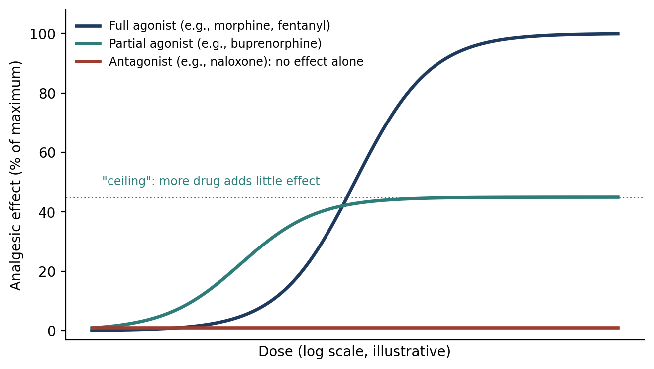
Figure 6.1. Dose–effect relationships for a full agonist, a partial agonist, and an antagonist at the mu opioid receptor (schematic).
Opioid Side Effects
Emesis, except in horses, rabbits, ruminants, rodents, and swine. It is rarely seen in the immediate post-operative period.
Hypothermia in most species, but hyperthermia may occur in cats, horses, swine, and ruminants.
Excitement and hyperactivity in cats, horses, goats, sheep, pigs, and cows.
Depression of the cough center and respiratory depression.
Mydriasis in species that show excitation, and miosis (pupil constriction) in species that become sedated.
Bradycardia through vagal stimulation.
In dogs, initial stimulation of defecation, followed by a tendency to ileus (reduced gut motility) and constipation.
Urinary retention.
Table 6.4. Species differences in opioid side effects.
| Effect | Species affected |
|---|---|
| Emesis | Most species EXCEPT horses, rabbits, ruminants, rodents, and swine |
| Hyperthermia (instead of hypothermia) | Cats, horses, swine, ruminants |
| Excitement and hyperactivity | Cats, horses, goats, sheep, pigs, cows |
| Mydriasis vs. miosis | Mydriasis in species that become excited; miosis in species that become sedated |
Individual Opioids
Morphine sulfate is the reference opioid against which others are compared. Its major pharmacologic effect is analgesia, and it is a useful analgesic in dogs, cats, horses, and rats. It induces a rapid and marked increase in serotonin synthesis; depresses the respiratory, cough, and vasomotor centers; decreases basal metabolic rate and body temperature; and stimulates the vomiting center. It does not affect motor function. In some species it causes excitement. Morphine is metabolized in the liver and eliminated in the urine, and is used infrequently in ruminants and swine in the clinical setting.
Meperidine hydrochloride (Demerol, pethidine) has about one-tenth the analgesic effect of morphine and is rapidly excreted, lasting less than 1 hour. It reduces salivary and respiratory secretions and does not cause vomiting. Rapid IV administration is not recommended because it may cause hypotension and convulsions; it is given SC 30 minutes before anesthesia. It has notable local anesthetic ability.
Methadone hydrochloride (Dolophine) is a synthetic opioid structurally unrelated to morphine that can be reversed with an opioid antagonist. Analgesia lasts 2–6 hours, and it decreases barbiturate doses by about 50%. It is described as stimulating respiratory rate and as not commonly used in North America in the peri-operative period.
⚠ Check current guidance
Methadone is now used more widely as a peri-operative analgesic in veterinary medicine than it once was, and most references describe it as causing respiratory depression like other mu agonists. Check current references.
Oxymorphone hydrochloride (Numorphan) is a synthetic opioid comparable to morphine in analgesic efficacy and duration but 10 times more potent. It decreases barbiturate doses by about 33–66% and can provide effective epidural analgesia.
Hydromorphone (Dilaudid) is a synthetic opioid with efficacy, potency, duration, and side effects similar to oxymorphone, but it is less expensive and more available. It is used as a sedative and restraining agent, analgesic, and pre-anesthetic, commonly in dogs, cats, and nonhuman primates. In cats it may cause hyperthermia, ataxia, hyperesthesia, and behavioral changes.
Fentanyl citrate is described as 250 times more potent than morphine. It has a rapid onset and short duration, with a peak at 30 minutes, so it is commonly given as a continuous infusion. Its respiratory depression may persist for hours. Cardiovascular stability is excellent, although it causes vagally mediated bradycardia unless countered with atropine. Combined with a benzodiazepine, it can induce general anesthesia in dogs with cardiovascular instability. It is also available as a transdermal patch.
⚠ Check current guidance
Published estimates of fentanyl's potency relative to morphine vary, and many references give a figure closer to 75–100 times. Confirm the value used in your exam references.
Carfentanil citrate is about 10,000 times more potent than morphine. It may be applied to the buccal or nasal mucosa and is used primarily for the capture of wild animals. Because of its extreme potency, accidental human exposure is life-threatening.
Sufentanil is a thienyl analog of fentanyl, 5–10 times as potent as fentanyl, with an elimination half-life of 2–2.5 hours. It may induce bradycardia. Used alone in dogs it produces unpredictable anesthesia, bradycardia, hypoventilation, and poor muscle relaxation, but combined with potent tranquilizers and glycopyrrolate it is an effective neuroleptanesthetic agent.
Alfentanil is one-fifth to one-tenth as potent as fentanyl, with a more rapid onset of action than fentanyl or sufentanil.
Etorphine is an oripavine derivative, described as 80 to 1,000 times more potent than morphine (SC). It can be antagonized with nalorphine or diprenorphine and is used primarily for the capture of wild animals.
⚠ Check current guidance
The oripavine structure, wildlife-capture use, and diprenorphine reversal are sometimes mistakenly attributed to alfentanil; they belong to etorphine (M99). The potency range for etorphine also varies between references. Confirm against your exam references.
Butorphanol tartrate (Torbugesic, Torbutrol) is a synthetic agonist-antagonist opioid, 3–5 times as potent as morphine, with less respiratory depression than morphine. It is commonly combined with xylazine, detomidine (cattle and horses), acepromazine, or midazolam. Because of its antagonist activity, it antagonizes the sedative effects of morphine and oxymorphone.
Buprenorphine (Buprenex) is a partial mu agonist, 25–30 times more potent than morphine, but with a maximum analgesic effect less than morphine's. It reaches a "ceiling" where additional doses have little effect. Its onset is relatively slow (20–30 minutes). It causes respiratory depression and is reversed by naloxone and naltrexone. IM administration lasts 6–12 hours and epidural administration 18–24 hours. It is highly protein-bound and mostly excreted unchanged in the feces.
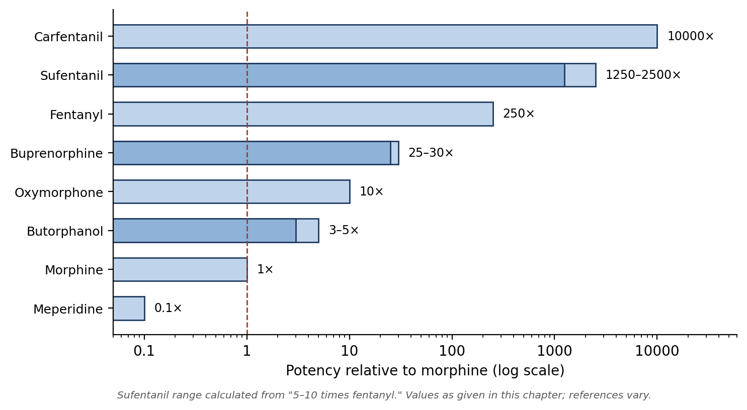
Figure 6.2. Relative potency of opioids compared with morphine. Higher potency means a smaller dose is needed, not necessarily greater maximum analgesia.
Table 6.5. Summary of opioids.
| Drug | Type | Potency vs. morphine | Key points |
|---|---|---|---|
| Morphine | Full agonist | 1× | Reference opioid; vomiting; used infrequently in ruminants and swine |
| Meperidine | Full agonist | 0.1× | < 1 hour; no vomiting; avoid rapid IV; local anesthetic action |
| Methadone | Full agonist | — | 2–6 hours; lowers barbiturate dose ~50% |
| Oxymorphone | Full agonist | 10× | Epidural use; lowers barbiturate dose ~33–66% |
| Hydromorphone | Full agonist | Similar to oxymorphone | Less expensive and more available; hyperthermia in cats |
| Fentanyl | Full agonist | 250× (as given here) | Short-acting; infusion or transdermal patch; excellent CV stability |
| Carfentanil | Full agonist | 10,000× | Wildlife capture; mucosal application |
| Sufentanil | Full agonist | 5–10× fentanyl | Half-life 2–2.5 h; neuroleptanesthesia with tranquilizers |
| Alfentanil | Full agonist | 1/5–1/10 fentanyl | Fastest onset |
| Etorphine | Full agonist | 80–1,000× (SC) | Oripavine; wildlife capture; reversed with diprenorphine or nalorphine |
| Butorphanol | Agonist-antagonist | 3–5× | Less respiratory depression; antagonizes morphine and oxymorphone sedation |
| Buprenorphine | Partial agonist | 25–30× | Ceiling effect; slow onset; IM 6–12 h, epidural 18–24 h |
Why it matters
Potency and efficacy are different things. Potency is how much drug is needed for an effect; efficacy is how large an effect the drug can produce at all. Buprenorphine is 25–30 times as potent as morphine, but its ceiling means its maximum analgesia is lower, which is why it may not be enough for severe pain.
6.4 Alpha-2-Adrenergic Agonists
Alpha-2-adrenergic agonists are the most widely used class of sedatives in veterinary medicine. By stimulating alpha-2 receptors, they reduce the release of norepinephrine in the brain and spinal cord, producing sedation, muscle relaxation, and analgesia. They can be used alone or with opioids to sedate animals for diagnostic and minor surgical procedures, and combined with ketamine they provide surgical anesthesia for brief, minor procedures. They are not addictive and they act as anticonvulsants.
Alpha-2 agonists have a wide range of drug interactions. When they are used, doses of barbiturates, inhalants, and dissociative anesthetics should be lowered. Their effects are reversible with alpha-2 antagonists such as yohimbine and atipamezole (Section 6.5).
Xylazine Hydrochloride (Rompun)
Xylazine was the first alpha-2 agonist used in veterinary medicine and remains the most common sedative-analgesic in horses and cattle. Combined with ketamine it acts as a short-term surgical anesthetic, and it may be combined with butorphanol to improve analgesia and sedation.
Timing. Maximum effect occurs in 10–15 minutes IM or 3–5 minutes IV. Analgesia lasts less than 1 hour, so painful procedures should be limited to 10–15 minutes after the onset of sedation. Higher doses prolong the effect but do not generally increase the degree of sedation. Excited animals (from anxiety or pain) may respond less well, because their high circulating catecholamines counteract the drug.
Cardiovascular effects. IV administration causes a brief period of hypertension and reflex bradycardia, followed by a longer-lasting decrease in cardiac output and arterial pressure. IM administration has less drastic effects. Xylazine can produce second-degree heart block, a relatively harmless arrhythmia originating at or below the AV node. IV overdose or accidental intra-arterial injection may cause seizures and collapse.
Other effects. Xylazine has poor efficacy in swine. It may cause emesis in cats and dogs, increases urine output in some species, and causes mydriasis. It has a significant local anesthetic effect, and epidural administration produces more profound and longer-lasting effects than lidocaine. It reduces insulin secretion from the pancreas, leading to hyperglycemia. This is not usually harmful, except in dehydrated patients, in whom it can cause a transient osmotic diuresis.
Detomidine
Detomidine was originally developed for use in horses and cattle and has cardiovascular effects similar to xylazine. It is commonly combined with opioids for enhanced sedation and analgesia, and may be used as a pre-anesthetic or combined with ketamine for anesthesia.
Medetomidine
Medetomidine is a sedative-analgesic developed for use in small animals, given IV or IM. It is more potent and longer-lasting than the other alpha-2 agonists, with a rapid onset of sedation, analgesia, and muscle relaxation after IM administration. It decreases injectable and inhalant anesthetic requirements, decreases urine specific gravity and increases urine production, and decreases blood pressure and respiratory rate in a dose-dependent manner.
Dexmedetomidine
Dexmedetomidine is the active form (isomer) of medetomidine. It is twice as potent as medetomidine and can be used at half the dose. It is used as a pre-anesthetic, providing sedation and analgesia, with side effects similar to medetomidine.
6.5 Alpha-2-Adrenergic Antagonists
Alpha-2 antagonists are used as reversal agents for alpha-2 agonists. Being able to reverse sedation quickly is a major safety advantage of the alpha-2 drugs.
Yohimbine reverses xylazine.
Tolazoline reverses the sedative and cardiovascular effects of xylazine in ruminants.
Atipamezole is a highly selective alpha-2 receptor antagonist, 200–300 times more selective than yohimbine. It reverses the effects of medetomidine in many species. It is given IM, although IV doses may be used in emergencies to reverse cardiovascular effects. It should not be used concurrently with anticholinergics, since both can cause dramatic increases in heart rate.
Table 6.6. Alpha-2 agonists and their reversal agents.
| Agonist | Primary use | Reversal agent |
|---|---|---|
| Xylazine | Horses and cattle (most common); with ketamine for short anesthesia | Yohimbine; tolazoline (ruminants); atipamezole |
| Detomidine | Horses and cattle | Atipamezole |
| Medetomidine | Small animals | Atipamezole |
| Dexmedetomidine | Small animals (half the medetomidine dose) | Atipamezole |
⚠ Check current guidance
Table 6.6 lists atipamezole for xylazine and detomidine because, as a non-specific alpha-2 antagonist, it reverses alpha-2 agonists generally; the text above specifically describes its use for medetomidine. Check species-specific product labels and current references.
6.6 Neuroleptanalgesics
A neuroleptanalgesic is a combination of a neuroleptic (a tranquilizer or sedative) and a potent opioid. The combination provides pronounced sedation and analgesia, sufficient for minor surgical procedures. The primary example is fentanyl citrate/droperidol (Innovar-Vet).
Neuroleptanalgesics produce intense analgesia of relatively short duration, along with sedation and immobilization, but also respiratory depression, hypotension, and bradycardia. Muscle relaxation is poor. They provide a wide margin of safety and an easy recovery, and they can be partially reversed with opioid antagonists (the opioid component is reversed; the tranquilizer is not).
⚠ Check current guidance
Innovar-Vet is no longer commercially available in the United States. Other opioid–sedative combinations are used for the same purpose. Check current references for neuroleptanalgesic combinations in use.
6.7 Muscle Relaxants & Neuromuscular Blocking Agents
Why it matters
These agents DO NOT provide analgesia or unconsciousness. A paralyzed animal that is not adequately anesthetized can be fully aware and in pain while appearing completely still. For this reason, The Guide expressly prohibits the use of neuromuscular blocking agents without an anesthetic (see Chapter 2).
Neuromuscular blocking agents (NMBAs) provide superior muscle relaxation as an adjunct to general anesthesia. They make intubation and ventilation easier, improve cardiovascular anesthetic management, prevent eye movement during ocular surgery, and reduce muscle resistance in long-bone orthopedic and abdominal surgery, where they reduce the anesthetic dose that would otherwise be needed to relax tense muscles.
NMBAs work at the neuromuscular junction, interfering with transmission of the signal from the motor neuron to the muscle (Figure 6.3). Because the diaphragm and intercostal muscles are paralyzed along with all other skeletal muscles, animals receiving NMBAs usually require mechanical ventilation.
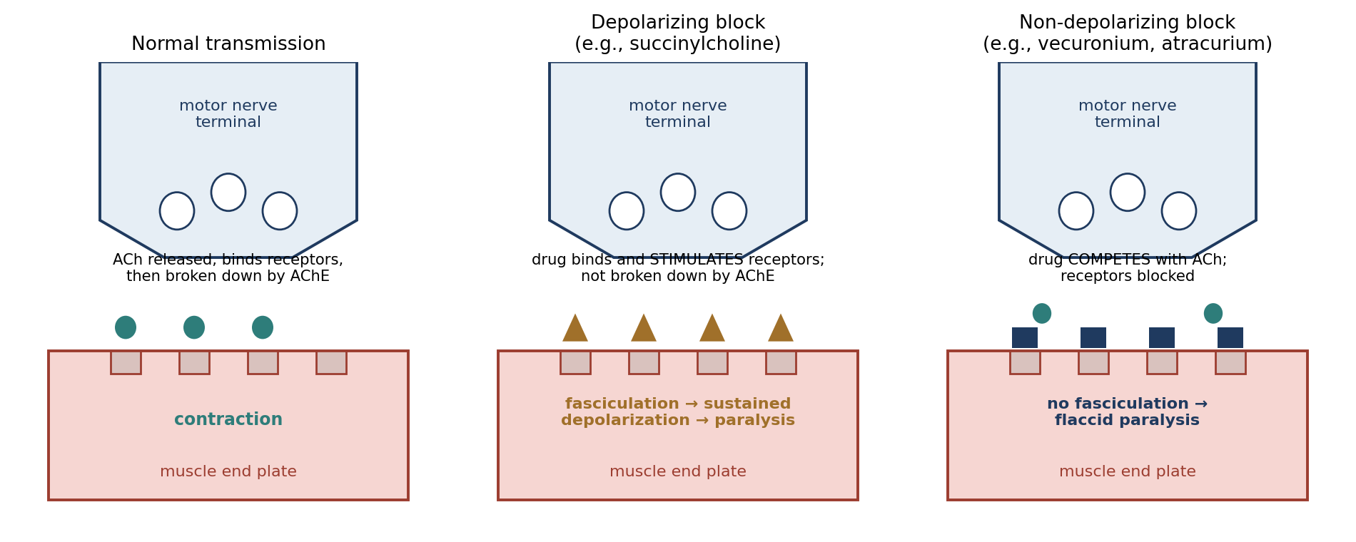
Figure 6.3. Normal neuromuscular transmission compared with depolarizing and non-depolarizing neuromuscular block. ACh, acetylcholine; AChE, acetylcholinesterase.
Monitoring the Paralyzed Patient
NMBAs make anesthesia management more difficult. The usual signs of anesthetic depth (twitching, muscle tone, and respiratory changes) are minimized or absent. The anesthetist must therefore monitor heart rate, blood pressure, ECG, and arterial blood gases to assess the animal's physiological state.
An inadequately anesthetized paralyzed animal will show tachycardia, arrhythmias, hypertension, and acidosis. Mydriasis and lacrimation may also be present, and horses will sweat if anesthesia is light. The anesthetist may also monitor the response of a peripheral nerve to an electrical stimulus to measure the degree of paralysis.
[ PHOTO PLACEHOLDER ] Suggested source: Wikimedia Commons (commons.wikimedia.org), search "peripheral nerve stimulator" or "train of four." Check the image's license and give the attribution it requires. |
|---|
Figure 6.4. A peripheral nerve stimulator used to assess neuromuscular block (for example, by train-of-four stimulation).
NMBAs interact with many drugs, including some antibiotics, lithium compounds, local anesthetics, barbiturates, quinidine, propranolol, calcium antagonists, diuretics, immunosuppressants, and corticosteroids. These interactions can intensify or prolong the block.
6.7.1 Centrally Acting Muscle Relaxants
Guaifenesin (glyceryl guaiacolate) acts in the central nervous system rather than at the neuromuscular junction. It is used as a muscle relaxant in large animals and has minimal effect on respiratory muscles, so animals continue to breathe. It may be combined with xylazine, ketamine, thiopental, and pentobarbital.
6.7.2 Depolarizing Neuromuscular Blocking Agents
Depolarizing agents have limited transfer across the placenta. They mimic the action of acetylcholine, binding to and stimulating the receptor and causing depolarization. Unlike acetylcholine, however, they are not broken down by acetylcholinesterase and are not antagonized by acetylcholine. They cause an initial depolarization of the motor end plate that lasts from minutes to hours, preventing the motor neurons from repolarizing and firing again. Increasing the amount of agent at the junction does not prolong the block. However, large, repeated, or prolonged doses may create a phase II block, which resembles the block produced by non-depolarizing agents.
Succinylcholine is the main example. It must be refrigerated. It has a rapid onset and provides excellent muscle relaxation, but causes marked fasciculation (muscle twitching) for about 30 seconds before the muscles relax. It is rapidly hydrolyzed in plasma by pseudocholinesterase, so only a small fraction of the injected dose reaches the neuromuscular junction; its effect is ended by diffusion away from the junction into the extracellular fluid.
Succinylcholine has several undesirable side effects:
The initial fasciculation and extensor rigidity may cause muscle pain, stiffness, and microscopic muscle damage.
Hypertension, tachycardia, sinus bradycardia, cardiac arrhythmias, and cardiac arrest.
Increased intraocular pressure and increased intracranial pressure (cats and dogs), even under thiopental or pentobarbital anesthesia.
Increased serum potassium.
Its action is prolonged by pregnancy and hepatic disease (which reduce pseudocholinesterase) and potentiated by inhalation and local anesthetics. Cats, swine, and ponies are resistant to it.
⚠ Check current guidance
Fasciculation after succinylcholine is generally brief, lasting seconds rather than minutes; its duration is sometimes misstated as 30 minutes. Confirm against your references.
6.7.3 Non-Depolarizing Neuromuscular Blocking Agents
Non-depolarizing agents compete with acetylcholine for its receptors, blocking them without stimulating them. They do not cause fasciculation; instead, muscle contractions weaken progressively until flaccid paralysis results. They have a slow onset of action. Their action is prolonged if hepatic or renal function is impaired, and large doses or prolonged administration can also prolong their effects.
They fall into two chemical categories:
Steroid analogs: pancuronium, vecuronium, pipecuronium, and rocuronium.
Benzylisoquinoliniums: curare (tubocurarine), metocurine, gallamine, atracurium, doxacurium, and mivacurium. (Gallamine is chemically distinct but is usually grouped here.)
Table 6.7. Non-depolarizing neuromuscular blocking agents.
| Agent | Duration | Elimination | Key points |
|---|---|---|---|
| Pancuronium | Long (40–60 min) | Mostly urine; some biliary | Increases HR (vagal block); difficult reversal after prolonged use; weak metabolites |
| Vecuronium | Shorter than pancuronium | Prolonged in renal failure | More potent; no CV or HR effects at normal doses; fast recovery; widely used |
| Pipecuronium | Long (2× pancuronium) | May be retained in kidneys for days | 2–4× pancuronium potency; rapid onset; no HR effect up to 100× therapeutic dose |
| Rocuronium | Rapid recovery | Mainly liver; small renal portion | 20% of vecuronium potency; rapid onset; higher doses speed onset but prolong duration |
| Curare (d-tubocurarine) | Long | Liver and kidneys; small biliary portion | Increases HR; histamine release → vasodilation, hypotension, tachycardia |
| Metocurine | — | Renal; 2% biliary | Much improved safety margin over curare |
| Gallamine | Long | Renal | Relatively impotent; consistent tachycardia; the only non-depolarizer that crosses the placenta |
| Atracurium | Intermediate | Hofmann elimination (spontaneous breakdown, pH- and temperature-dependent) | Must be refrigerated; not prolonged by renal or hepatic disease; widely used |
| Doxacurium | Long | — | No autonomic effects; no histamine release |
| Mivacurium | Slightly longer than succinylcholine; ½ of vecuronium | — | No autonomic effects |
Atracurium deserves particular attention. Its molecule is unstable and breaks down by itself at body temperature through a pH- and temperature-dependent process called Hofmann elimination. Because this does not depend on the liver or kidneys, atracurium's action is not prolonged in animals with renal or hepatic disease.
⚠ Check current guidance
Several agents in Table 6.7 (such as pipecuronium, doxacurium, metocurine, gallamine, and curare) are rarely or no longer used clinically, but they may still appear in exam references.
6.7.4 Reversal Agents
Reversal agents for non-depolarizing blockers work best when spontaneous recovery of muscle strength has already begun; they have little effect if a large dose of a non-depolarizing agent has just been given.
Anticholinesterases (edrophonium, neostigmine, and pyridostigmine) inhibit acetylcholinesterase, so acetylcholine accumulates and outcompetes the blocking agent. Because acetylcholine also increases at parasympathetic sites throughout the body, these drugs may cause bradycardia, sinus arrest, bronchospasm, miosis, intestinal hyperperistalsis, and salivation. An anticholinergic should be given first to prevent these effects.
4-Aminopyridine and guanidine increase acetylcholine release. They cause CNS stimulation (restlessness, confusion, and convulsions) and are best combined with an anticholinesterase.
Table 6.8. Summary of reversal agents in this chapter.
| Reversal agent | Reverses |
|---|---|
| Flumazenil | Benzodiazepines (diazepam, midazolam) |
| Naloxone, naltrexone | Opioids (including buprenorphine); partially reverses neuroleptanalgesics |
| Diprenorphine, nalorphine | Etorphine |
| Yohimbine | Xylazine |
| Tolazoline | Xylazine in ruminants |
| Atipamezole | Medetomidine (and other alpha-2 agonists) |
| Edrophonium, neostigmine, pyridostigmine (with an anticholinergic) | Non-depolarizing neuromuscular blockers |
| 4-Aminopyridine, guanidine | Non-depolarizing neuromuscular blockers (best with an anticholinesterase) |
Chapter Summary
Premedication reduces stress, provides analgesia and muscle relaxation, controls secretions and reflexes, prevents vomiting, reduces anesthetic requirements, and smooths induction and recovery. Anticholinergics (atropine, glycopyrrolate) reduce secretions and treat vagal bradycardia; glycopyrrolate lasts longer and works in rabbits. Tranquilizers calm the animal but provide no analgesia; phenothiazines cause vasodilation, and benzodiazepines relax muscle and are reversed by flumazenil.
Opioids are the cornerstone of pre-emptive analgesia; they differ in potency, receptor action (full, partial, mixed), duration, and species-specific side effects, and are reversed by naloxone. Alpha-2 agonists are the most widely used veterinary sedatives, providing sedation, muscle relaxation, and analgesia, with marked cardiovascular effects and reversal by yohimbine, tolazoline, or atipamezole. Neuroleptanalgesics combine a tranquilizer and an opioid. Neuromuscular blockers provide paralysis without analgesia or unconsciousness, require ventilation and careful autonomic monitoring, and are reversed (non-depolarizing types) by anticholinesterases given after an anticholinergic.
Key Terms
Agonist-antagonist: An opioid that activates some opioid receptors while blocking others (e.g., butorphanol).
Anticholinergic: A drug that blocks muscarinic acetylcholine receptors (e.g., atropine, glycopyrrolate).
Anticholinesterase: A drug that inhibits acetylcholinesterase, increasing acetylcholine (e.g., neostigmine).
Ceiling effect: The point beyond which increasing the dose produces little additional effect.
Cycloplegia: Paralysis of the eye's focusing (ciliary) muscle.
Depolarizing block: Neuromuscular block caused by sustained stimulation of acetylcholine receptors (e.g., succinylcholine).
Fasciculation: Brief, uncoordinated muscle twitching.
Hofmann elimination: Spontaneous, pH- and temperature-dependent breakdown of atracurium in the body.
Miosis: Constriction of the pupil.
Mydriasis: Dilation of the pupil.
Neuroleptanalgesia: Sedation and analgesia produced by combining a neuroleptic and a potent opioid.
Non-depolarizing block: Neuromuscular block caused by competitive blockade of acetylcholine receptors.
Partial agonist: A drug that only partially activates its receptor (e.g., buprenorphine).
Phase II block: A non-depolarizing-like block that can develop after large or prolonged doses of a depolarizing agent.
Potency: The amount of drug needed to produce a given effect.
Pre-emptive analgesia: Analgesia given before a painful stimulus to prevent sensitization of the nervous system.
Pseudocholinesterase: A plasma enzyme that rapidly breaks down succinylcholine.
Review Questions
1. Anticholinergics are the treatment of choice for bradycardia caused by:
A. Hypothermia only
B. Opioids, xylazine, and vagal reflexes
C. Hyperkalemia
D. Atipamezole
2. Why may atropine be ineffective in some rabbits?
A. Rabbits lack muscarinic receptors
B. Some rabbits have atropine esterase, which destroys atropine
C. Atropine cannot be given to rabbits by injection
D. Rabbits metabolize atropine into glycopyrrolate
3. Which statement about tranquilizers is correct?
A. They provide strong analgesia
B. They have no analgesic effects
C. They are reversed by naloxone
D. They increase anesthetic requirements
4. Which drug reverses the sedative effects of benzodiazepines?
A. Naloxone
B. Flumazenil
C. Atipamezole
D. Neostigmine
5. Which species are generally NOT prone to opioid-induced emesis?
A. Dogs and cats
B. Horses, rabbits, ruminants, rodents, and swine
C. Nonhuman primates only
D. All species vomit after opioids
6. Buprenorphine is best described as:
A. A full mu agonist with no ceiling effect
B. A partial mu agonist that reaches a ceiling where additional doses have little effect
C. An opioid antagonist
D. An alpha-2 agonist
7. Which drug is used to reverse medetomidine and dexmedetomidine?
A. Yohimbine
B. Atipamezole
C. Flumazenil
D. Edrophonium
8. Neuromuscular blocking agents provide:
A. Analgesia and unconsciousness
B. Muscle relaxation only, with no analgesia or unconsciousness
C. Sedation and analgesia
D. Unconsciousness without muscle relaxation
9. Which sign suggests that a paralyzed animal is inadequately anesthetized?
A. Bradycardia and hypotension
B. Tachycardia, arrhythmias, hypertension, and acidosis
C. Muscle twitching
D. Increased respiratory rate
10. Why should an anticholinergic be given before an anticholinesterase reversal agent such as neostigmine?
A. To speed the onset of paralysis
B. To prevent muscarinic side effects such as bradycardia, bronchospasm, and salivation
C. To provide analgesia
D. To deepen anesthesia
Answer Key
1. B. Anticholinergics block the vagal (acetylcholine-mediated) slowing of the heart, which is the mechanism of opioid-, xylazine-, and vagal-reflex-induced bradycardia.
2. B. Some rabbits produce atropine esterase, which destroys large amounts of atropine. Glycopyrrolate is effective in rabbits.
3. B. Tranquilizers relieve anxiety and reduce anesthetic requirements, but they have no analgesic effects, so painful procedures still require analgesics.
4. B. Flumazenil is a specific benzodiazepine antagonist. Naloxone reverses opioids, atipamezole reverses alpha-2 agonists, and neostigmine reverses non-depolarizing neuromuscular blockers.
5. B. Emesis is a common opioid side effect except in horses, rabbits, ruminants, rodents, and swine.
6. B. Buprenorphine is a partial mu agonist. Its maximum analgesic effect is less than morphine's, and beyond a certain dose more drug adds little effect.
7. B. Atipamezole is a highly selective alpha-2 antagonist that reverses medetomidine (and dexmedetomidine) in many species.
8. B. Neuromuscular blockers paralyze skeletal muscle but provide no analgesia or unconsciousness. The Guide prohibits their use without anesthesia.
9. B. Because a paralyzed animal cannot move or breathe on its own, autonomic signs such as tachycardia, arrhythmias, hypertension, and acidosis (and possibly mydriasis and lacrimation) are the main indicators of light anesthesia.
10. B. Anticholinesterases increase acetylcholine everywhere, not only at the neuromuscular junction, causing bradycardia, sinus arrest, bronchospasm, miosis, hyperperistalsis, and salivation. An anticholinergic blocks these effects.
Chapter 7: Anesthesia Basics
Learning objectives
After studying this chapter, you should be able to:
Define the key terms used to describe analgesia, anesthesia, and states of CNS depression.
Describe the types of anesthesia and the properties of injectable and inhalant general anesthetics, including MAC.
Explain how metabolic rate, body size, fat, age, sex, and disease affect anesthetic requirements.
Recognize the signs of each stage and plane of general anesthesia.
Explain the basic pharmacokinetics of anesthetic drugs, and describe anesthetic considerations for cardiovascular, pulmonary, neurologic, renal, hepatic, gastrointestinal, and endocrine disease.
Before learning individual anesthetic drugs (Chapters 8–11), it helps to understand what anesthesia is, how anesthetics reach and leave their site of action, how anesthetic depth is recognized, and how disease changes the way an animal responds. These principles apply to every anesthetic and every species.
7.1 Definitions
Anesthesia uses a precise vocabulary. Many of these terms describe different degrees of central nervous system (CNS) depression or different extents of pain relief, and they are easily confused.
Pain Relief and Anesthesia
Analgesia: freedom from, or absence of, pain.
Local analgesia (anesthesia): loss of sensation in a defined (circumscribed) body area.
Regional analgesia: loss of sensation in a larger, though still limited, body area than local anesthesia (for example, a paralumbar nerve block).
Anesthesia: from the Greek anaisthesia, meaning insensibility. It describes loss of sensation to all or part of the body: a state of controllable, reversible insensibility in which both sensory perception and motor responses are markedly depressed.
General anesthesia: drug-induced unconsciousness characterized by controlled, reversible depression of the CNS, with analgesia. The patient is not aroused by noxious stimuli, and sensory, motor, and autonomic reflexes are attenuated.
Balanced anesthesia: anesthesia induced by a multiple-drug approach, in which each drug targets a specific component of the anesthetic state (consciousness, analgesia, muscle relaxation, and autonomic reflexes).
Dissociative anesthesia: a form of general anesthesia, though not necessarily complete unconsciousness, induced by drugs (such as ketamine) that functionally dissociate the thalamocortical and limbic systems. It is characterized by catalepsy, catatonia, and amnesia: the eyes remain open and the swallowing reflex stays intact, and skeletal muscle hypertonus persists unless a strong sedative, a muscle relaxant, or another concurrent drug is given (Chapter 8).
Surgical anesthesia: the stage or plane of general anesthesia that provides unconsciousness, muscle relaxation, and analgesia sufficient for painless surgery.
Why it matters
Balanced anesthesia is now the standard approach. Because each drug only has to do one job, lower doses of each can be used, which reduces the side effects (especially cardiovascular and respiratory depression) that come with high doses of any single agent.
States of CNS Depression
Four terms describe states short of general anesthesia. They are easiest to remember as points along a spectrum of awareness (Table 7.1).
Table 7.1. States of CNS depression short of general anesthesia.
| Term | Definition | Awareness | Arousal |
|---|---|---|---|
| Tranquilization | Behavioral change in which anxiety is relieved and the patient is relaxed; may appear indifferent to minor pain | Aware of surroundings | — |
| Sedation | Central depression with drowsiness | Generally unaware of surroundings | Responds to painful manipulation |
| Hypnosis | Artificially induced sleep or trance from moderate CNS depression | Asleep | Readily aroused |
| Narcosis | Drug-induced deep sleep; may or may not be accompanied by analgesia | Asleep | Not easily aroused |
Why it matters
Two details in these definitions have practical consequences. A tranquilized animal may appear indifferent to minor pain without actually being pain-free, and narcosis may or may not include analgesia. Neither state should be assumed to provide pain relief; analgesics are still required for painful procedures (Chapter 13).
7.2 Types of Anesthesia
Anesthesia can be produced in many ways. Inhalation and injectable methods are the most common in research, and local techniques are increasingly used as part of balanced anesthesia (Chapter 11).
Table 7.2. Types of anesthesia.
| Type | Description |
|---|---|
| Inhalation | Anesthetic gases or vapors inhaled in combination with oxygen |
| Injectable | Agents given intravenously (IV), intramuscularly (IM), subcutaneously (SC), intraperitoneally (IP), or intrathecally (IT) |
| Oral and rectal | Agents given into the gastrointestinal tract (liquid anesthetics or suppositories) |
| Local | Agents applied topically or injected into or around a surgical site, or around a large nerve trunk supplying a region |
| Electronarcosis | An electric current passed through the cerebrum to induce deep narcosis |
| Transcutaneous electrical nerve stimulation (TENS, TNS, TES) | Local analgesia from low-intensity, high-frequency electrical stimulation of the skin via surface electrodes |
| Acupuncture | An ancient Chinese system of analgesia using fine needles inserted at defined locations |
| Hypothermia | Local or general lowering of body temperature to supplement anesthesia and decrease analgesic drug use (primarily in neonates and cardiovascular procedures) |
7.3 General Anesthesia
General anesthesia is a reversible process. It results from the action of the anesthetic on the brain and spinal cord, and its ultimate effect depends on the drug's ability to cross the blood-brain barrier.
Anesthetic doses are based on the "average, normal, healthy" animal, and are then modified based on experience and the individual animal's response. An individual's response depends on the metabolism, uptake, and distribution (pharmacokinetics) of the anesthetic; the animal's genetics, sex, and age; and any pre-existing disease or pathology. To choose the best agent, the anesthetist must know each agent's mechanism of action, dangers, and equipment requirements, as well as the animal's condition, the desired experimental outcome, and the effects of the surgical procedure.
The Perfect General Anesthetic
An ideal general anesthetic would:
Not depend on metabolism to end its action and be eliminated.
Permit rapid induction, quick changes in depth, and rapid recovery.
Not depress cardiopulmonary function.
Not irritate tissue.
Be inexpensive, stable, non-flammable, and non-explosive.
Require no special equipment.
The perfect anesthetic agent does not exist. Every real agent compromises on at least some of these properties, which is one reason balanced anesthesia combines drugs.
Injectable and Inhalation Anesthetics
General anesthetics fall into two categories, which differ fundamentally in how they are controlled (Table 7.3).
Injectable anesthetics enter the bloodstream for transport to their target tissues and require redistribution to end their effect. They are generally detoxified in the liver and excreted by the kidneys, following first-order kinetics, in which a constant fraction of the drug is metabolized in a given period. Once injected, they cannot be withdrawn, so they offer less control: anesthesia lasts until the drug is redistributed, metabolized, or reversed.
Inhalation anesthetics enter the bloodstream from the lungs and are eliminated primarily through the lungs. Their uptake and elimination depend on partial pressures and pressure gradients (Section 7.5). Because depth responds quickly to changes in the inhaled concentration, they offer more control over the anesthetic.
Table 7.3. Injectable versus inhalation anesthetics.
| Property | Injectable | Inhalation |
|---|---|---|
| Entry to blood | Injection | Lungs |
| Termination of effect | Redistribution, then liver metabolism and renal excretion | Primarily exhaled |
| Kinetics | First-order (constant fraction per unit time) | Partial pressure gradients |
| Control of depth | Less (lasts until metabolized or reversed) | More (responds quickly to changes) |
Minimum Alveolar Concentration (MAC)
The potency of inhalant anesthetics is compared using MAC, the minimum (sometimes called median) alveolar concentration. MAC is the end-tidal concentration of an inhaled anesthetic, at 1 atmosphere, that produces immobility in 50% of subjects exposed to a noxious stimulus such as an incision. A lower MAC means a more potent anesthetic. Because about half of animals will still move at 1 MAC, surgical anesthesia usually requires somewhat more than 1 MAC.
MAC is used to study physiological reactions to stimuli and the effects of other compounds on susceptibility to anesthetic agents. It is affected by age, hypothermia, anemia, disease, and the administration of other depressant drugs, all of which tend to lower the amount of inhalant needed. Chapter 9 gives MAC values for individual agents.
Effects of Metabolism on General Anesthesia
As a general rule, the higher the metabolic rate, the more anesthetic is required. Several factors affect metabolic rate (Table 7.4).
Table 7.4. Factors affecting basal metabolic rate (BMR) and anesthetic requirement.
| Factor | Effect on BMR | Anesthetic requirement |
|---|---|---|
| Smaller body size | Higher BMR per unit of body weight | Larger dose per unit of body weight |
| More body fat | Lower (fat is relatively non-metabolizing) | Less (but fat may store some agents and prolong recovery) |
| Higher activity | Higher | More |
| Disease or pathology | Lower | Less |
| Increasing age | Lower (except newborns, which have lower BMRs than adolescents and young adults) | Less |
| Male sex | About 7% higher than equivalent females | Slightly more |
| Recent feeding | May increase | More |
Why it matters
Fat has two opposing effects. Because it is relatively non-metabolizing, an obese animal needs less anesthetic per kilogram than a lean one, so dosing on total body weight can cause overdose. But fat can also absorb some anesthetic agents and act as a reservoir, slowly releasing the drug back into the blood and prolonging recovery.
7.4 Stages of General Anesthesia
As anesthetic depth increases, the CNS is depressed progressively, from higher brain centers down to the vital centers in the medulla. The physiological effects are traditionally divided into four stages, with Stage III subdivided into planes. Figure 7.1 summarizes the signs. In practice, modern drug combinations can blur these classic signs, so they are best used together with physiological monitoring (Chapter 12).
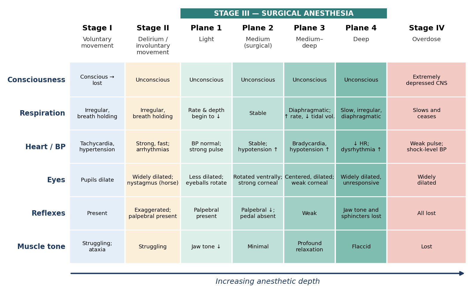
Figure 7.1. Signs of the stages and planes of general anesthesia.
Stage I: Voluntary Movement
Stage I lasts from the initial administration of anesthetic to the loss of consciousness. Epinephrine release may cause tachycardia, hypertension, and irregular or increased respirations, and the patient may hold its breath. The pupils dilate, struggling may be present, and progressive ataxia is seen. Some analgesic effects may be present at the transition from Stage I to Stage II.
Stage II: Delirium or Involuntary Movement
Stage II lasts from the onset of unconsciousness until a regular breathing pattern resumes. Voluntary control is lost as the CNS is depressed, and reflexes become primitive and exaggerated. External stimuli can provoke struggling, breath holding, tachypnea, and hyperventilation. Continued catecholamine release causes a strong, fast heartbeat, and cardiac arrhythmias may occur. The pupils dilate widely, and nystagmus is common in horses. The eyelash and palpebral reflexes are present. Vocalization, excessive salivation, vomiting, and, in susceptible species, laryngeal spasm may occur.
Stimulation of any kind should be avoided during Stage II. A smooth, rapid induction moves the animal through this stage quickly, which is one goal of premedication (Chapter 6).
Stage III: Surgical Anesthesia
Stage III is characterized by unconsciousness with progressive depression of the reflexes. Ventilation becomes slow and regular, then progressively decreases in rate and depth. Muscles relax, the swallowing and vomiting reflexes are lost, and bradycardia progresses. Depending on the reference, Stage III is divided into three or four planes:
Plane 1 (light) persists until eyeball movement ceases. Blood pressure returns to normal and the pulse is strong. Respiratory rate and depth begin to decrease, the pupils become less dilated, and the eyeballs may rotate. The eyelash and palpebral reflexes are present, jaw tone is decreased, and there is a slight reaction to surgical manipulation.
Plane 2 (medium) is the plane of surgical anesthesia, acceptable for most surgical procedures. Respiration and pulse rate are stable, laryngeal reflexes are absent, and muscle relaxation and analgesia are adequate. Hypotension increases and capillary refill time begins to slow. The palpebral reflex diminishes, the corneal reflex is strong, and the eyeball rotates ventrally. Abdominal muscle tone and jaw tone are minimal, the pedal reflex is absent, and the probability of cardiac dysrhythmia is low.
Plane 3 (medium–deep) is deep surgical anesthesia. Intercostal muscle function decreases, so diaphragmatic breathing appears, with increased respiratory rate and decreased tidal volume. The corneal reflex is weak, muscle relaxation is profound, and the pupils are centered and dilated. Bradycardia intensifies and hypotension continues to increase.
Plane 4 (deep) brings an increasing probability of dysrhythmia. Respirations are slow, irregular, and diaphragmatic, and heart rate falls. Cyanosis may be seen. The pupils are widely dilated and unresponsive to light, muscle tone is flaccid, and jaw tone and sphincter control are lost.
Stage IV: Overdose
In Stage IV the CNS is extremely depressed. Respirations slow and cease, the pulse is weak or imperceptible, and blood pressure falls to shock levels. Capillary refill time is greatly increased, mucous membranes are pale, the pupils are widely dilated, and all reflexes and muscle tone are lost. The probability of cardiac dysrhythmia is at its highest. Death is imminent unless corrective measures are taken:
Withdraw the anesthetic.
Provide oxygen.
Initiate artificial respiration.
Chapter 18 covers emergency procedures, including cardiopulmonary resuscitation.
Why it matters
The eye signs are useful because they change in a predictable order, but they can mislead. Pupils are dilated in Stage II and again in deep Plane 3–4 and Stage IV, so a dilated pupil alone does not tell you whether the animal is too light or too deep. Always interpret eye signs together with reflexes, jaw tone, respiration, and cardiovascular parameters.
7.5 Anesthesia Pharmacokinetics
Pharmacokinetics describes what the body does to a drug: how it is absorbed, distributed, metabolized, and eliminated. General anesthesia is produced by the anesthetic's action on the brain and spinal cord, and that action depends on the plasma concentration of the agent. Unless an agent is given directly into the cerebrospinal fluid, it is carried to the CNS by the blood.
Body Compartments
For pharmacokinetic purposes, the body can be divided into compartments that differ in blood supply and in their tissue–blood partition coefficients (how readily the drug dissolves in that tissue compared with blood):
Vessel-rich: brain, liver, heart, and kidney. These receive a large share of cardiac output and take up drug quickly.
Intermediate: muscle and skin.
Vessel-poor: adipose and residual tissue. These take up drug slowly but can hold a great deal of it.
The concentration of a drug at its site of action is also affected by binding to plasma proteins, which keeps the drug from penetrating cell membranes, and by absorption into tissues that store, metabolize, and excrete drugs. After entering the bloodstream, a drug enters the various tissues according to their perfusion, their capacity for the drug, and the partial pressure gradient between blood and tissue.
Partial Pressure Gradients
The concentration of drug in blood and tissues is generally governed by partial pressure gradients (Figure 7.2). Molecules move randomly. Where there are more molecules on one side of a boundary than the other, more of them happen to cross from the crowded side to the less crowded side than the reverse. This movement is passive, not active. Eventually the system reaches equilibrium, with equal numbers of molecules moving in each direction. However, differences in permeability or solubility between two areas can result in a higher concentration on one side of a membrane than the other at equilibrium.
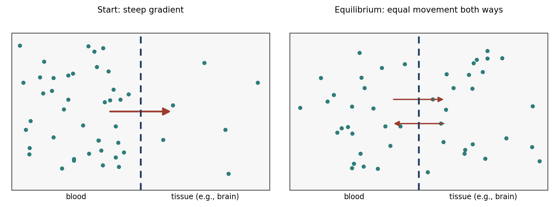
Figure 7.2. Diffusion down a partial pressure gradient. Net movement stops at equilibrium, although molecules continue to cross in both directions.
The Blood-Brain Barrier
The ultimate effect of any general anesthetic depends on its ability to cross the blood-brain barrier, which has permeability characteristics similar to cell membranes. Penetration by drugs that are non-lipophilic, ionized, or protein-bound is limited. This is why the best anesthetics are lipid-soluble and why, as Chapter 6 noted, charged molecules such as glycopyrrolate have limited CNS effects.
7.5.1 Effects of Anesthetic Route on Pharmacokinetics
Intravenous (IV) administration eliminates the absorption phase, so the onset of action is quicker than by any other route. The plasma concentration falls rapidly as the drug is taken up by vessel-rich tissues such as the brain, producing a quick onset of anesthesia. Then, following the pressure gradient, the drug re-enters the blood from these tissues and is redistributed to muscle, fat, and vessel-poor tissues (Figure 7.3). This redistribution, not metabolism, is what ends the effect of a single IV dose. The duration of action is therefore shorter than with other injectable routes, but the drug remains in the body longer than an inhalant does.
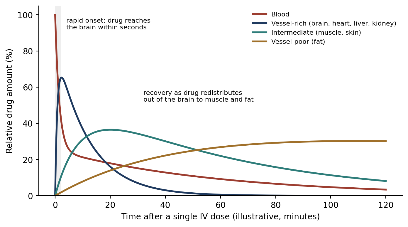
Figure 7.3. Redistribution of an injectable anesthetic after a single IV dose (schematic). The brain is part of the vessel-rich group.
Other injectable routes (IM, SC, IP, and so on) require absorption into the plasma. The gradual rise in plasma levels causes a more gradual flow of drug into the brain, so onset is delayed but the effect lasts longer than with IV administration. These routes typically require a higher dose than IV, and redistribution and metabolism begin while the drug is still being absorbed.
Inhalation anesthetics are volatile organic compounds with molecules smaller than injectable agents. They permeate the bloodstream, tissues, and blood-brain barrier quickly, and they are primarily exhaled rather than biotransformed.
7.5.2 Elimination
Circulation carries drugs to vessel-rich organs that can metabolize, eliminate, or excrete them. The liver is primarily responsible for biotransformation (metabolism), the kidneys for excretion, and the lungs for elimination of gases. The rate of biotransformation is determined by the drug concentration at the site of metabolism.
Most drug metabolism follows first-order kinetics: a constant fraction of the drug is metabolized in a given period. If the metabolic pathways become saturated, metabolism follows zero-order kinetics: a constant amount is eliminated per unit time, regardless of how much drug is present (Figure 7.4). Species variation exists in biotransformation and metabolism, which is one reason doses cannot simply be transferred from one species to another.
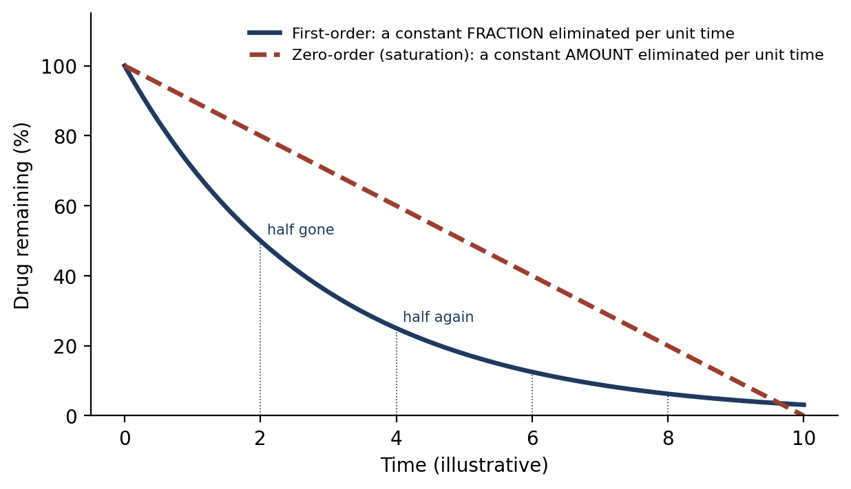
Figure 7.4. First-order and zero-order elimination.
Several factors modify distribution and elimination:
Fear, struggling, fever, and increased cardiac output change how quickly a drug circulates and equilibrates between blood and tissues.
Shock preserves blood flow to the brain while reducing flow elsewhere. This decreases the potential for redistribution, dilution, distribution, and excretion of the drug, causing a rapid induction and prolonged recovery.
Hypercarbia and hypocarbia (high and low blood carbon dioxide) also alter drug distribution.
⚠ Check current guidance
The effects of fear, struggling, fever, and cardiac output on drug distribution are described differently in different references. Check how your exam references explain these modifying factors.
7.6 Anesthetic Issues with Disease & Pathology
Disease affects both how an animal handles anesthetic drugs and how well it tolerates their side effects. The pre-anesthetic evaluation (Chapter 5) identifies these conditions so that drugs and techniques can be chosen to minimize risk. Table 7.5 at the end of this section summarizes the key points.
7.6.1 Cardiovascular (CV) Dysfunction
Most pre-anesthetic and anesthetic agents cause cardiovascular depression. Animals with CV dysfunction are more prone to fluid overload and arrhythmias, and because they often have poor oxygenation to begin with, they should be pre-oxygenated for 5–7 minutes before induction. Drugs that may induce tachycardia or large changes in vascular resistance should be used with extreme care and only when there is no alternative. Alpha-2-adrenergic agonists should be avoided in patients with impaired cardiac output.
7.6.2 Pulmonary Dysfunction
Most pre-anesthetic and anesthetic agents also cause respiratory depression. Pulmonary dysfunction may be caused by diaphragmatic hernia, pneumothorax, hydrothorax, pneumonia, pulmonary edema, atelectasis, or airway obstruction.
The anesthetist must balance lowering doses, especially of pre-anesthetic drugs, to limit further respiratory compromise, against preventing anxiety, which itself increases oxygen demand. Useful approaches include:
Low-dose acepromazine combined with butorphanol to provide enough sedation. Phenothiazine tranquilizers have minimal effect on ventilation, and mixed agonist-antagonist opioids cause less respiratory depression than pure agonists.
Remembering that atropine and glycopyrrolate decrease airway resistance but increase the viscosity of airway secretions.
Regional anesthesia, which is gaining popularity in these cases when possible.
Pre-oxygenating for 5–7 minutes before induction, if the animal's condition allows the extra time.
If general anesthesia is necessary, intubation and ventilation are essential. A rapid induction following sedation may be needed to gain quick control of the airway with intubation and positive pressure ventilation; this is best achieved with IV thiopental, propofol, etomidate, or ketamine. Mask induction may cause excessive struggling. Anesthesia is best maintained with inhalants and positive pressure ventilation. Use nitrous oxide with caution, because it can increase the severity of a pneumothorax, and discontinue it if cyanosis is present.
7.6.3 Neurologic Disease
Neurologic disease in this context usually refers to spinal cord disorders and, to a lesser extent, intracranial disorders. In normal, conscious animals, cerebral blood flow (CBF) and intracranial pressure (ICP) are tightly regulated, but anesthetics can interfere with that regulation. In head trauma patients, ICP and CBF are already increased, so a further loss of regulation can cause additional brain damage.
Isoflurane, sevoflurane, etomidate, and barbiturates provide some cerebral protection.
The direct effects of opioids on CBF and ICP are minimal, but end-tidal CO₂ or blood gases should be monitored and the patient ventilated if needed to prevent hypercapnia, because CO₂ dilates cerebral vessels and raises ICP.
Hyperventilation to end-tidal CO₂ levels of 30–35 mmHg often eliminates volatile-anesthetic-induced increases in CBF.
Nitrous oxide causes the greatest increases and should be avoided in neurosurgery.
Monitor blood pressure closely before and during anesthesia. If it is elevated before anesthesia, do not allow it to drop during anesthesia, because the brain may depend on that pressure for perfusion.
Restrict fluids to the minimum necessary to maintain adequate circulating volume and cardiac output, to avoid worsening brain swelling.
7.6.4 Renal Disease
All anesthetic agents are likely to decrease glomerular filtration rate by decreasing renal blood flow, and renal ischemia may occur during anesthesia because of systemic hypotension or renal vasoconstriction. Although the direct effects of anesthesia may end when surgery ends, some patients do not fully regain the ability to regulate urine production for several days. Key considerations are:
Lower doses of barbiturates and other injectable drugs may be required because of acidosis, which increases the proportion of active, unbound drug.
Potassium may be elevated (hyperkalemia). Patients with serum potassium higher than 5.5–6 mEq/L should not be anesthetized until levels are lowered. Hyperkalemia appears on the ECG as peaked T waves, a prolonged PR interval, wide QRS complexes, and loss of P waves.
Succinylcholine can raise potassium to life-threatening levels.
Monitor for post-operative oliguria (low urine output).
Chronic renal failure can cause anemia; patients undergoing anesthesia should receive a transfusion if hematocrit is below 18–20%.
Avoid nephrotoxic drugs, such as NSAIDs.
Maintain mean arterial pressure (MAP) above 70–80 mmHg to preserve renal blood flow.
Monitor central venous pressure (CVP) to prevent volume overload and assess myocardial function, maintaining it at 3–5 cm H₂O.
7.6.5 Hepatic Disease
The liver receives blood from both the hepatic artery and the portal vein, and anesthetics can affect hepatic blood flow by changing the tone of either. Halothane decreases portal vein flow but has little effect on hepatic artery flow. Isoflurane decreases portal vein flow but increases hepatic artery flow, for a net increase in overall flow. Sevoflurane and desflurane are associated with decreased total hepatic blood flow. Decreased blood flow may delay drug elimination.
Acepromazine, thiobarbiturates, droperidol, and alpha-2-adrenergic agonists should be avoided in patients with moderate to severe liver disease, and phenothiazine (acepromazine) and butyrophenone (droperidol) tranquilizers may cause hypotension. Propofol, ketamine, and inhalation anesthetics are generally the safest choices.
7.6.6 Gastrointestinal (GI) Disease
A damaged GI tract may release toxins into the bloodstream. Conditions such as bloat and gastric dilatation-volvulus can decrease cardiac function and ventilation by compressing the large veins and the diaphragm. Metabolic alkalosis may occur from gastric sequestration of hydrogen ions; in advanced disease, metabolic acidosis may develop as decreased cardiac output and poor ventilation increase lactate production, which can lead to cardiac arrhythmias.
Measure serum electrolytes, pH, and bicarbonate before surgery, and use fluid therapy to correct acid-base imbalances.
Avoid large doses of arrhythmogenic agents (such as thiobarbiturates and halothane) and alpha-2 agonists.
Xylazine may decrease gastroesophageal sphincter pressure and increase GI reflux, decrease intestinal motility, and decrease cardiac output.
Nitrous oxide is contraindicated before gastric decompression, because it increases intragastric volume and pressure.
Good induction choices include neuroleptanalgesic combinations, propofol, and diazepam/ketamine. Maintenance with isoflurane or sevoflurane is recommended.
Why it matters
Nitrous oxide is far more soluble in blood than nitrogen. When it is given, it diffuses into gas-filled spaces (a distended stomach, a pneumothorax, an obstructed bowel) much faster than nitrogen can leave them, so the space expands and its pressure rises. This is the common reason nitrous oxide is avoided in pneumothorax, GI distension, and neurosurgery.
7.6.7 Endocrine Disorders
Common examples are diabetes, Addison's disease, Cushing's syndrome, hypothyroidism, and hyperthyroidism. The specific anesthetic regimen is less important than proper treatment of the condition itself. The patient should be stabilized before anesthesia whenever possible, and agents should be selected for the shortest recovery time and/or easiest reversibility.
Table 7.5. Summary of anesthetic considerations in disease.
| System | Key concerns | Key recommendations |
|---|---|---|
| Cardiovascular | CV depression; fluid overload; arrhythmias; poor oxygenation | Pre-oxygenate 5–7 min; avoid drugs causing tachycardia or large changes in vascular resistance; avoid alpha-2 agonists if cardiac output is impaired |
| Pulmonary | Respiratory depression; anxiety increases demand | Low-dose ace + butorphanol; regional anesthesia; pre-oxygenate; rapid IV induction; intubate and ventilate; caution with N₂O |
| Neurologic | Loss of CBF/ICP regulation; hypercapnia raises ICP | Isoflurane, sevoflurane, etomidate, barbiturates; ventilate to EtCO₂ 30–35 mmHg; avoid N₂O; maintain BP; restrict fluids |
| Renal | Reduced renal blood flow; acidosis; hyperkalemia; anemia | Don't anesthetize if K⁺ > 5.5–6 mEq/L; avoid succinylcholine and NSAIDs; transfuse if Hct < 18–20%; MAP > 70–80 mmHg; CVP 3–5 cm H₂O |
| Hepatic | Reduced hepatic blood flow delays elimination | Avoid acepromazine, thiobarbiturates, droperidol, alpha-2 agonists; propofol, ketamine, inhalants safest |
| Gastrointestinal | Toxins; impaired cardiac function and ventilation; acid-base imbalance | Check electrolytes, pH, bicarbonate; avoid arrhythmogenic agents, alpha-2 agonists, and N₂O before decompression; neuroleptanalgesics, propofol, or diazepam/ketamine; maintain on isoflurane or sevoflurane |
| Endocrine | Underlying disease | Stabilize first; choose agents for short recovery and easy reversal |
⚠ Check current guidance
Thiopental (a thiobarbiturate) and halothane are no longer commercially available in the United States, but they appear frequently in anesthesia references and exam materials.
Chapter Summary
Anesthesia ranges from local and regional analgesia to general anesthesia, a reversible, drug-induced unconsciousness with analgesia. Balanced anesthesia uses several drugs, each targeting one component. Tranquilization, sedation, hypnosis, and narcosis are lesser states of CNS depression. Injectable anesthetics end their effect by redistribution and metabolism and offer less control; inhalants are exhaled and offer more control, with potency compared by MAC. Higher metabolic rates (small size, activity, males, recent feeding) increase anesthetic requirements; fat, disease, and age decrease them.
Anesthesia progresses through Stage I (voluntary movement), Stage II (delirium; avoid stimulation), Stage III (surgical anesthesia, with Plane 2 suitable for most surgery), and Stage IV (overdose; withdraw anesthetic, give oxygen, ventilate). Drugs move along partial pressure gradients, must cross the blood-brain barrier, and after IV injection are redistributed from vessel-rich to vessel-poor tissues; most are eliminated by first-order kinetics. Cardiovascular, pulmonary, neurologic, renal, hepatic, GI, and endocrine disease each call for specific adjustments in drug choice and monitoring.
Key Terms
Analgesia: Freedom from, or absence of, pain.
Balanced anesthesia: Anesthesia using multiple drugs, each targeting a component of the anesthetic state.
Basal metabolic rate (BMR): The rate of energy use by the body at rest.
Blood-brain barrier: The barrier between blood and brain tissue that limits passage of non-lipophilic, ionized, or protein-bound drugs.
Dissociative anesthesia: Anesthesia in which drugs such as ketamine dissociate the thalamocortical and limbic systems, producing a cataleptoid state with open eyes, intact swallowing, and muscle hypertonus.
First-order kinetics: Elimination of a constant fraction of drug per unit time.
General anesthesia: Drug-induced, reversible unconsciousness with analgesia, in which the patient is not aroused by noxious stimuli.
Hyperkalemia: Elevated blood potassium.
Hypnosis: Artificially induced sleep from which the patient is readily aroused.
MAC: Minimum (median) alveolar concentration: the end-tidal inhalant concentration that prevents movement to a noxious stimulus in 50% of subjects.
Narcosis: Drug-induced deep sleep from which the patient is not easily aroused; may or may not include analgesia.
Partial pressure gradient: A difference in concentration that drives passive movement of molecules from higher to lower concentration.
Pharmacokinetics: The absorption, distribution, metabolism, and elimination of drugs.
Redistribution: Movement of a drug from vessel-rich tissues (including the brain) to muscle and fat, ending its effect.
Sedation: Central depression with drowsiness; the patient is unaware of surroundings but responds to pain.
Tranquilization: Relief of anxiety; the patient is relaxed but aware.
Zero-order kinetics: Elimination of a constant amount of drug per unit time, occurring when metabolism is saturated.
Review Questions
1. Which term describes a state in which anxiety is relieved and the patient is relaxed but still aware of its surroundings?
A. Sedation
B. Narcosis
C. Tranquilization
D. Hypnosis
2. Balanced anesthesia is best described as:
A. Using one drug at a high dose
B. Using multiple drugs, each targeting a component of the anesthetic state
C. Alternating between injectable and inhalant agents
D. Anesthesia with equal amounts of two drugs
3. MAC is defined as the end-tidal concentration of an inhalant, at 1 atmosphere, that:
A. Kills 50% of subjects
B. Produces immobility in 50% of subjects exposed to a noxious stimulus
C. Produces unconsciousness in all subjects
D. Causes apnea in 50% of subjects
4. Compared with a larger animal, a smaller animal generally requires:
A. A smaller dose per unit of body weight
B. A larger dose per unit of body weight
C. The same dose per unit of body weight
D. No anesthetic
5. During which stage of anesthesia should all stimulation be avoided because of exaggerated reflexes, struggling, and possible arrhythmias?
A. Stage I
B. Stage II
C. Stage III, Plane 2
D. Stage IV
6. Which plane of Stage III is acceptable for most surgical procedures?
A. Plane 1
B. Plane 2
C. Plane 3
D. Plane 4
7. After a single IV dose of an injectable anesthetic, the animal wakes primarily because the drug:
A. Is exhaled
B. Redistributes from the brain to muscle, fat, and other tissues
C. Is instantly metabolized by the liver
D. Is excreted unchanged by the kidneys within seconds
8. First-order kinetics means that:
A. A constant amount of drug is eliminated per unit time
B. A constant fraction of drug is eliminated per unit time
C. The drug is never eliminated
D. Elimination occurs only in the lungs
9. A patient's serum potassium is 6.5 mEq/L. What should be done?
A. Anesthetize normally
B. Give succinylcholine to aid intubation
C. Lower the potassium level before anesthesia
D. Restrict fluids
10. Why should nitrous oxide be avoided before gastric decompression and in patients with pneumothorax?
A. It causes hypertension
B. It increases the volume and pressure of gas-filled spaces
C. It is metabolized by the liver
D. It causes bradycardia
Answer Key
1. C. Tranquilization relieves anxiety while the patient remains aware. A sedated patient is unaware of its surroundings but responds to painful manipulation.
2. B. Balanced anesthesia uses several drugs, each aimed at a specific component (consciousness, analgesia, muscle relaxation, or autonomic reflexes), so lower doses of each can be used.
3. B. MAC is the end-tidal concentration that prevents movement in response to a noxious stimulus, such as an incision, in 50% of subjects.
4. B. Smaller animals have a higher basal metabolic rate per unit of body weight, so they require a larger dose per kilogram.
5. B. Stage II is the stage of delirium or involuntary movement. Reflexes are exaggerated, and stimulation can trigger struggling, breath holding, vomiting, laryngospasm, or arrhythmias.
6. B. Plane 2 provides stable respiration and pulse, adequate muscle relaxation and analgesia, an absent pedal reflex, and a low probability of dysrhythmia.
7. B. Following the pressure gradient, the drug moves back out of the brain into the blood and is redistributed to muscle, fat, and vessel-poor tissues, lowering brain concentration.
8. B. In first-order kinetics a constant fraction is metabolized in a given period. Zero-order kinetics (a constant amount per unit time) occurs when metabolic pathways are saturated.
9. C. Patients with serum potassium higher than 5.5–6 mEq/L should not be anesthetized until levels are lowered. Succinylcholine can raise potassium to life-threatening levels.
10. B. Nitrous oxide diffuses into gas-filled spaces faster than nitrogen leaves them, increasing intragastric volume and pressure and the severity of a pneumothorax.
Chapter 8: Injectable Anesthetic Agents
Learning objectives
After studying this chapter, you should be able to:
Explain the general principles of injectable anesthesia, including dosing to effect and the need for drug combinations.
Classify barbiturates by duration of action, and describe their effects, administration, and the factors that alter their action.
Compare the oxybarbiturates and thiobarbiturates in common use.
Describe the properties and uses of neurosteroids, propofol, etomidate, and other non-barbiturate injectable agents.
Describe the effects of dissociative anesthetics, including ketamine and tiletamine-zolazepam.
Injectable anesthetics are widely used in research, alone or as induction agents before inhalant maintenance. Several principles apply to them all:
Each agent has relatively specific effects within the CNS, so a combination of drugs is generally needed to affect all necessary body systems adequately.
IV agents other than ketamine provide only mental depression. Analgesics and/or inhalant anesthetics are needed to provide all the components of general anesthesia.
The advantage of this is that drugs can be chosen to target specific physiological effects.
Responses to injectable agents vary between individuals, more than responses to inhalants do.
Doses are a guide, not an absolute. When given IV, injectable agents should be given to effect.
Opioids are NOT anesthetics, but they reduce the anesthetic dose required.
⚠ Check current guidance
Many injectable agents in this chapter (including thiopental, thiamylal, Althesin, chloral hydrate, and metomidate) are no longer commercially available in the United States or are rarely used, but they still appear in references and exam materials. Doses and availability should always be confirmed with a veterinarian and current formularies.
8.1 Barbiturates
Barbiturates were first prepared by Conrad and Gutzeit in 1882. They contain a pyrimidine nucleus; the parent compound, barbituric acid, has no hypnotic activity itself, and its activity comes from the side groups added to it. Barbiturates act directly on CNS neurons in a manner similar to gamma-aminobutyric acid (GABA), the brain's main inhibitory neurotransmitter, and they inhibit the synaptic actions of some excitatory neurotransmitters. They provide poor analgesia at safe doses.
Barbiturates are divided into four groups by duration of action (Figure 8.1). The short- and ultra-short-acting barbiturates are used for clinical anesthesia; the intermediate- and long-acting barbiturates are used for sedation and seizure control.
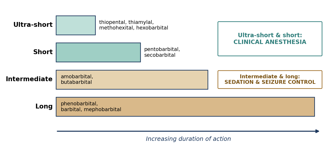
Figure 8.1. Classification of barbiturates by duration of action, with representative agents.
Table 8.1. Barbiturate groups.
| Group | Agents | Primary use |
|---|---|---|
| Ultra-short-acting | Hexobarbital, Kemithal, thiamylal, thiopental (also methohexital, Section 8.1.3) | Clinical anesthesia (induction) |
| Short-acting | Cyclobarbital, Cyclopal, pentobarbital, secobarbital | Clinical anesthesia |
| Intermediate-acting | Allylbarbituric acid, amobarbital, aprobarbital, butabarbital, butallylonal, butethal, hexethal, probarbital, propallylonal, vinbarbital | Sedation; seizure control |
| Long-acting | Barbital, diallylbarbituric acid, mephobarbital, phenobarbital | Sedation; seizure control |
8.1.1 Effects of Administration
Barbiturate effects depend on dose. At hypnotic doses, there is little effect on respiration or basal metabolic rate. At anesthetic doses:
Respiration is depressed.
There is cardiovascular depression, both central and peripheral: cardiac output and stroke volume fall, heart rate increases (as a reflex), and blood pressure decreases.
Basal metabolic rate and body temperature are lowered.
The drug crosses cell membranes and the placenta.
Barbiturates bind to plasma protein. Cerebrospinal fluid contains less protein than plasma, so at equilibrium the CSF has a lower barbiturate concentration.
Blood pH strongly affects barbiturate action (Figure 8.2). Barbiturates are weak acids, and only the un-ionized form crosses cell membranes readily. In acidosis (pH below 7.4), more of the drug is un-ionized, so more penetrates cells and anesthesia deepens. In alkalosis, caused by hyperventilation or alkalinizing agents, more is ionized, effectiveness falls, and anesthesia lightens.
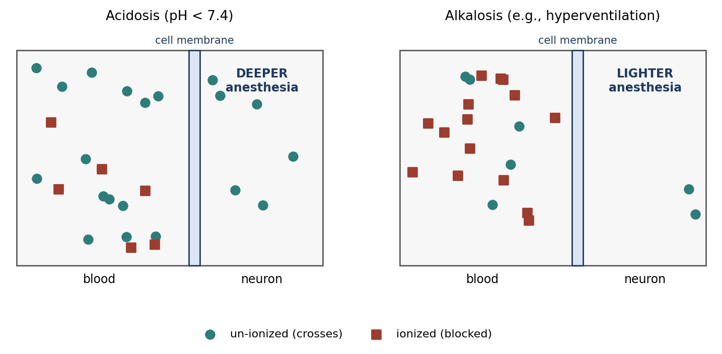
Figure 8.2. Effect of blood pH on barbiturate penetration into nerve cells.
Ultra-short-acting barbiturates are redistributed faster, NOT metabolized faster, than short-acting barbiturates. Their brief action comes from rapid movement out of the brain into muscle and fat (Chapter 7, Figure 7.3).
The "Glucose Effect"
During recovery from barbiturate anesthesia, giving glucose can cause re-anesthetization in some species. Susceptibility varies widely (Table 8.2), which matters when choosing post-operative fluids.
Table 8.2. Species susceptibility to the glucose effect.
| Susceptibility | Species |
|---|---|
| Susceptible | Guinea pigs, chickens, pigeons, rabbits, hamsters |
| Intermediate | Dogs |
| Not susceptible | Mice, rats, goldfish, tadpoles |
8.1.2 Administration
IV administration is the preferred route, because it allows dosing to effect (Figure 8.3):
Rapidly inject the first 1/2 to 1/3 of the calculated dose. Slow injection may allow the animal to pass slowly through the excitement stage.
Give the remainder slowly, to effect, assessing anesthetic depth as you go.
Administration through a catheter is recommended, because barbiturate solutions are highly alkaline and irritating.
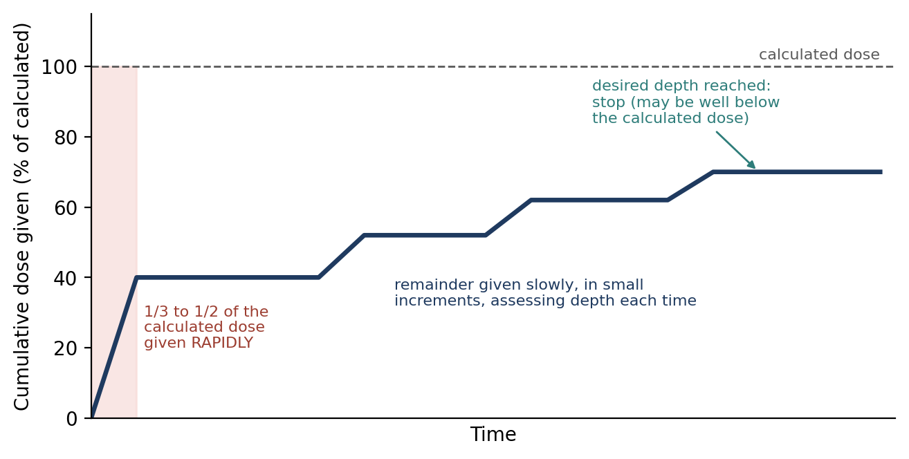
Figure 8.3. Giving an IV anesthetic to effect. The total dose given depends on the animal's response, not only on the calculated dose.
Perivascular injection (outside the vein) results in "barbiturate slough", a tissue injury that takes 2–4 weeks to heal and will scar. If it occurs:
Infiltrate the area with lidocaine or 2% procaine (1–2 mL) to prevent vasospasm, aid dilution and absorption of the barbiturate, and change the acidity to help minimize damage.
Saline may also be infused to further dilute the barbiturate.
Corticosteroids, NSAIDs, and hot packs may also help.
IP and IM administration are not widely used except in rodents. They do not allow dosing to effect and can be quite painful. IM or intrathecal injection may be indicated for wild animals. Oral administration is variable and is usually used for sedation.
8.1.3 Oxybarbiturates
Oxybarbiturates have an oxygen atom at a key position in the molecule; thiobarbiturates (Section 8.1.4) have a sulfur atom there instead, which makes them more lipid-soluble and faster-acting.
Phenobarbital sodium is a long-acting, effective anticonvulsant that is considerably cheaper than newer drugs. It is excreted slowly in the urine and tends to be cumulative, so an oral loading dose is given first, followed by a daily maintenance dose. Overdose causes loss of motor coordination. In treating strychnine poisoning, it is given IV to effect until muscle relaxation is seen.
Pentobarbital sodium was in widespread use in cats and dogs by 1940, but has since been replaced by inhalant and balanced anesthesia for most clinical purposes. Its characteristics include:
Induction: sub-anesthetic doses often cause CNS stimulation and excitement, so it is given to effect. After the initial IV dose, arterial blood pressure decreases and heart rate increases for 10–20 minutes.
Cardiovascular and respiratory effects: anesthetic doses decrease systolic blood pressure, stroke volume, pulse pressure, central venous pressure, PaO₂, pH, and body temperature. Heart rate, PaCO₂, and peripheral resistance increase after 1.5 hours, and cardiac output decreases. MAP falls during induction but returns to awake values in about 30 minutes. Deep anesthesia depresses renal function, and the effects of high doses may closely resemble shock.
Placenta: it crosses freely, and high doses may cause high mortality among newborns.
IP injection: part of the drug is absorbed into the portal system and destroyed immediately in the liver, which is one reason IP doses are less predictable.
Recovery usually takes 6–18 hours but may last as long as 72 hours. Whining, shivering, running motions, and thrashing (delirium) may occur, so tranquilizers are advised during recovery.
It is no longer used in North America for small companion animals, cattle, and horses because of its prolonged recovery and marked respiratory depression.
⚠ Check current guidance
Pentobarbital remains widely used in research (including for euthanasia and for some rodent and non-survival procedures). Its use as a sole anesthetic for survival surgery is increasingly discouraged because of its poor analgesia. Check your institution's policies.
Methohexital sodium (Brevital) is an ultra-short-acting oxybarbiturate that contains no sulfur atom. Its short duration is due more to redistribution than to metabolism. Its lethal dose is about 2.5 times the median anesthetic dose, and the main danger of overdose is respiratory failure. The solution is stable for as long as 6 months at room temperature. Recovery is quick but difficult, with muscle tremors or violent excitement, even with pre-anesthetic sedation; dogs are ambulatory about 30 minutes after injection. It is a good induction drug but causes transient apnea, so be ready to intubate and support respiration. It is best followed by inhalant anesthesia for maintenance.
8.1.4 Thiobarbiturates
Thiopental sodium was the first thiobarbiturate to become popular as an anesthetic agent for animals. Key points:
Storage: store refrigerated. It is unstable in aqueous solution; as solutions age they become turbid and crystals precipitate, causing a loss of activity but not increased toxicity.
Metabolism: it produces about 12 metabolic products excreted in the urine; 86% is excreted within 4 days. Its ultra-short action is due to rapid redistribution and localization in fat, not rapid metabolism.
Cumulative effect: repeated doses accumulate (as fat becomes saturated), leading to possible prolonged anesthesia.
Cardiopulmonary effects: it initially causes marked respiratory depression. About five minutes after administration, heart rate, aortic pressure, peripheral vascular resistance, and left ventricular pressure increase. Arrhythmias are accentuated by xylazine, halothane, methoxyflurane, and epinephrine. Cardiopulmonary depression is reduced if it is given with a lidocaine bolus (11 mg/kg).
Other effects: pronounced hyperglycemia occurs during prolonged thiopental anesthesia.
Contraindications: neonates, and feline porphyria.
Horses: commonly mixed with 5% guaifenesin for equine anesthesia.
Thiamylal sodium has an anesthetic potency in dogs about 1.5 times that of thiopental. Normal saline is suggested as a diluent, and solutions are stable for up to 14 days. It is less cumulative than thiopental and tolerance with repeated injections has not been noted. It is more arrhythmogenic than thiopental but has fewer cardiovascular effects. A single bolus provides about 15 minutes of surgical anesthesia. It may be used alone or with guaifenesin in horses and cattle, may produce apnea, and is described as very safe and nontoxic.
Table 8.3. Comparison of barbiturates used in anesthesia.
| Agent | Type / duration | Key points |
|---|---|---|
| Phenobarbital | Oxybarbiturate; long | Anticonvulsant; cumulative; loading dose then maintenance; strychnine poisoning |
| Pentobarbital | Oxybarbiturate; short | Excitement at sub-anesthetic doses; recovery 6–18 h (up to 72 h); crosses placenta; IP partly destroyed by liver |
| Methohexital | Oxybarbiturate; ultra-short | No sulfur; LD ≈ 2.5× median anesthetic dose; transient apnea; rough recovery; good induction agent |
| Thiopental | Thiobarbiturate; ultra-short | Refrigerate; redistribution into fat; cumulative; arrhythmias with xylazine, halothane, epinephrine; avoid in neonates and feline porphyria |
| Thiamylal | Thiobarbiturate; ultra-short | 1.5× thiopental potency; less cumulative; more arrhythmogenic; ~15 min per bolus |
Why it matters
Because barbiturates end their effect mainly by redistribution, the first dose wears off quickly. But each additional dose adds to the drug already stored in fat and muscle, so later doses wear off more and more slowly. This is why repeated barbiturate doses can produce unexpectedly long recoveries.
8.2 Non-Barbiturate Anesthetic Drugs
8.2.1 Neurosteroids
Althesin is a combination of the steroids alphaxalone and alphadolone acetate. Like barbiturates, neurosteroids work by enhancing GABA-mediated neurodepression, and Althesin should not be used along with barbiturates. Its characteristics include:
An exceptionally high therapeutic index with little cumulative effect; additional doses are not cumulative.
Anesthetic duration that varies among species; onset in 6–12 minutes and a 15-minute duration.
Neutral pH, so perivascular injection does not cause damage or pain. IM administration produces variable results.
Good muscle relaxation.
Side effects including urination, defecation, muscle tremors, paddling, salivation, and hyperesthesia.
In cats, possible edema of the feet, ears, and muzzle; in dogs, possible allergic reactions with decreased blood pressure and wheals at the injection site. These are usually transient.
Violent recovery in horses, prevented by giving xylazine beforehand.
Alfaxan-CD is a newer neurosteroid formulation that does not cause histamine release. The allergic reactions seen with Althesin were caused largely by its solvent (Cremophor EL); Alfaxan uses a cyclodextrin carrier instead.
⚠ Check current guidance
Althesin has been withdrawn from the market. Alfaxalone (Alfaxan) is now approved in the United States and other countries for use in dogs and cats and is used in many research species. Check current product information.
8.3 Miscellaneous Injectable Anesthetics
Chloral Hydrate, U.S.P.
Chloral hydrate may be given orally or, as a solution, IV or IP. Oral administration may cause vomiting, and perivascular administration is irritating. It depresses the cerebrum; sub-anesthetic doses do not affect motor and sensory nerves. It is a good hypnotic but a poor anesthetic: the amount needed for anesthesia is close to the lethal dose. It produces deep sleep lasting several hours, with weak analgesic action, and decreases respiration and blood pressure. A small amount is excreted unchanged in the urine. Doses vary extensively, and it is no longer used often because safer and more effective drugs are available.
Chloralose
Alpha-chloralose is produced by heating anhydrous glucose with trichloroacetaldehyde in a water bath. It produces minimal cardiovascular depression and better preserves active reflexes, with less depression of cortical neuronal function than pentobarbital. These properties make it valuable for long-duration, non-survival experiments, especially studies of reflexes and nervous system function.
Magnesium Sulfate
Magnesium sulfate globally depresses the CNS. It may be used for euthanasia only if given AFTER the animal has been rendered unconscious with another agent, because it is not itself an anesthetic and causes distress in a conscious animal.
Metomidate (Hypnodil)
Metomidate is a hypnotic with muscle-relaxant properties that induces sleep without analgesia. General anesthesia can be produced by combining it with neuroleptics or analgesics. Apnea may occur with rapid IV injection. It is often used as a sedative-anesthetic for fish.
Etomidate
Etomidate enhances the action of GABA. A single injection produces a relatively brief, dose-related hypnosis. Its outstanding feature is cardiovascular and respiratory stability: it does not depress cardiovascular or respiratory centers or cause histamine release, and it does not trigger malignant hyperthermia in swine.
Etomidate decreases the cerebral metabolic rate of oxygen consumption, has anticonvulsant properties, and may protect the brain after episodes of global ischemia, which makes it a good induction drug for neurosurgical procedures. It may be a preferred induction agent for traumatized patients and for those with cardiovascular or respiratory difficulties, cirrhosis, intracranial lesions, or requiring C-sections. Venous pain during injection is common (in humans). Long-term infusion is not recommended, because etomidate suppresses the adrenal glands' production of cortisol.
Propofol
Propofol is not related to barbiturates or steroid anesthetics. It is supplied as a white emulsion in soybean oil, glycerol, and egg lecithin. Because this emulsion supports microbial growth and endotoxin production, the original formulation must be discarded 6 hours after opening; a formulation with preservatives can be kept for 28 days.
[ PHOTO PLACEHOLDER ] Suggested source: Wikimedia Commons (commons.wikimedia.org), search "propofol." Check the image's license and give the attribution it requires. |
|---|
Figure 8.4. Propofol, showing its characteristic milky white emulsion.
Pharmacokinetics. Rapid uptake into the CNS produces a rapid induction. Propofol is rapidly redistributed from the brain and metabolized from the blood, and its lipophilic nature gives it a large volume of distribution. Recovery is quick and smooth.
Analgesia. Propofol is a sedative-hypnotic with minimal analgesic activity. An animal will respond to painful stimuli even under propofol anesthesia unless it has been pre-treated with an analgesic. Acepromazine on board decreases the required dose.
Other properties and cautions. Propofol decreases intracranial pressure and causes minimal fetal depression when used for C-sections. Perivascular injection causes no tissue damage, though pain on IV injection is common in humans. Repeated doses in cats may injure red blood cells (oxidative damage). It is not recommended in rabbits because of its low margin of safety, and it is relatively expensive.
⚠ Check current guidance
Propofol is now widely used for induction in many species, and newer formulations are available. Its main side effects in most species are dose-related apnea and hypotension, particularly with rapid injection. Check current references for species-specific guidance.
Tricaine Methanesulfonate (MS-222)
MS-222 is used to anesthetize amphibians and fish by bathing, gill spraying, or injection. It may be autoclaved. Solutions are acidic and are commonly buffered (for example, with sodium bicarbonate) before use in fish.
Table 8.4. Summary of non-barbiturate injectable agents.
| Agent | Mechanism / class | Key points |
|---|---|---|
| Althesin / Alfaxan-CD | Neurosteroid; enhances GABA | High therapeutic index; neutral pH; non-cumulative; Alfaxan-CD lacks histamine release |
| Chloral hydrate | Hypnotic | Good hypnotic, poor anesthetic; anesthetic dose near lethal; weak analgesia; irritating |
| Chloralose | Hypnotic | Minimal CV depression; preserves reflexes; long non-survival experiments |
| Magnesium sulfate | CNS depressant | Euthanasia only after unconsciousness |
| Metomidate | Hypnotic | Sleep without analgesia; fish sedation |
| Etomidate | Enhances GABA | CV and respiratory stability; neurosurgery; no malignant hyperthermia trigger; no long infusions |
| Propofol | Sedative-hypnotic | Rapid, smooth induction and recovery; minimal analgesia; discard in 6 h (28 days with preservative); RBC injury in cats; avoid in rabbits |
| MS-222 | Local/general anesthetic for aquatic species | Fish and amphibians; bath, gill spray, or injection |
8.4 Dissociative Anesthetics
Dissociative anesthetics work differently from other general anesthetics. Instead of producing generalized depression of brain centers, they interrupt ascending transmission from the unconscious to the conscious parts of the brain, functionally "dissociating" them. Ketamine does this largely by blocking NMDA receptors for the excitatory neurotransmitter glutamate.
The result is a cataleptoid state: the eyes remain open with a slow nystagmic gaze, muscle tone is increased (hypertonus), and muscle movements occur that are unrelated to surgical stimulation. Because the eyes stay open, they must be protected with ophthalmic lubricant.
Dissociatives are cerebral vasodilators that increase cerebral blood flow and intracranial pressure, making them undesirable for patients with head trauma, brain tumors, ocular lesions, or CNS disease. Use caution in animals with significant renal or hepatic dysfunction. They provide intense but short somatic analgesia but poor visceral analgesia.
Ketamine
Ketamine is a Schedule III controlled substance, the most common dissociative injectable, and the least potent dissociative. It produces dose-related unconsciousness and analgesia.
Onset and duration: an IV bolus rapidly crosses the blood-brain barrier, giving a rapid onset. One IV bolus lasts about 15–20 minutes, ended by rapid redistribution.
Tissue irritation: ketamine's pH is 3.5, so IM injection may cause some tissue irritation, and SC injection may cause severe irritation and tissue trauma.
Site of action: primarily the thalamoneocortical projection system.
Seizures: it does not induce seizures except in susceptible species or individuals, and may be anticonvulsant at low doses.
Analgesia: greater for somatic than visceral pain.
Cardiovascular and CNS effects: increases cerebral blood flow, intracranial pressure, CSF pressure, heart rate, MAP, and cardiac output. This sympathetic stimulation is why the survival rate for "shocky" animals is better than with halothane.
Respiration: causes a transient decrease in respiratory rate.
Recovery: hallucinatory behavior may be seen, especially in nonhuman primates.
Metabolism: dogs and horses metabolize it extensively in the liver, and repeated doses can lead to hepatic damage. In cats it is eliminated mostly unchanged by the kidneys, so sleep time is prolonged in animals with renal insufficiency.
Muscle tone: skeletal muscle tone is not reduced and may be increased in some species, so ketamine is often combined with a tranquilizer or sedative (such as a benzodiazepine or alpha-2 agonist) to provide muscle relaxation.
Telazol (Tiletamine-Zolazepam)
Telazol is a Schedule III controlled substance combining a dissociative (tiletamine) with a benzodiazepine (zolazepam), which counteracts the dissociative's muscle rigidity. Its characteristics include:
Supply: a lyophilized powder; after reconstitution, stable for 4 days at room temperature and 2 weeks refrigerated.
Onset: about 6–8 minutes IM and 60–90 seconds IV.
Duration: about 20–30 minutes in dogs and 40–50 minutes in cats.
A wide margin of safety, with rapid, smooth induction and recovery.
Retained reflexes: swallowing, coughing, pedal, corneal, and vomiting reflexes are retained, so reflexes cannot be used to judge depth in the usual way.
Good muscle relaxation and a lingering analgesic effect.
Cardiovascular effects: may increase heart rate and respiratory rate. Systolic pressure rises initially, drops slightly within five minutes, and stays at that lower level for the rest of the anesthetic period, while diastolic pressure increases throughout.
May be combined with ketamine and/or xylazine.
Table 8.5. Comparison of ketamine and Telazol.
| Property | Ketamine | Telazol |
|---|---|---|
| Composition | Dissociative alone | Tiletamine (dissociative) + zolazepam (benzodiazepine) |
| Schedule | III | III |
| Duration | ~15–20 min per IV bolus | ~20–30 min (dogs); ~40–50 min (cats) |
| Muscle tone | Not reduced; may increase | Good muscle relaxation |
| Tissue effects | pH 3.5; IM irritation; SC severe irritation | — |
| Reflexes | Eyes open, nystagmus | Swallowing, cough, pedal, corneal, vomiting retained |
| Typical combinations | Tranquilizers, benzodiazepines, alpha-2 agonists | Ketamine and/or xylazine |
⚠ Check current guidance
Reconstituted Telazol stability and storage recommendations depend on the current product label, which may differ from the values given here. Check the label before use.
Why it matters
Dissociatives are popular in research because a single IM injection can produce restraint and analgesia in species that are hard to handle or catheterize. But because reflexes, open eyes, and muscle tone persist, anesthetic depth must be judged with extra care, and visceral procedures need additional analgesia.
Chapter Summary
Injectable anesthetics are given to effect, vary between individuals, and (except ketamine) provide mental depression without analgesia, so they are combined with other drugs. Barbiturates act like GABA, provide poor analgesia, and are classified from ultra-short to long-acting; their action deepens with acidosis and lightens with alkalosis, ends mainly by redistribution, and can be prolonged by repeated doses or, in some species, by glucose. Give the first 1/3–1/2 rapidly IV, then the rest to effect, and treat perivascular injection promptly to limit slough.
Neurosteroids (Althesin, Alfaxan) have a high therapeutic index. Etomidate offers cardiovascular and respiratory stability. Propofol gives rapid, smooth induction and recovery but minimal analgesia and supports microbial growth. Chloral hydrate, chloralose, magnesium sulfate, metomidate, and MS-222 have specialized uses. Dissociatives (ketamine, Telazol) produce a cataleptoid state with good somatic but poor visceral analgesia, increase cerebral blood flow and intracranial pressure, and are usually combined with tranquilizers or sedatives.
Key Terms
Barbiturate slough: Tissue injury and sloughing caused by perivascular injection of a barbiturate.
Cataleptoid state: A trance-like state with open eyes, increased muscle tone, and movements unrelated to stimulation, produced by dissociatives.
Dissociative anesthetic: An anesthetic that interrupts transmission between unconscious and conscious parts of the brain (e.g., ketamine).
GABA: Gamma-aminobutyric acid, the main inhibitory neurotransmitter of the brain.
Glucose effect: Re-anesthetization caused by giving glucose during recovery from barbiturates in susceptible species.
Neurosteroid: A steroid anesthetic that enhances GABA-mediated inhibition (e.g., alphaxalone).
Oxybarbiturate: A barbiturate with an oxygen atom at the key position (e.g., pentobarbital, methohexital).
Perivascular: Outside a blood vessel (an injection that misses the vein).
Thiobarbiturate: A barbiturate with a sulfur atom at the key position (e.g., thiopental, thiamylal).
Therapeutic index: The ratio between a drug's toxic dose and its effective dose; a high index means a wide margin of safety.
To effect: Dosing in increments until the desired response is reached, rather than giving a fixed calculated dose.
Review Questions
1. Which injectable agent, unlike other IV agents, provides analgesia as well as mental depression?
A. Propofol
B. Thiopental
C. Ketamine
D. Etomidate
2. Ultra-short-acting barbiturates have a short duration of action because they are:
A. Metabolized faster
B. Redistributed faster
C. Excreted unchanged by the lungs
D. Not lipid-soluble
3. How does acidosis affect barbiturate anesthesia?
A. It lightens anesthesia
B. It has no effect
C. It increases the amount of barbiturate that penetrates cells, deepening anesthesia
D. It speeds metabolism
4. The "glucose effect" refers to:
A. Hypoglycemia during barbiturate anesthesia
B. Re-anesthetization when glucose is given during recovery from barbiturates
C. Glucose speeding recovery from propofol
D. Hyperglycemia caused by xylazine
5. When giving an IV barbiturate "to effect," the first portion of the calculated dose should be:
A. 1/10, given slowly
B. 1/3 to 1/2, given rapidly
C. The entire dose, given rapidly
D. 1/2, given over 30 minutes
6. Which statement about methohexital is correct?
A. It is a long-acting oxybarbiturate
B. Recovery is smooth and quiet
C. It is ultra-short-acting, and recovery may involve muscle tremors or violent excitement
D. It contains a sulfur atom
7. Which injectable agent does not depress cardiovascular or respiratory centers and is a good choice for patients with cardiovascular disease or intracranial lesions?
A. Pentobarbital
B. Etomidate
C. Ketamine
D. Chloral hydrate
8. Why must unpreserved propofol be discarded 6 hours after opening?
A. It loses potency
B. It becomes toxic to the liver
C. Its lipid emulsion supports microbial growth and endotoxin production
D. It precipitates
9. Dissociative anesthetics are undesirable in patients with head trauma because they:
A. Lower blood pressure
B. Increase cerebral blood flow and intracranial pressure
C. Cause seizures in all species
D. Have no analgesic effect
10. Telazol is a combination of:
A. Ketamine and diazepam
B. Tiletamine and zolazepam
C. Alphaxalone and alphadolone
D. Fentanyl and droperidol
Answer Key
1. C. IV agents other than ketamine provide only mental depression and require analgesics and/or inhalants to complete general anesthesia.
2. B. Ultra-short-acting barbiturates are redistributed faster, not metabolized faster, than short-acting barbiturates.
3. C. Lower pH increases the un-ionized fraction of barbiturate, which crosses cell membranes and produces deeper anesthesia. Alkalosis has the opposite effect.
4. B. In susceptible species (guinea pigs, chickens, pigeons, rabbits, hamsters), giving glucose during barbiturate recovery can cause the animal to become re-anesthetized.
5. B. The first 1/3 to 1/2 is given rapidly to carry the animal past the excitement stage; the remainder is given slowly to effect.
6. C. Methohexital is an ultra-short-acting oxybarbiturate (no sulfur). Its recovery is quick but can be difficult, even with sedation.
7. B. Etomidate does not depress CV or respiratory centers or cause histamine release, and it decreases cerebral oxygen consumption.
8. C. Propofol's soybean oil, glycerol, and egg lecithin emulsion supports microbial growth. A preserved formulation can be kept for 28 days.
9. B. Dissociatives are cerebral vasodilators that increase cerebral blood flow and intracranial pressure.
10. B. Telazol combines the dissociative tiletamine with the benzodiazepine zolazepam.
Chapter 9: Inhalation Anesthesia
Learning objectives
After studying this chapter, you should be able to:
Explain the chemistry and physical properties of inhalant anesthetics, including vapor pressure, solubility, and partition coefficients.
Describe the pharmacokinetics of inhalants and the meaning of MAC.
Compare historical and modern inhalant anesthetics, including nitrous oxide, halothane, isoflurane, sevoflurane, and desflurane.
Trace gas flow through an anesthesia machine and breathing circuit, and describe vaporizers, ventilators, CO₂ absorbers, and scavenging systems.
Compare breathing systems and their flow rates, and handle compressed gas cylinders safely.
Inhalation anesthetics differ from all other anesthetic agents because they are administered through, and mostly eliminated from, the lungs unchanged. Because metabolism is not needed to end their action, they allow rapid and predictable changes in anesthetic depth: turning the vaporizer down lightens anesthesia within minutes. The price of this control is that inhalants require specialized equipment for delivery, and that equipment must be understood and maintained to be safe.
9.1 Characteristics
All inhalant anesthetics except nitrous oxide are organic compounds. They are subdivided into:
Aliphatic hydrocarbons, such as halothane.
Ethers (two organic radicals connected by an oxygen atom), such as enflurane, methoxyflurane, isoflurane, sevoflurane, and desflurane.
It was discovered that the lack of an ether group increases the risk of cardiac arrhythmias. For this reason, all modern inhalants (newer than halothane) are ethers. It was also found that halogenation, the addition of fluorine, chlorine, or bromine, increases potency. Fluorinated compounds vary greatly in safety, reactivity, and potency.
Inhaled anesthetics exist in two physical forms:
A gas exists in gaseous form at room temperature and sea-level atmospheric pressure. Nitrous oxide (N₂O) is the only anesthetic gas.
A vapor is the gaseous state of a substance that is a liquid at ambient temperature and pressure. All the other inhalants are vapors.
Inhalant anesthetics are made of molecules moving at high speed. The impact of these molecules against the walls of their container, and against each other, creates pressure. Quantities of inhalants are commonly expressed as a concentration: the percentage of the agent in the whole gas mixture.
9.1.1 Governing Laws of Physics
Table 9.1. Gas laws relevant to inhalation anesthesia.
| Law | Statement | Relevance |
|---|---|---|
| Boyle's law | Decreasing the volume of a given amount of gas increases its pressure | Compressed gas cylinders; ventilation |
| Charles's law | Increasing temperature without increasing volume increases pressure | Cylinders exposed to heat; vaporizer temperature |
| Dalton's law of partial pressure | The total pressure of a gas mixture equals the sum of the partial pressures of each gas | Each inhalant acts according to its own partial pressure in the mixture |
9.1.2 Factors Affecting Inhalant Anesthetics
Vapor pressure. All molecules in a liquid are in constant random motion, and some in the surface layer break free and enter the vapor phase (vaporization or evaporation). In a closed container at constant temperature, this process reaches equilibrium, and the vapor molecules exert force like a gas. At any given temperature there is a maximum amount of vapor that can exist above a liquid: the saturated vapor pressure. This is the key difference between a gas and a vapor: a gas can mix with another gas in concentrations up to 100%, but a vapor can only exist in concentrations up to the ceiling imposed by its vapor pressure.
Temperature. Heat gives a liquid more energy, allowing more molecules to escape, so vaporization increases with temperature. The boiling point is the temperature at which vapor pressure equals atmospheric pressure, and the liquid becomes a gas.
Solubility. Gas molecules randomly enter a liquid until the amount outside and inside the liquid is at equilibrium. Solubility matters because the agent's solubility in body fluids and tissues determines its potency, effectiveness, and pharmacokinetics. Solubility is expressed as a partition coefficient: the ratio of the agent's concentration in one medium to that in another at equilibrium, such as blood/gas, brain/blood, or blood/lipid.
9.1.3 Pharmacokinetics
The goal is to place an adequate partial pressure of anesthetic in the brain to cause the desired level of CNS depression. The agent moves down a series of partial pressure gradients to the brain and to other tissues (Figure 9.1).
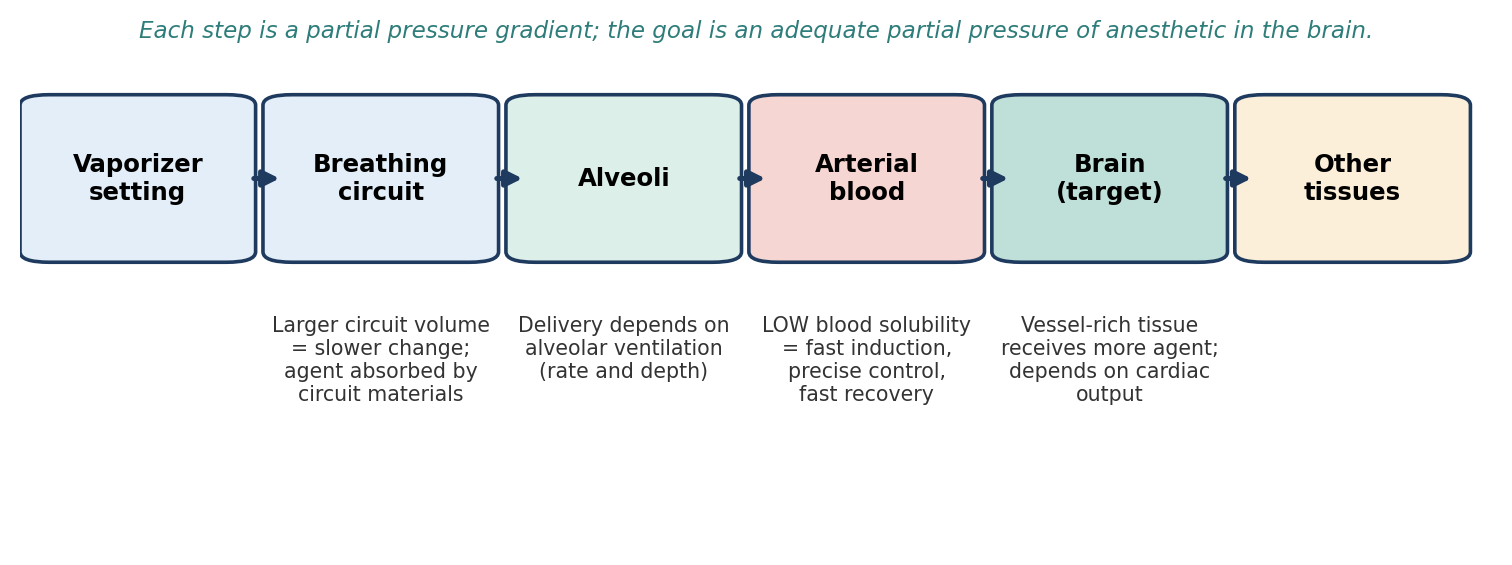
Figure 9.1. The path of an inhalant from vaporizer to brain, and the main factors that affect each step.
Circuit. The agent is delivered in oxygen to the alveoli, but an appropriate inspired concentration must first build up in the breathing circuit. The larger the circuit volume, the longer it takes for a change at the vaporizer to be reflected in the animal. Solubility of the agent in circuit materials (such as rubber) also has an effect.
Alveoli. The greater the alveolar ventilation (a function of respiratory rate and depth), the more agent is delivered to the alveoli.
Blood. An agent with low blood solubility needs only a small amount to reach equilibrium with the blood, after which it readily passes on to other tissues. Low blood solubility means rapid induction, more precise control of anesthetic depth, and rapid recovery.
Tissues. From the blood, the agent travels to target tissues. The more vascular a tissue, the more blood is available to transfer agent to it, so delivery is a function of cardiac output.
Why it matters
It seems backwards that a less soluble agent acts faster, but think of the blood as a sponge between the lungs and the brain. A highly soluble agent soaks into the sponge, and the partial pressure (which is what drives the agent into the brain) rises slowly. A poorly soluble agent fills the sponge quickly, so its partial pressure in the blood, and then in the brain, rises fast.
9.1.4 Biotransformation
Although most of an inhalant is exhaled unchanged, some is metabolized, and the amount varies greatly between agents. With older agents, metabolism may contribute to recovery. However, metabolism can also produce acute and chronic toxicity in certain organs, especially the liver and kidneys. Modern agents are metabolized very little, which is one reason they are safer.
9.1.5 Minimum Alveolar Concentration (MAC)
As introduced in Chapter 7, MAC is the amount of agent required to produce immobility in 50% of healthy subjects exposed to a noxious stimulus. MAC is inversely proportional to potency: the lower the MAC, the more potent the agent. MAC refers to the percentage of agent in the alveoli, not the setting on the vaporizer or the concentration in the circuit.
9.2 Historical Inhalant Anesthetics
Several inhalants are no longer used because they were flammable or explosive, or toxic (Table 9.2).
Table 9.2. Historical inhalant anesthetics and why they were abandoned.
| Agent | Problem |
|---|---|
| Chloroform | May cause liver failure |
| Cyclopropane | Explosive |
| Fluroxene | Hepatotoxic |
| Trichloroethylene | Hepatotoxic, cardiotoxic, and neurotoxic when used with soda lime |
| Diethyl ether | Flammable and explosive |
Diethyl ether was the principal inhaled anesthetic before the development of nonflammable agents, and it was replaced because of its flammability. Anesthetic concentrations are explosive, and patients can exhale enough ether to be flammable or explosive even after death. Ether is highly irritating to the respiratory tract, can cause laryngospasm, and can turn pre-existing, chronic, subclinical respiratory disease into an acute, severe infection.
9.3 Modern Inhalant Anesthetics
Nitrous Oxide (N₂O)
Nitrous oxide is not a potent anesthetic by itself but is useful as an adjunct to other inhalants: it decreases induction time and the amount of the accompanying anesthetic needed. Its MAC is greater than 100% (about 200% in dogs), so it cannot produce anesthesia alone at normal pressure. To prevent hypoxia, no more than 75% of the inspired gas can be N₂O (a 3:1 ratio with oxygen).
N₂O has a rapid onset because of its low blood solubility. Its cardiovascular and respiratory effects are minimal, and it has little or no effect on liver and kidney function, although care is needed in patients whose function is already compromised. It interferes with CO₂ monitoring.
Two hazards are characteristic of N₂O:
Expansion of gas spaces. N₂O transfers into gas-filled organs and body spaces, which can worsen a pneumothorax, enlarge a gas embolus, or increase pressure in the middle ear (see Chapter 7).
Diffusion hypoxia. When N₂O is stopped, it moves rapidly from the blood into the lungs, displacing oxygen. To prevent this, continue oxygen after N₂O is stopped.
N₂O cylinders hold the agent as a liquid: new cylinders contain about 95% liquid. As gas escapes, more liquid vaporizes to replace it, so pressure does not drop until all the liquid is gone, at which point the tank is only about 25% full. Usage should therefore be tracked by weight (Section 9.4.8).
⚠ Check current guidance
Nitrous oxide has well-documented analgesic properties at sub-anesthetic concentrations. It is described below (Section 9.4.8) as having an anesthetic effect but no analgesic effect; check how your exam references describe it.
Halothane
Halothane is no longer distributed in North America, but it appears frequently in references. It is susceptible to decomposition, so thymol is added for stability; thymol is less volatile than halothane and accumulates in vaporizers, causing malfunction unless they are regularly cleaned. Halothane is highly volatile, with moderate induction and recovery times. It does not have strong analgesic properties.
Up to 50% of inspired halothane is metabolized, and it may be hepatotoxic. It causes respiratory depression, depresses the myocardium and sensitizes it to catecholamines (raising the risk of arrhythmias), and moderately relaxes vascular smooth muscle. It increases cerebral blood flow and has been implicated in malignant hyperthermia.
Methoxyflurane
Methoxyflurane was popular from 1960 to 1990 but is no longer available in North America. It has low volatility and high blood solubility, which made it safe in non-precision vaporizers but gave it a slow onset and recovery. It has potent analgesic effects, with some post-operative analgesia. It is extensively metabolized (about 70%) and can cause renal damage with prolonged anesthesia. It is a respiratory depressant.
Enflurane
Enflurane is rarely used in veterinary clinical or laboratory animal anesthesia but remains in limited use elsewhere. It produces cardiac and respiratory depression similar to halothane, lowers the seizure threshold, and causes malignant hyperthermia in susceptible species. It is metabolized only slightly, so it is less toxic than halothane.
Isoflurane
Isoflurane combines low solubility with high potency, giving rapid induction and recovery. It has fewer cardiovascular effects and a greater safety margin than halothane, and it is a potent coronary vasodilator. Hyperventilation before administration limits increases in cerebral blood flow. Because minimal metabolism occurs, it is not very toxic to the kidneys or liver. It is irritating to mucous membranes and the respiratory system, can cause ocular irritation when an induction chamber is used, and some species become apneic during induction or when anesthesia is light.
Desflurane
Desflurane has effects similar to isoflurane but only limited use in veterinary medicine. It has lower blood solubility than isoflurane, halothane, or methoxyflurane, which allows rapid equilibrium, quick and precise changes in depth, and very rapid induction and recovery. It may cause tachycardia and respiratory irritation. Its low boiling point (near room temperature) means it requires a special heated vaporizer. It is expensive.
Sevoflurane
Sevoflurane has effects similar to isoflurane and, like desflurane, lower blood solubility than isoflurane, halothane, or methoxyflurane, with rapid equilibrium, precise control, and very rapid induction and recovery. With the exception of desflurane, it has the fastest induction and recovery of the inhalants. It is less of a respiratory irritant than desflurane and isoflurane, which makes it well suited to mask or chamber induction. It can raise intracranial pressure and cause respiratory depression.
⚠ Check current guidance
Sevoflurane is sometimes described as "the most volatile anesthetic." By vapor pressure, desflurane is the most volatile of the modern agents, which is why it needs a heated vaporizer. Check how your exam references describe sevoflurane's volatility.
Table 9.3. Comparison of modern and recent inhalant anesthetics.
| Agent | Speed of induction/recovery | Metabolism | Key features and cautions |
|---|---|---|---|
| Nitrous oxide | Rapid | Minimal | Adjunct only (MAC > 100%); max 75% of inspired gas; expands gas spaces; diffusion hypoxia; weigh cylinders |
| Halothane | Moderate | Up to 50% | Not distributed in North America; thymol buildup; sensitizes heart to catecholamines; hepatotoxic; malignant hyperthermia |
| Methoxyflurane | Slow | ~70% | Not available in North America; potent analgesia; renal damage |
| Enflurane | — | Small | Lowers seizure threshold; malignant hyperthermia; rarely used |
| Isoflurane | Rapid | Minimal | Greater safety margin than halothane; coronary vasodilator; airway and eye irritant; apnea in some species |
| Desflurane | Very rapid (fastest) | — | Lowest solubility; heated vaporizer; tachycardia; airway irritant; expensive |
| Sevoflurane | Very rapid (second only to desflurane) | — | Less irritating; raises ICP; respiratory depression |
9.4 Equipment
9.4.1 Anesthetic Delivery Systems
Inhalant agents usually require a vaporizer, which converts the liquid anesthetic into vapor and adds it to the carrier gas at a controlled concentration. There are three methods of vaporization (Figure 9.2):
Flow-over: carrier gas is directed over the surface of the liquid anesthetic. Wicks may be used to increase the surface area.
Bubble-through: carrier gas is passed below the surface of the liquid through a diffuser, which disperses bubbles through the liquid anesthetic.
Injection: a known amount of liquid anesthetic or pure vapor is injected into a known volume of gas, delivering accurate concentrations.
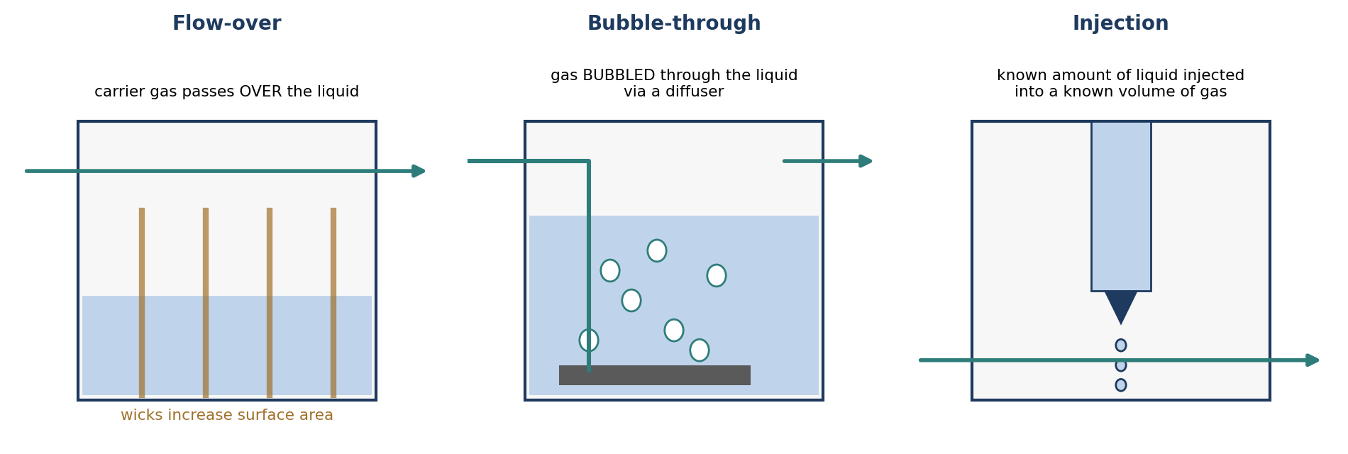
Figure 9.2. The three methods of vaporization.
Ether, methoxyflurane, and chloroform may be given with a soaked cotton mask or gauze, or by placing small animals in a chamber with anesthetic-soaked gauze. This open-drop method is dangerous, because it is difficult to control the dose and depth of anesthesia, and it exposes personnel to the agent.
Animals may also be placed in an induction chamber into which anesthetic gas is introduced from an anesthesia machine. The chamber should be attached to a scavenging system, or the animal should be anesthetized in a ventilated fume hood. Ideally, the animal is induced with an injectable anesthetic and maintained on gas.
9.4.2 Location of Vaporizers
Vaporizers can be located out of the breathing circuit (VOC) or in the circuit (VIC).
Vaporizer out of circuit (VOC) is the most common style of anesthesia machine. It allows more precise control over the concentration of inhalant delivered to the animal. Figure 9.3 shows the path of gas through a VOC circle system:
Gas, typically oxygen, is delivered from a cylinder (or pipeline) to the anesthesia machine through a regulator.
The oxygen passes through a flowmeter, which adjusts the flow rate and pressure through the system.
The oxygen passes through the vaporizer, where anesthetic is added in a known concentration.
The oxygen–anesthetic mixture passes through a unidirectional inspiratory valve into the inspiratory breathing tube. An air intake valve allows room air to enter the system if oxygen flow is disrupted.
A reservoir bag meets peak inspiratory demand, accommodates exhalation, and allows assisted or controlled ventilation. With a ventilator, the bag is replaced by a tube leading to the ventilator bellows.
The mixture enters the animal by spontaneous inspiration or ventilator pressure, and exhaled gas returns through the expiratory breathing tube.
Excess gas leaves through the pop-off valve (adjustable pressure-limiting, or APL, valve) to the scavenging system. The pop-off valve allows system pressure to be adjusted. With a ventilator, outflow goes to the ventilator, which has its own scavenging connection.
The remaining gas passes through the CO₂ absorber, where carbon dioxide is removed, and the gas continues around the circle.
A manometer measures the pressure in the breathing system.
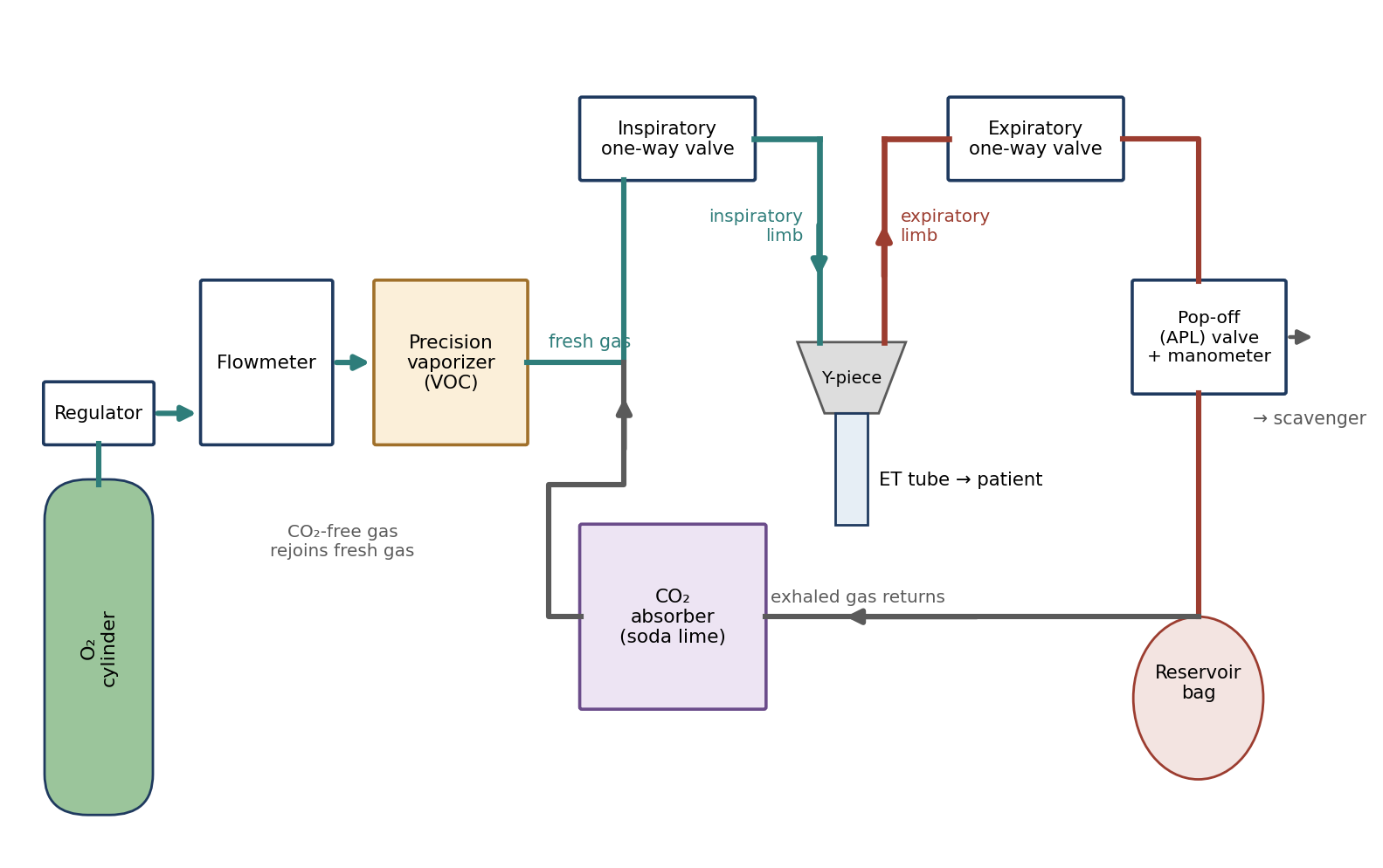
Figure 9.3. Simplified schematic of gas flow through a circle breathing system with a vaporizer out of circuit (VOC). Component positions vary between machines.
Breathing-system pressures above 20–25 mmHg are potentially dangerous because they may prevent CO₂ absorption, and pressures above 25–30 mmHg may cause lung trauma.
⚠ Check current guidance
Many current references express breathing-system pressures in cm H₂O rather than mmHg, and recommended limits vary. Be sure you know which unit your manometer displays (see also Chapter 5, cuff inflation).
Vaporizer in circuit (VIC) systems place the vaporizer inside the breathing circle, where the patient's own breathing draws gas through it. Anesthetic is absorbed into the gas stream more passively, and the output is unpredictable: the faster the animal breathes (spontaneously or with positive pressure ventilation), the more anesthetic it receives. As an animal begins to recover and breathes faster, it receives more anesthetic without any adjustment by the operator. Gas passes through a VIC system as follows: oxygen from the regulator and flowmeter passes through a unidirectional inspiratory valve and then through the vaporizer, then through the inspiratory tube to the patient. Exhaled gas passes through the expiratory tube and a unidirectional expiratory valve into the reservoir bag; excess gas leaves through the exhaust, and the rest passes through the soda lime (Sodasorb) canister before being reused.
Table 9.4. Comparison of vaporizer out of circuit and in circuit.
| Vaporizer out of circuit (VOC) | Vaporizer in circuit (VIC) | |
|---|---|---|
| Use | Most common | Less common |
| Control | Precise; set concentration | Imprecise; unpredictable output |
| Effect of faster breathing | No change in delivered concentration | More anesthetic delivered |
| Typical vaporizer | Precision, high-resistance | Non-precision, low-resistance (e.g., Stephens) |
9.4.3 Vaporizers
Vaporizers can be agent-specific or multipurpose. Their design is governed by the physics of vaporization:
Heat energy is required for vaporization. The latent heat of vaporization is the number of calories needed to change 1 gram of liquid to vapor.
Because of this heat requirement, the anesthetic cools as it vaporizes, and since vapor pressure varies with temperature, cooling reduces output. Uncontrolled cooling limits the vaporizer's output.
Vaporizers are therefore built from materials with high specific heat, which supply heat to the liquid and slow cooling, and high thermal conductivity, which passes heat from the room to the liquid. Copper and bronze are most commonly used. The vaporizer body acts as a heat sink. Some vaporizers, such as those for desflurane, are actively heated. Precision vaporizers must compensate for flow, temperature, and pressure, and various methods of back-pressure compensation are used.
[ PHOTO PLACEHOLDER ] Suggested source: Wikimedia Commons (commons.wikimedia.org), search "isoflurane vaporizer" or "anaesthetic vaporizer." Check the image's license and give the attribution it requires. |
|---|
Figure 9.4. An agent-specific precision vaporizer (for example, an isoflurane vaporizer) mounted on an anesthesia machine.
Specific vaporizer types are summarized in Table 9.5:
Tec precision vaporizers were designed specifically for halothane or isoflurane. They are VOC, concentration-calibrated, variable-bypass, flow-over, thermocompensated, agent-specific, and high-resistance. Examples include the Fluotec Mark 3, Pentec Mark 2, and Isotec 3. Their output is nearly linear across the concentrations and flow rates typically used. The Tec 6 and Tec 6 Plus are precision vaporizers made specifically for desflurane: dual-circuit, injection, supplied heat, agent-specific, and high-resistance.
The Ohio calibrated vaporizer is variable-bypass, flow-over with wick, automatically temperature-compensated, agent-specific, VOC, and high-resistance. It was an earlier model relative to the Tec 3 and Tec 4, and was made for isoflurane, halothane, and sevoflurane. Tilting up to 20° in use, or 45° when not in use, does not cause problems. It may cause discoloration of the liquid anesthetic.
The Siemens vaporizer is a concentration-calibrated, injection-type, non-thermocompensated, agent-specific, plenum (high-resistance) unit designed to couple with a Siemens ventilator. It is not used extensively in veterinary anesthesia.
Measured-flow vaporizers (Verni-Trol and Copper Kettle) are flowmeter-controlled vaporizers formerly popular in human anesthesia. They are measured-flow, bubble-through, high-resistance, VOC, temperature-compensated, and multipurpose, and require calculations to set flow rates. They are infrequently used now because of imprecision, though some remain in facilities.
The Stephens simple vaporizer is variable-bypass, flow-over, non-temperature-compensated, VIC, low-resistance, and multipurpose. It is non-precision and non-calibrated, with a glass vaporization chamber. It is suitable for methoxyflurane or ether, but not for isoflurane or halothane. With no temperature control, its output depends heavily on ambient temperature. Its major advantage is low cost; its major disadvantage is unknown, variable output.
Table 9.5. Vaporizer types.
| Vaporizer | Classification | Agents |
|---|---|---|
| Tec (Fluotec 3, Pentec 2, Isotec 3) | VOC, concentration-calibrated, variable bypass, flow-over, thermocompensated, agent-specific, high resistance | Halothane or isoflurane |
| Tec 6 / Tec 6 Plus | Dual circuit, injection, supplied heat, agent-specific, high resistance | Desflurane |
| Ohio calibrated | Variable bypass, flow-over with wick, temperature-compensated, agent-specific, VOC, high resistance | Isoflurane, halothane, sevoflurane |
| Siemens | Concentration-calibrated, injection, non-thermocompensated, agent-specific, plenum | Agent-specific; couples with Siemens ventilator |
| Verni-Trol / Copper Kettle | Measured flow, bubble-through, high resistance, VOC, temperature-compensated, multipurpose | Multipurpose (requires calculations) |
| Stephens | Variable bypass, flow-over, non-compensated, VIC, low resistance, multipurpose | Methoxyflurane or ether (NOT isoflurane or halothane) |
Why it matters
A precision vaporizer filled with the wrong agent can deliver a dangerously high or low concentration, because each is calibrated for one agent's vapor pressure. Agent-specific filling systems (keyed fillers) exist to prevent this error.
9.4.4 Mechanical Ventilators
An anesthesia ventilator replaces the reservoir bag and the anesthetist's hand. In a typical bellows ventilator, driving gas enters the housing around the bellows and compresses it, forcing breathing gas along the circuit into the patient. Overflow gas from the patient system exits through the ventilator's pop-off valve to the scavenger, and the tidal volume is set with a tidal volume control.
Bellows may be ascending or descending, named for their movement during expiration (Figure 9.5). Ascending bellows are safer, because they collapse if the circuit is opened or has a slow leak, while a descending bellows will continue moving and the operator may not know there is a problem.
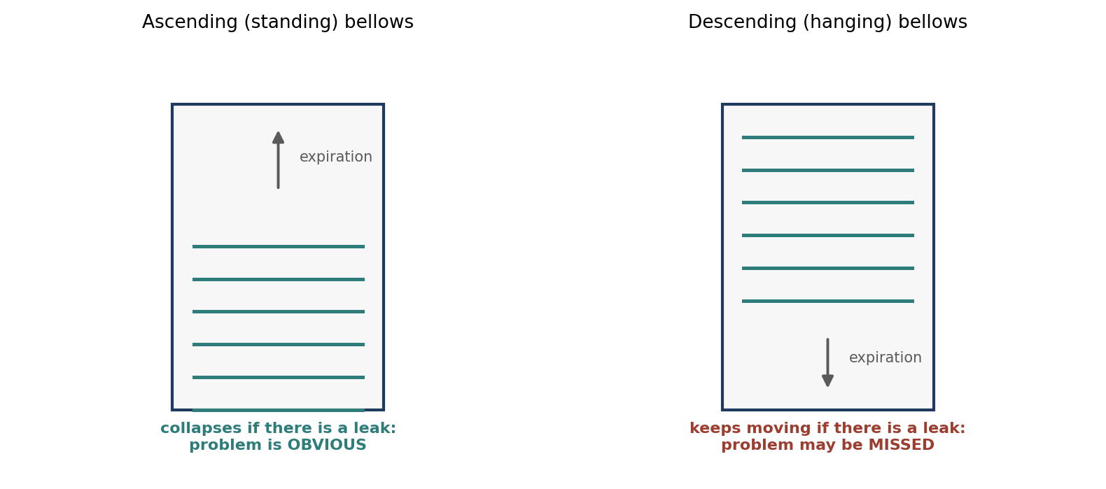
Figure 9.5. Ascending and descending ventilator bellows.
Table 9.6. Modes of mechanical ventilation.
| Mode | Description |
|---|---|
| Intermittent positive pressure ventilation (IPPV) | Airway pressure is above ambient during inspiration and falls to ambient to allow expiration |
| Conventional positive pressure ventilation (CPPV) | A form of IPPV delivering a preset tidal volume at a preset frequency |
| Positive end-expiratory pressure (PEEP) | Pressure at the end of expiration is kept above ambient, which helps keep alveoli open |
| Continuous positive airway pressure (CPAP) | Airway pressure is kept above ambient during spontaneous breathing |
| Intermittent mandatory ventilation (IMV) | Spontaneous breathing is allowed while assisted breaths (with reduced tidal volume and/or rate) are given; used when weaning from a ventilator. Periodic "sigh bagging" by the anesthetist is also a form of IMV |
9.4.5 CO₂ Absorber
The CO₂ absorber removes carbon dioxide before gas passes back to the patient side of a rebreathing circuit. The canister should be large enough to hold a gas volume around the granules equal to or greater than the patient's maximum tidal volume. It is commonly filled with soda lime or barium hydroxide lime.
The absorbent is gradually expended and stops absorbing CO₂. How long a canister lasts depends on flow rates and on how much time the system is used with spontaneous breathing versus mechanical ventilation. Absorbent may contain an indicator that changes color as the granules are expended. However, the color can fade back overnight even though the absorbent is still exhausted, so it is necessary to keep track of hours of use and change the absorbent before saturation. Working absorbent also heats up (a "heat line"), because absorbing CO₂ is a chemical reaction that produces heat.
Why it matters
If the absorbent is exhausted, exhaled CO₂ is rebreathed. The patient becomes hypercapnic, which causes acidosis, tachycardia, and arrhythmias, and can be mistaken for light anesthesia. Rising inspired CO₂ on a capnograph is a sign that the absorbent needs changing (Chapter 12).
9.4.6 Breathing Systems
Breathing systems are classified by whether exhaled gas is rebreathed (after CO₂ removal) and by the fresh gas flow used (Table 9.7).
Circle (rebreathing) systems use a CO₂ absorber and one-way valves so that exhaled gas can be reused:
Closed circle systems use a low flow of gases, with oxygen flow approximately equal to the patient's oxygen consumption, which varies with metabolic rate, body weight, surface area, temperature, anesthetic depth, and type of anesthetic. In practice, the reservoir bag is watched to adjust flow: under-inflation means flow should be increased; over-inflation means flow should be decreased. Flow must still be sufficient for the vaporizer to work properly. Nitrous oxide is generally not used, because of the risk of a hypoxic gas mixture at low oxygen inflows. Closed systems are completely dependent on CO₂ absorption. They are economical, retain heat and humidity, and are less likely to expose personnel to waste gas.
Low-flow circle systems use an oxygen flow greater than the patient's oxygen consumption but less than 22 mL/kg/min; a suggested rate for small animals is 10–15 mL/kg/min. They are economical, reduce waste gas, and retain heat and moisture. However, a concentration-calibrated, variable-bypass vaporizer may deliver too little anesthetic during mask induction or during the transition from a short-acting injectable induction to inhalant maintenance. For this reason, higher flows are used for the first 15–30 minutes, followed by a change to low flow.
Semi-closed circle systems use a fresh gas flow that exceeds the patient's uptake of oxygen and anesthetic, traditionally 22–44 mL/kg/min, or about three times the patient's oxygen consumption. A significant amount of excess gas leaves through the pop-off valve. Nitrous oxide can be used safely. For spontaneously breathing patients, use the system that produces the least resistance to gas flow.
The to-and-fro system places the CO₂ absorbent canister between the endotracheal tube connector and a reservoir bag, so gas moves back and forth through it. It is suitable for both large and small animals, but it is cumbersome and carries a greater risk of the patient inhaling alkaline dust from the absorbent.
Mapleson systems do not use chemical CO₂ absorbent; they rely on high fresh gas flow to wash exhaled CO₂ out of the system. They are commonly called non-rebreathing systems, although some rebreathing may occur, and they have no valves to prevent it. They require more gas and promote hypothermia and drying of the respiratory tract, but they are useful for small species in which the dead space and resistance of a standard circle circuit would be too high. The two main styles are:
The Magill system (Mapleson A), which is efficient during spontaneous ventilation. Fresh gas flow should approximate the patient's minute volume, and the volume of the tubing and bag should be equal to or greater than the patient's tidal volume.
The Bain coaxial system (a modified Mapleson D), a tube within a tube (Figure 9.6). The inner tube supplies fresh gas and the outer tube removes exhaled gas, with a pop-off valve incorporated at the bag. The recommended flow rate is 100–150 mL/kg/min.
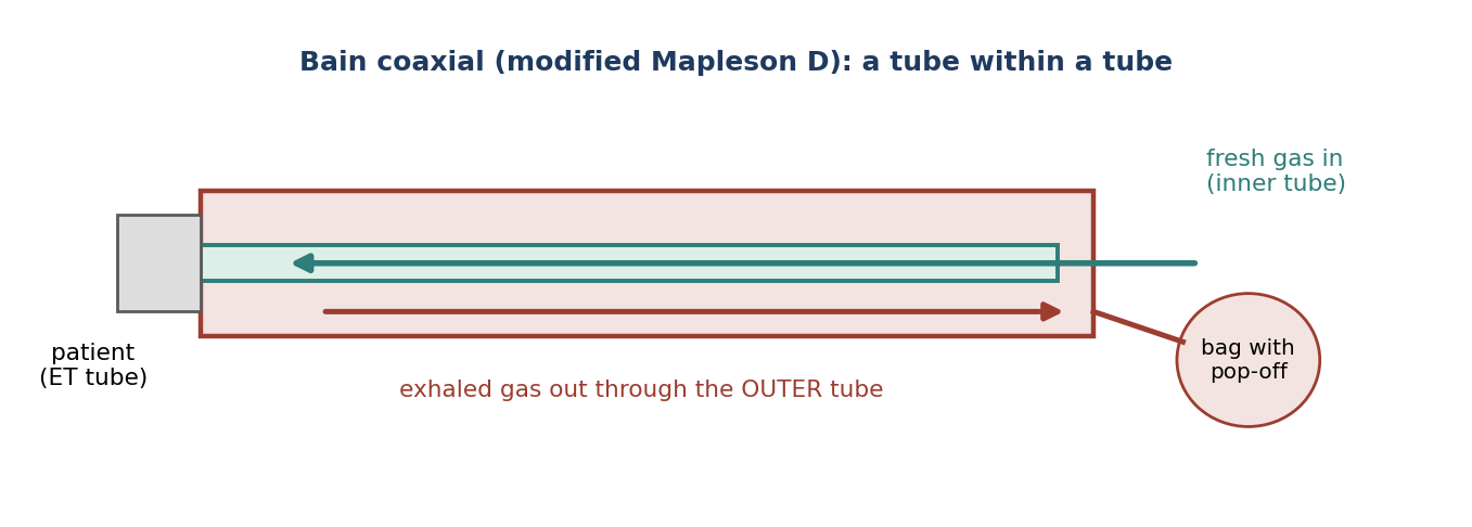
Figure 9.6. The Bain coaxial non-rebreathing system.
⚠ Check current guidance
Recommended fresh gas flows for the Bain system vary between references, and many give higher values (for example, 200–300 mL/kg/min). Check your exam references.
Ayre's T-piece and Norman mask elbow systems are classified as Mapleson F systems, or Mapleson E systems when there is no reservoir bag. One end attaches to the endotracheal tube connector and the other to an expiratory tube. When a reservoir bag is present, the arrangement is called a Jackson-Rees system.
Systems with non-rebreathing valves, such as the Stephens-Slater system, are mainly historical. They contain two one-way valves, which tended to stick, but have minimal dead space and resistance. Resuscitation bags (self-inflating bags) facilitate resuscitation and transport of apneic or anesthetized patients; they let the patient inhale room air and exhale through an exhalation port.
Table 9.7. Comparison of breathing systems.
| System | CO₂ removal | Typical fresh gas flow | Notes |
|---|---|---|---|
| Closed circle | Absorbent | ≈ patient O₂ consumption | Most economical; watch bag; avoid N₂O; risk of hypoxic mixture |
| Low-flow circle | Absorbent | > O₂ consumption but < 22 mL/kg/min (10–15 mL/kg/min small animals) | Higher flows for first 15–30 min |
| Semi-closed circle | Absorbent | 22–44 mL/kg/min (≈ 3 × O₂ consumption) | Excess gas to pop-off; N₂O safe |
| To-and-fro | Absorbent (canister at patient) | — | Cumbersome; risk of inhaling alkaline dust |
| Magill (Mapleson A) | High flow | ≈ minute volume | Efficient with spontaneous breathing |
| Bain (modified Mapleson D) | High flow | 100–150 mL/kg/min (as given here) | Coaxial; good for small patients |
| Ayre's T-piece / Norman elbow (Mapleson E/F) | High flow | — | Jackson-Rees when a bag is added |
9.4.7 Scavenging Systems
Exposure to waste anesthetic gases is a significant concern and a potential health risk for personnel. Scavenging systems collect waste gas from the breathing system, usually the gas leaving the pop-off valve. An interface prevents pressure changes in the scavenging system from being transmitted to the breathing system, which could otherwise over-pressurize or empty the patient's circuit.
Passive systems rely on the flow of gas in the breathing circuit to push waste gas into the scavenger.
Active systems use fans or vacuum to apply a low negative pressure that draws waste gas into the scavenger. They are generally safer and release less anesthetic gas into the room.
Either type may vent waste gas to the outside, or pass it through activated charcoal, which absorbs anesthetic vapors. Activated charcoal does not absorb nitrous oxide. Charcoal canisters must be weighed before first use and after each use; a canister is saturated when it reaches the weight specified by the manufacturer, and failing to track this can allow anesthetic gas to vent into the room.
9.4.8 Gas Cylinders
Gas in cylinders is under high pressure, and the pressure of a full tank varies with the gas. Cylinders require regulators, which are specific for each gas and cannot be interchanged because of their thread patterns. Regulators reduce the pressure to a level acceptable to the anesthesia machine, 37–50 psi.
Gas from large tanks reaches the anesthesia machine either through a pipeline ending in a gas-specific threaded or pinned connector, or directly through high-pressure hoses. Machines also have hanger yokes for small cylinders (E-tanks) of one or two gases, such as oxygen and nitrous oxide. E-tanks connect through a pin-index safety system, in which the arrangement of pins differs for each gas, preventing accidental interchange.
[ PHOTO PLACEHOLDER ] Suggested source: Wikimedia Commons (commons.wikimedia.org), search "pin index safety system" or "E cylinder." Check the image's license and give the attribution it requires. |
|---|
Figure 9.7. An E-size cylinder valve showing the pin-index safety holes, and a cylinder secured to a cart.
Safety concerns:
Oxygen and nitrous oxide support combustion.
Sudden release of gas from a broken cylinder can cause injury or propel other equipment. Tanks are unbalanced with a small base; if one falls and its valve breaks, it can become a rocket-like projectile.
Chain tanks to a wall or cart at all times; never leave them unsecured.
Cap valve stems when not attached to a regulator or gas line.
Store tanks away from emergency exits and high-traffic areas.
Tank colors are not universally standardized and may vary between manufacturers (Table 9.8). Do not rely on color to identify unlabeled tanks; unlabeled tanks should not be used.
Table 9.8. Typical U.S. cylinder colors (not universally standardized).
| Color | Gas |
|---|---|
| Green | Oxygen |
| Blue | Nitrous oxide |
| Yellow | Medical air |
| Black | Nitrogen |
| Grey | Carbon dioxide |
Oxygen (O₂). Anesthetized patients often have depressed respiration, so room air (compressed medical-grade air) cannot maintain adequate oxygenation. Oxygen may be supplied as a gas or as a highly pressurized liquid. A full oxygen cylinder of any size has a pressure of about 2,200 psi. A full E-tank holds about 700 L and an H-tank about 7,000 L. Because oxygen is stored as a gas, pressure is proportional to volume: an E-tank at 1,100 psi (half the full pressure) contains about 350 L.
Why it matters
Knowing the volume in an oxygen tank lets you estimate how long it will last. At 2 L/min, an E-tank reading 1,100 psi (about 350 L) will last roughly 175 minutes. Checking this before a long procedure prevents running out of oxygen mid-surgery.
Carbon dioxide (CO₂) is usually used for rodent immobilization or euthanasia, and may be mixed with oxygen to reduce irritation.
Nitrogen (N₂) is used to power drills and other equipment. Medical-grade air is a mixture of gases similar in composition to normal room air.
Nitrous oxide (N₂O) is used primarily mixed with oxygen to decrease the MAC of the inhalant being used. It is described here as having an anesthetic effect but no analgesic effect. A full N₂O cylinder has a pressure of about 750 psi at room temperature and contains both liquid and gas, with about 95% of the volume liquid. As the liquid vaporizes, the cylinder cools and frost may form. Because pressure is not proportional to volume, contents must be determined by weight.
Table 9.9. Oxygen and nitrous oxide cylinders compared.
| Property | Oxygen | Nitrous oxide |
|---|---|---|
| Physical state in cylinder | Gas | Liquid (~95%) and gas |
| Full pressure | ~2,200 psi | ~750 psi |
| Pressure vs. contents | Proportional | Not proportional (constant until liquid is gone) |
| How to measure contents | Pressure gauge | Weight |
| Full E-tank volume | ~700 L | — |
Chapter Summary
Inhalants are given and eliminated mainly through the lungs, allowing rapid, predictable changes in depth. All except nitrous oxide are organic; modern agents are halogenated ethers. Vapors are limited by their vapor pressure, which rises with temperature. Inhalants move down partial pressure gradients from circuit to alveoli to blood to brain; low blood solubility gives fast induction and recovery, and MAC (inversely related to potency) measures alveolar concentration. Historical agents were abandoned for flammability or toxicity. Isoflurane and sevoflurane are the mainstays, desflurane is fastest but needs a heated vaporizer, and nitrous oxide is only an adjunct, with risks of gas-space expansion and diffusion hypoxia.
Precision VOC vaporizers deliver set concentrations and compensate for temperature, flow, and pressure; VIC vaporizers deliver more agent as breathing speeds up. Ascending ventilator bellows reveal leaks. CO₂ absorbent must be tracked by hours of use. Circle systems (closed, low-flow, semi-closed) rebreathe gas through absorbent; Mapleson systems (Magill, Bain, T-piece) rely on high flow and suit small patients. Active scavenging protects personnel; charcoal does not absorb N₂O. Cylinders must be secured, identified by label, and fitted with gas-specific regulators; oxygen contents are read by pressure, nitrous oxide by weight.
Key Terms
APL (pop-off) valve: Adjustable pressure-limiting valve that releases excess gas from the breathing system to the scavenger.
Blood/gas partition coefficient: A measure of an inhalant's solubility in blood; lower values mean faster induction and recovery.
Diffusion hypoxia: Hypoxia caused by nitrous oxide flooding into the alveoli when it is discontinued.
Gas: A substance in gaseous form at room temperature and sea-level pressure (e.g., N₂O).
Latent heat of vaporization: Calories required to change 1 g of liquid to vapor.
Mapleson system: A non-rebreathing breathing system that relies on high fresh gas flow to remove CO₂.
Partition coefficient: The ratio of an agent's concentration in two media at equilibrium.
PEEP: Positive end-expiratory pressure.
Pin-index safety system: Gas-specific pin arrangement on small cylinders that prevents connection to the wrong yoke.
Precision vaporizer: An agent-specific, calibrated vaporizer that delivers a set concentration regardless of flow and temperature.
Saturated vapor pressure: The maximum vapor pressure of a liquid at a given temperature.
Scavenging system: Equipment that collects and removes waste anesthetic gases.
Soda lime: A CO₂ absorbent used in rebreathing circuits.
Vapor: The gaseous state of a substance that is liquid at room temperature and pressure.
VIC / VOC: Vaporizer in circuit / vaporizer out of circuit.
Review Questions
1. Why are all modern inhalant anesthetics (newer than halothane) ethers?
A. Ethers are cheaper
B. The lack of an ether group was found to increase the risk of cardiac arrhythmias
C. Ethers are non-volatile
D. Ethers cannot be halogenated
2. An inhalant with LOW blood solubility will produce:
A. Slow induction and slow recovery
B. Rapid induction, precise control of depth, and rapid recovery
C. No anesthetic effect
D. Higher MAC in all cases
3. MAC is inversely proportional to:
A. Potency
B. Body weight
C. Vaporizer temperature
D. Tidal volume
4. Why should the contents of a nitrous oxide cylinder be tracked by weight rather than pressure?
A. The gauge is inaccurate
B. The cylinder contains liquid, so pressure stays constant until the liquid is gone, when the tank is only about 25% full
C. Nitrous oxide has no pressure
D. Pressure rises as the tank empties
5. What is diffusion hypoxia, and how is it prevented?
A. Hypoxia from too little N₂O; give more N₂O
B. N₂O rapidly leaving the blood into the lungs and displacing oxygen when it is stopped; continue oxygen after stopping N₂O
C. Hypoxia caused by isoflurane; switch to sevoflurane
D. Hypoxia from a closed circuit; open the pop-off valve
6. Why is a vaporizer out of circuit (VOC) preferred over a vaporizer in circuit (VIC)?
A. It is cheaper
B. It allows precise control of the delivered concentration, independent of the patient's breathing
C. It requires no oxygen
D. It increases anesthetic delivery as the patient breathes faster
7. Why is an ascending bellows considered safer than a descending bellows?
A. It delivers larger tidal volumes
B. It collapses if the circuit is opened or leaks, making the problem obvious
C. It does not need a scavenger
D. It works without oxygen
8. The color indicator in a CO₂ absorbent has faded back overnight. The absorbent:
A. Has regenerated and is fully usable
B. Is still exhausted; track hours of use to change it before saturation
C. Must be heated before use
D. Should be rinsed with water
9. Mapleson (non-rebreathing) systems prevent rebreathing by:
A. Chemical CO₂ absorption
B. One-way valves
C. High fresh gas flow rates
D. A closed circuit
10. Which statement about activated charcoal scavenging canisters is correct?
A. They absorb nitrous oxide
B. They never need to be replaced
C. They must be weighed before first use and after each use, and do not absorb nitrous oxide
D. They provide active suction
Answer Key
1. B. Halothane, an aliphatic hydrocarbon, sensitizes the heart to arrhythmias. The discovery that lacking an ether group increases arrhythmia risk led to all newer agents being ethers.
2. B. A poorly soluble agent saturates the blood quickly and readily moves on to the brain, giving fast induction, precise control, and fast recovery.
3. A. A lower MAC means less agent is needed to prevent movement, so the agent is more potent.
4. B. As gas is used, liquid N₂O vaporizes to replace it, keeping pressure constant until all the liquid is gone. By then the tank is only about 25% full.
5. B. When N₂O is discontinued, it floods from the blood into the alveoli and dilutes oxygen. Continuing 100% oxygen afterward prevents hypoxia.
6. B. A VOC vaporizer delivers a set concentration. With a VIC, faster breathing increases anesthetic delivery without any operator adjustment, so output is unpredictable.
7. B. An ascending bellows collapses with a leak or disconnection. A descending bellows may keep moving, so the operator may not notice the problem.
8. B. The color change can fade overnight, but the lime remains expended. Keep track of hours used to change it before saturation.
9. C. Mapleson systems have no absorbent and no valves to prevent rebreathing; they rely on high flow to wash out exhaled CO₂.
10. C. Charcoal canisters are saturated when they reach the manufacturer's specified weight, so they must be weighed regularly. They do not absorb nitrous oxide.
Chapter 10: Physical Methods of Anesthesia
Learning objectives
After studying this chapter, you should be able to:
Explain how hypothermia reduces metabolism and organ oxygen demand, and why it can be used to support anesthesia and surgery.
Describe the risks of hypothermia, including the temperature at which ventricular fibrillation becomes likely.
Compare the three methods used to induce hypothermia.
Describe electronarcosis, its effects, and the reasons it is considered ethically questionable.
Most anesthesia is produced with drugs. A small number of techniques instead use physical means, such as cold or electrical current, to depress the nervous system or reduce the body's needs. These methods are now used mainly in specialized situations, but they illustrate important physiological principles and still appear in research and exam references.
This chapter covers the two principal physical methods: hypothermia and electronarcosis. Two other physical approaches, TENS and acupuncture, were introduced briefly in Chapter 7 (Table 7.2). Hypothermia as a complication of anesthesia, and how to prevent it, was covered in Chapter 5 (Section 5.5).
10.1 Hypothermia
Induced hypothermia is the deliberate lowering of body temperature to supplement anesthesia, reduce the need for analgesic drugs, or protect organs during procedures that interrupt their blood supply. It is used primarily in neonates and in cardiovascular procedures. Hypothermia may be general (the whole body is cooled) or local (only a region or organ is cooled).
How Hypothermia Works
Cooling the body lowers metabolism. Because cells need less energy, they also need less oxygen: the colder an animal becomes, the less oxygen a given organ requires. As a result, the heart, brain, liver, and some other vital organs can survive for longer periods without part or all of their blood supply than they could at normal temperature. This is the principle that allows surgeons to stop blood flow to an organ, or to the whole body, long enough to perform delicate repairs.
With the use of cardioplegic solutions (solutions that stop the heart in a relaxed state), the heart may be stopped for about 30 minutes without damage. Small laboratory animals can be cooled to about 0 °C and recover. Figure 10.1 summarizes these landmarks.
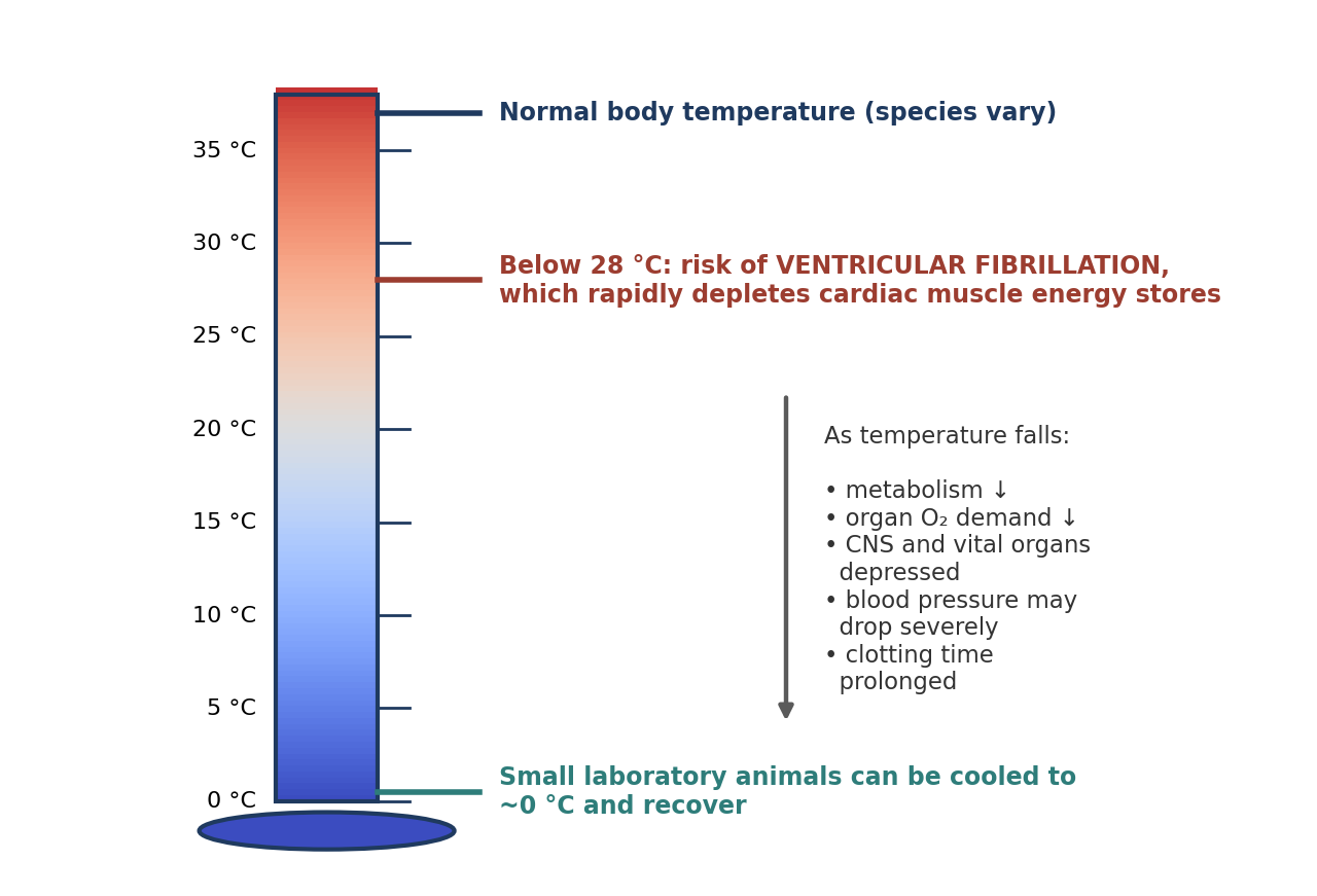
Figure 10.1. Key temperature landmarks in induced hypothermia, and the physiological effects of cooling.
Shivering must be controlled first with another anesthetic agent. Shivering is the body's main defense against cold: it generates heat and greatly increases oxygen consumption, which would defeat the purpose of cooling.
Why it matters
The value of hypothermia is the same as its danger. Lowering metabolism protects organs from lack of oxygen, but it also depresses the brain, heart, and blood vessels that keep the animal alive. Induced hypothermia is therefore always a controlled, monitored procedure, unlike the accidental hypothermia that good peri-operative care tries to prevent.
Risks of Hypothermia
Profound depression of the CNS and vital organs.
A severe drop in blood pressure.
Ventricular fibrillation below 28 °C. Below this temperature, heart muscle may fibrillate, and fibrillation rapidly depletes the energy stores of the cardiac muscle.
Prolonged clotting time, because the enzymes and platelets involved in clotting work poorly at low temperatures. This increases the risk of bleeding during and after surgery.
Methods of Inducing Hypothermia
There are three methods of cooling a patient (Figure 10.2 and Table 10.1):
Immersion (surface cooling): the patient is placed in ice water or wrapped in an ice-water recirculating blanket.
Body-cavity cooling: iced saline is poured slowly into a body cavity.
Extracorporeal cooling: blood is circulated outside the body through a heat exchanger, as in cardiopulmonary bypass, and then returned to the body.
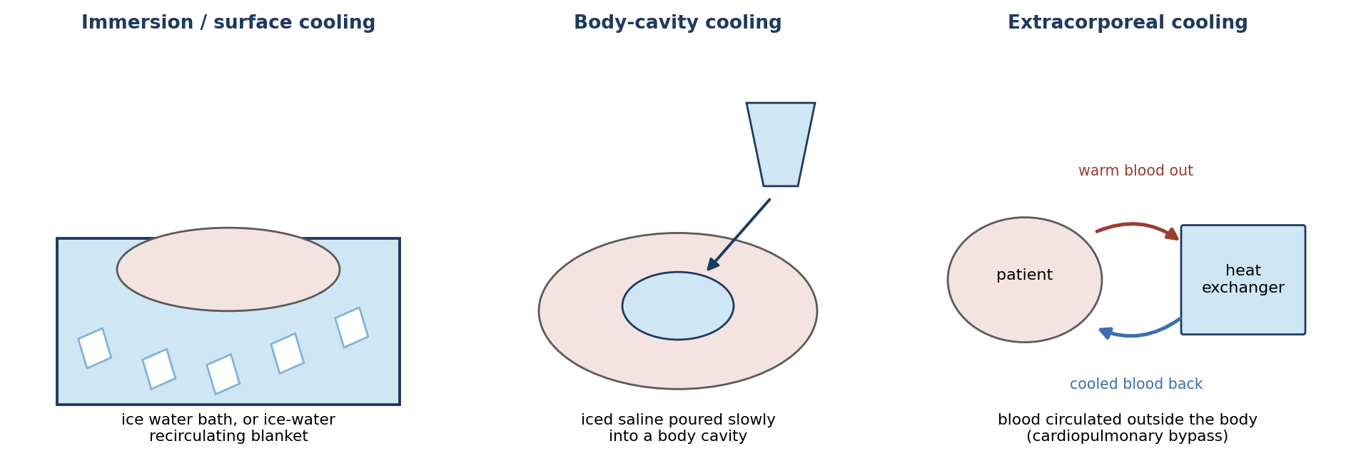
Figure 10.2. The three methods of inducing hypothermia.
Table 10.1. Comparison of methods for inducing hypothermia.
| Method | How it works | Considerations |
|---|---|---|
| Immersion / surface | Ice water bath or ice-water recirculating blanket | Simple; cools from the outside in, so it is slower and less precise |
| Body cavity | Iced saline poured slowly into a body cavity | Cools internal organs directly; requires an open cavity |
| Extracorporeal | Blood circulated through a heat exchanger and returned (cardiopulmonary bypass) | Fastest and most controllable; requires specialized equipment and personnel |
[ PHOTO PLACEHOLDER ] Suggested source: Wikimedia Commons (commons.wikimedia.org), search "heart-lung machine" or "cardiopulmonary bypass machine." Check the image's license and give the attribution it requires. |
|---|
Figure 10.3. A heart-lung (cardiopulmonary bypass) machine with its heat exchanger, as used for extracorporeal cooling.
Hypothermia in Neonatal Rodents
In research, a common application of hypothermia is anesthesia of neonatal rodents for brief procedures. Very young pups tolerate cooling well, and inhalant or injectable anesthetics can be difficult to dose safely in such small animals. To avoid frostbite or tissue injury, the pup should not be placed in direct contact with ice; a barrier such as a latex sleeve or gauze is used. The pup must be rewarmed gradually and monitored until it is moving normally before it is returned to the dam.
⚠ Check current guidance
Whether hypothermia provides adequate anesthesia and analgesia, and the age limits for its use, are addressed by institutional policies and current guidance (such as the AVMA euthanasia guidelines and IACUC-approved protocols). Many institutions limit hypothermia anesthesia to pups under about 7 days old. Check your institution's policies and current references.
10.2 Electronarcosis
In electronarcosis, electrodes are applied to the head and deliver electrical current through the cerebrum, producing deep narcosis. The current is thought to activate opioid and/or non-opioid control centers in the brain.
Electronarcosis has significant drawbacks:
Induction is characterized by convulsions unless paralytics are administered.
Endotracheal intubation should always be performed, to protect the airway and allow ventilation.
Hyperthermia is commonly seen, caused by disturbance of the brain's thermoregulatory center.
Skin burns and brain lesions have been found after its use.
It produces severe stress.
It is difficult to monitor and ethically questionable.
Why it matters
Electronarcosis is a useful reminder that the absence of movement is not the same as the absence of pain or distress. If paralytics are needed to stop the convulsions it causes, the anesthetist loses many of the usual signs of anesthetic depth, the same problem described for neuromuscular blocking agents in Chapter 6. Combined with the stress, burns, and brain lesions it can cause, this makes electronarcosis very difficult to justify when drug-based anesthesia is available.
Table 10.2. Comparison of hypothermia and electronarcosis.
| Hypothermia | Electronarcosis | |
|---|---|---|
| Mechanism | Lowers metabolism and organ O₂ demand | Electrical current through the cerebrum activates opioid and/or non-opioid centers |
| Main uses | Neonates; cardiovascular procedures; organ protection | Rarely used |
| Preparation | Control shivering with another anesthetic | Paralytics to prevent convulsions; always intubate |
| Temperature effect | Deliberately lowered | Hyperthermia common |
| Main risks | CNS and organ depression; hypotension; VF below 28 °C; prolonged clotting | Convulsions; burns; brain lesions; severe stress; difficult to monitor |
Chapter Summary
Hypothermia lowers metabolism so that organs need less oxygen and can tolerate reduced blood supply. It may be general or local, is used mainly in neonates and cardiovascular procedures, and requires shivering to be controlled first with another agent. With cardioplegic solutions the heart can be stopped for about 30 minutes, and small laboratory animals can be cooled to about 0 °C and recover. Its risks include profound CNS and organ depression, severe hypotension, ventricular fibrillation below 28 °C, and prolonged clotting. Cooling is achieved by immersion, body-cavity cooling with iced saline, or extracorporeal circulation through a heat exchanger.
Electronarcosis passes current through the cerebrum. It causes convulsions unless paralytics are given, requires intubation, commonly causes hyperthermia, can cause burns and brain lesions, produces severe stress, and is difficult to monitor and ethically questionable.
Key Terms
Cardioplegic solution: A solution used to stop the heart in a relaxed state during cardiac surgery.
Cardiopulmonary bypass: Circulation of blood outside the body through a machine that oxygenates it and, if needed, cools or warms it.
Electronarcosis: Deep narcosis produced by passing electrical current through the cerebrum.
Extracorporeal: Outside the body.
Heat exchanger: A device that cools or warms blood as it passes through.
Induced hypothermia: Deliberate lowering of body temperature to support anesthesia or protect organs.
Ventricular fibrillation: Chaotic, ineffective electrical activity of the ventricles, in which the heart does not pump blood.
Review Questions
1. Why can organs survive longer without blood supply when an animal is cooled?
A. Cooling increases blood flow
B. The colder an organ becomes, the less oxygen it requires
C. Cooling dissolves blood clots
D. Cooling raises blood pressure
2. Before body temperature is lowered for hypothermic anesthesia, what must be controlled with another anesthetic agent?
A. Sweating
B. Shivering
C. Vomiting
D. Urination
3. With cardioplegic solutions and hypothermia, the heart may be stopped without damage for about:
A. 30 seconds
B. 5 minutes
C. 30 minutes
D. 6 hours
4. Below what body temperature does the heart become at risk of ventricular fibrillation?
A. 35 °C
B. 32 °C
C. 28 °C
D. 10 °C
5. Which is a recognized risk of hypothermia?
A. Shortened clotting time
B. Severe drop in blood pressure and prolonged clotting time
C. Increased metabolism
D. Hypertension
6. Which cooling method circulates blood outside the body through a heat exchanger?
A. Immersion
B. Body-cavity cooling
C. Extracorporeal cooling
D. Surface blanket cooling
7. What is the basis of electronarcosis?
A. Freezing the skin
B. Passing electrical current through the cerebrum via electrodes on the head
C. Stimulating peripheral nerves through the skin
D. Injecting an anesthetic into the spinal canal
8. Unless paralytics are given, induction of electronarcosis is characterized by:
A. Smooth sleep
B. Convulsions
C. Bradycardia only
D. Hypothermia
9. Which temperature change is commonly seen during electronarcosis?
A. Hypothermia, from vasodilation
B. Hyperthermia, from disturbance of the thermoregulatory center
C. No change
D. Hypothermia, from shivering
10. Why is electronarcosis considered ethically questionable?
A. It is too expensive
B. It produces severe stress, is difficult to monitor, and can cause burns and brain lesions
C. It requires a veterinarian
D. It cannot be reversed
Answer Key
1. B. Hypothermia lowers metabolism, so organs such as the heart, brain, and liver need less oxygen and tolerate reduced or absent blood supply for longer.
2. B. Shivering is the body's defense against cooling. It generates heat and increases oxygen demand, so it must be controlled initially with another anesthetic agent.
3. C. With cardioplegic solutions, the heart may be stopped for about 30 minutes without damage.
4. C. Below 28 °C, heart muscle may develop ventricular fibrillation, which rapidly depletes cardiac muscle energy stores.
5. B. Hypothermia profoundly depresses the CNS and vital organs, may cause a severe drop in blood pressure, and prolongs clotting time.
6. C. Extracorporeal cooling, as in cardiopulmonary bypass, circulates blood through a heat exchanger and returns it to the body.
7. B. Electronarcosis uses electrodes on the head to pass current through the cerebrum, activating opioid and/or non-opioid control centers in the brain.
8. B. Induction of electronarcosis is characterized by convulsions unless paralytics are administered, and endotracheal intubation should always be performed.
9. B. Electronarcosis commonly causes hyperthermia by disturbing the brain's thermoregulatory center.
10. B. Electronarcosis produces severe stress, is difficult to monitor, and has been associated with skin burns and brain lesions.
Chapter 11: Local & Regional Anesthesia
Learning objectives
After studying this chapter, you should be able to:
Explain how local anesthetics block nerve conduction, and how potency, onset, and duration relate to their chemistry.
Classify local anesthetics as ester- or amide-linked and compare common agents.
Describe topical anesthetics and their cautions, including methemoglobinemia.
Describe infiltration, field, nerve and plexus, ring, IV regional (Bier), epidural, intercostal, intrapleural, and muscle blocks.
Recognize correct and incorrect needle placement during epidural injection.
Local and regional anesthesia block pain at its source, before the signal reaches the brain. Used alone, they allow minor procedures in sedated or even conscious animals. Used with general anesthesia, they are a core part of balanced anesthesia (Chapter 7): they reduce the amount of general anesthetic needed, blunt the body's stress response to surgery, and can provide pain relief that lasts into recovery (Chapter 13).
How Local Anesthetics Work
Local anesthetics are a group of chemically related compounds that reversibly bind sodium channels and block impulse conduction in nerve fibers (Figure 11.1). A nerve impulse travels by opening sodium channels in sequence along the fiber; when the channels in a segment of nerve are blocked, the impulse cannot pass, and the sensation is never transmitted to the brain. When the drug diffuses away or is broken down, the channels recover and normal sensation returns.
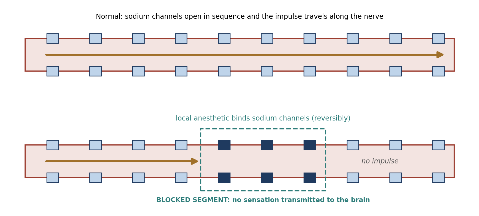
Figure 11.1. Local anesthetics reversibly bind sodium channels, preventing impulse conduction through the blocked segment of nerve.
Small, unmyelinated pain fibers are generally blocked more easily than large motor fibers, which is why sensation is often lost before (or without) loss of movement.
Local anesthetics are available as solutions, gels, creams, and aerosols for selective application by several routes: topical application, infiltration of an incision site, plexus nerve block, epidural, regional nerve block, and spinal anesthesia.
Classification
Local anesthetics are classified by several properties:
Potency, which is affected by the size of the molecule and how lipophilic (fat-soluble) it is.
Speed of onset, which is associated with lipid solubility and acid dissociation (pKa). More lipophilic agents have a quicker onset, and agents whose pKa is closer to tissue pH reach their active form faster.
Duration of effect, which increases with lipid solubility.
Rate of vascular absorption, which varies with the vascularity of the injection site and the physicochemical and pharmacological properties of the drug. The faster a drug is absorbed into the blood, the shorter its local effect and the greater the risk of systemic toxicity.
Chemically, local anesthetics are either ester-linked or amide-linked (Figure 11.2 and Table 11.1):
Ester-linked agents have short half-lives (they are broken down quickly, largely by plasma enzymes), and allergic reactions may occur. Procaine and chloroprocaine are low-potency and short-acting; tetracaine is high-potency and long-acting.
Amide-linked agents are very stable and rely on enzymatic degradation in the liver. Articaine is intermediate-potency and short-acting; lidocaine, mepivacaine, and prilocaine are intermediate in potency and duration; ropivacaine is intermediate-potency and long-acting; and bupivacaine, levobupivacaine, and etidocaine are high-potency and long-acting.
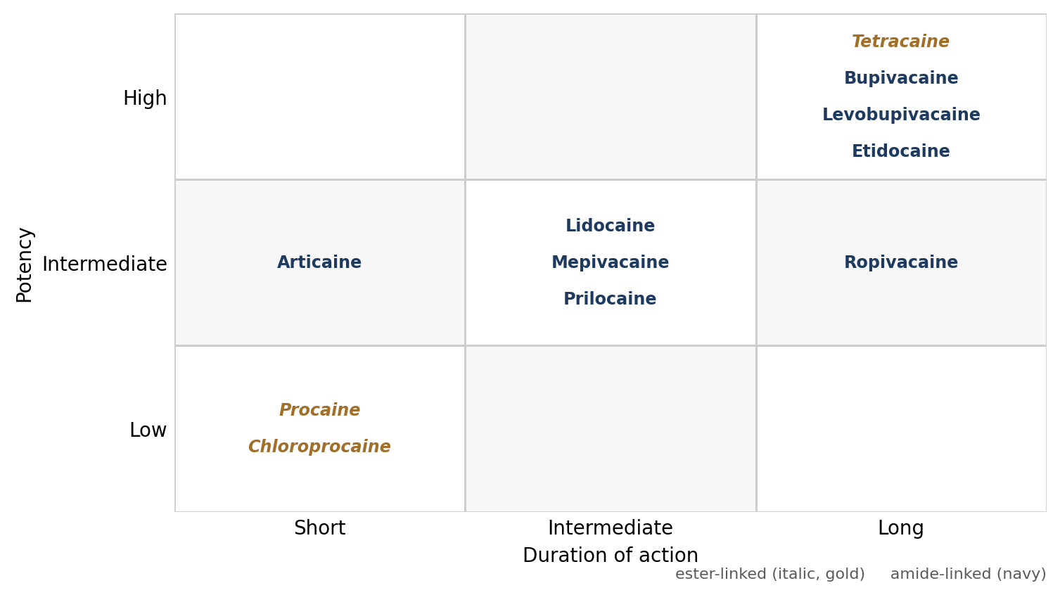
Figure 11.2. Local anesthetics arranged by potency and duration of action.
Table 11.1. Classification of local anesthetics.
| Linkage | Potency / duration | Agents | Characteristics |
|---|---|---|---|
| Ester | Low / short | Procaine, chloroprocaine | Short half-lives; allergic reactions possible |
| Ester | High / long | Tetracaine | |
| Amide | Intermediate / short | Articaine | Very stable; metabolized by the liver |
| Amide | Intermediate / intermediate | Lidocaine, mepivacaine, prilocaine | |
| Amide | Intermediate / long | Ropivacaine | |
| Amide | High / long | Bupivacaine, levobupivacaine, etidocaine |
Why it matters
A simple memory aid: amide-linked local anesthetics have an "i" in their name before "-caine" (lidocaine, bupivacaine, mepivacaine, ropivacaine), while esters do not (procaine, tetracaine). Because amides are cleared by the liver, liver disease can prolong their effects and increase the risk of toxicity.
⚠ Check current guidance
All local anesthetics can cause systemic toxicity if too much is absorbed or the drug is injected into a vessel, typically beginning with CNS signs (sedation, muscle twitching, seizures) and progressing to cardiovascular depression; bupivacaine is particularly cardiotoxic. Maximum safe doses differ by agent and species (cats are especially sensitive). Calculate the total dose before injecting and check current references.
11.1 Topical Anesthetics
Topical anesthetics are effective when applied to mucous membranes, such as the eye, larynx, and mouth. Many are available as injectables but are also supplied in topical formulations: creams, ointments, gels, powders, and aerosols. Common agents and concentrations are listed in Table 11.2.
Table 11.2. Topical anesthetics.
| Agent | Concentration | Notes |
|---|---|---|
| Lidocaine | 2–5% | Preferred over benzocaine to aid intubation |
| Proparacaine | 0.5% | Recommended local ocular anesthetic |
| Tetracaine | 0.5–2% | — |
| Butacaine | 2% | — |
| Cocaine | 4–10% | — |
| Benzocaine (e.g., Cetacaine gels and sprays) | 14–20% | Implicated in methemoglobinemia; use sparingly |
| Ethyl chloride | Spray | Freezes small areas of intact skin; large areas risk frostbite |
| EMLA cream (lidocaine + prilocaine) | — | Requires 45–60 minutes to penetrate skin |
| Procaine cream | — | Penetrates skin |
Benzocaine products (14–20%, such as Cetacaine gels and sprays) have been implicated in causing methemoglobinemia, a condition in which hemoglobin is oxidized and can no longer carry oxygen. They are used to aid intubation but should be used sparingly; lidocaine is a better choice. Cats are particularly susceptible (Chapter 5). Recent literature suggests that prilocaine may also cause methemoglobinemia.
Ethyl chloride sprayed on intact skin freezes a small area and causes loss of sensation. It should only be used on small areas, because freezing large areas can cause frostbite. Proparacaine (0.5%) is recommended as a local ocular anesthetic. Procaine cream penetrates skin, and EMLA cream (a mixture of lidocaine and prilocaine) requires 45–60 minutes to penetrate intact skin, so it must be applied well in advance, for example before placing an IV catheter.
11.2 Infiltration
In infiltration anesthesia, local anesthetic is injected directly into the tissue to be anesthetized. It is the most reliable and safest method of local anesthesia. Lidocaine is the most common local anesthetic for this purpose.
It is injected subcutaneously (SC) or intradermally (ID) at the intended incision site before cutting (a "line block").
It may also be applied to muscles exposed at the surgical site.
Lidocaine has been shown to have a dose-dependent inhibition of bacterial growth.
It is used frequently with epinephrine in dental extractions, because epinephrine constricts local vessels, decreasing bleeding and slowing absorption of the anesthetic so that it lasts longer.
Mepivacaine or procaine may also be used. Solutions without epinephrine are chosen where vasoconstriction could cause local ischemia and necrosis, such as in extremities or tissue with poor circulation.
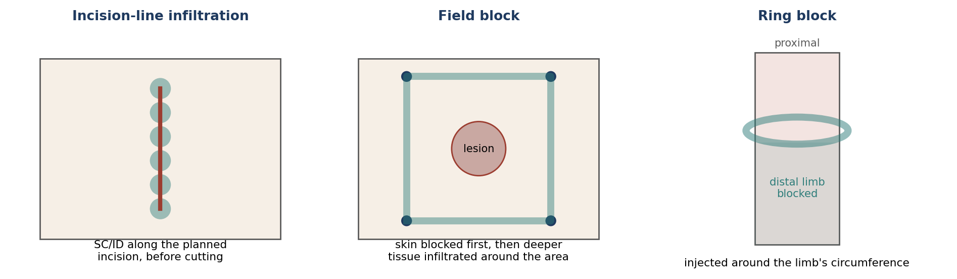
Figure 11.3. Patterns of local infiltration: incision-line infiltration, field block, and ring block.
11.2.1 Field Block
In a field block, the skin is blocked first, and then anesthetic is infiltrated into the deeper tissues around the area, creating a wall of anesthesia around the site rather than injecting into it. Field blocks are used to anesthetize large areas of skin, for skin biopsies and for removal of small growths within the outer layers of skin.
11.2.2 Local Nerve Block / Plexus Block
A nerve or plexus block is an injection given in the vicinity of a main nerve (or network of nerves) serving a region, such as a forelimb, hind limb, face, or chest. Lidocaine is commonly used. Nerve blocks are useful for minor procedures and for providing post-operative analgesia. Animals should be sedated, both for their comfort and so they remain still while the needle is placed near the nerve.
11.2.3 Ring Block
In a ring block, local anesthetic is injected around the circumference of a limb, blocking all the nerves that pass that point and providing analgesia for the distal portion of the limb.
11.2.4 IV Regional Anesthesia (Bier Block)
IV regional anesthesia anesthetizes an extremity for procedures lasting less than 90 minutes. Local anesthetic is injected into a vein of a limb that has been isolated from the circulation by a tourniquet, so the drug fills the limb's vessels and diffuses into its tissues. The steps are:
Place an IV catheter in a vessel of the limb.
Place a tourniquet proximal to the surgical site.
Exsanguinate the limb by elevating it and wrapping it tightly with an Esmarch bandage, which drives blood out and prevents blood from flowing back in.
Tighten the tourniquet, then remove the bandage.
Inject lidocaine into the IV catheter.
After the tourniquet is removed, sensation returns in 5–15 minutes, and analgesia lasts up to 30 minutes. Durations under 90 minutes appear safe; longer tourniquet times can lead to ischemic injury.
Why it matters
The tourniquet does two jobs: it keeps the anesthetic in the limb, where it is needed, and keeps it out of the general circulation, where a large IV dose of local anesthetic could cause systemic toxicity. For this reason the tourniquet should not be released too soon after injection.
11.2.5 Epidural
In epidural anesthesia, local anesthetic is injected into the epidural space, the space outside the dura mater that surrounds the spinal cord and nerve roots. The lumbosacral space (L7–S1) is the most common site of administration (Figure 11.4).
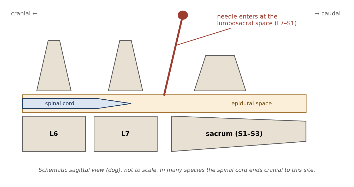
Figure 11.4. Lumbosacral epidural injection.
Epidural anesthesia is excellent for procedures caudal to the umbilicus, such as hind limb, perineal, and lower abdominal surgery. It is useful for cesarean section, because it does not depress the newborns' body systems as general anesthetics crossing the placenta would, and it allows a conscious mother to care for her young.
Before injecting, check the needle hub for cerebrospinal fluid (CSF) or blood (Table 11.3):
Blood indicates puncture of the venous plexus in the vertebral canal; injecting here would deliver the drug into the bloodstream.
CSF indicates subarachnoid puncture: the needle has passed through the dura into the space around the cord, and a full epidural dose given here could cause an excessively high, extensive block.
When the needle enters the epidural space, a drop of saline placed in the hub of the spinal needle should be "sucked in" by the negative pressure there (the "hanging drop" technique).
Table 11.3. Interpreting what appears at the needle hub during epidural injection.
| Observation | Interpretation | Action |
|---|---|---|
| Hanging drop of saline drawn into the hub | Needle tip is in the epidural space | Inject |
| Blood | Puncture of the venous plexus | Do not inject; reposition |
| Cerebrospinal fluid | Subarachnoid puncture | Do not give a full epidural dose; reposition or adjust the dose |
Lidocaine or bupivacaine is commonly used. Drugs may also be infused continuously through a catheter threaded into the epidural space. Opioids and alpha-2 agonists can also be given epidurally (Chapter 6).
[ PHOTO PLACEHOLDER ] Suggested source: Wikimedia Commons (commons.wikimedia.org), search "spinal needle" or "Tuohy needle." Check the image's license and give the attribution it requires. |
|---|
Figure 11.5. A spinal needle with stylet, as used for lumbosacral epidural injection.
⚠ Check current guidance
Where the spinal cord and dural sac end differs between species (for example, they extend further caudally in cats than in dogs), which affects the risk of subarachnoid puncture at the lumbosacral site. Check species-specific references before performing an epidural.
11.2.6 Intercostal Nerve Block
An intercostal nerve block anesthetizes the chest wall, for example for a thoracotomy or rib injury. Each intercostal nerve runs along the caudal border of its rib, together with the intercostal artery and vein. Inject at the caudal border of the rib near the intervertebral foramen, where the nerve emerges from the spine. Because the areas supplied by neighboring intercostal nerves overlap, the block must include at least two adjacent intercostal spaces (Figure 11.6).
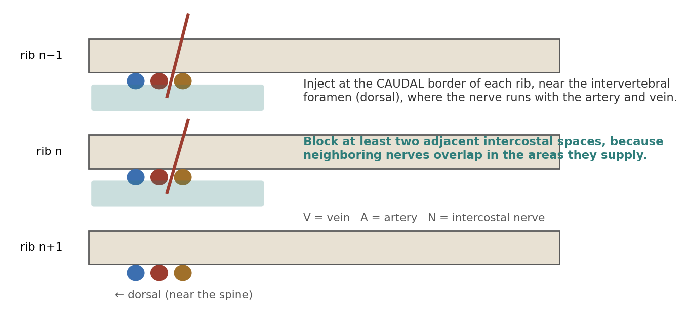
Figure 11.6. Intercostal nerve block. The nerve, artery, and vein run along the caudal border of each rib.
Why it matters
Because the intercostal artery and vein lie right beside the nerve, aspirate before injecting to make sure the needle is not in a vessel. Accidental intravascular injection delivers the drug straight into the circulation.
11.2.7 Intrapleural Regional Analgesia
In intrapleural analgesia, local anesthetic is injected into the pleural space (between the lung and chest wall), either intermittently or continuously through a catheter, where it bathes the intercostal nerves from inside the chest. It is useful for reducing pain after rib trauma or a thoracotomy, and can also treat acute and chronic pain originating from thoracic and upper abdominal structures. It is ineffective if the drug is mistakenly given into an adjacent space.
11.2.8 General Muscle Block
A general muscle block is an injection of local anesthetic into muscles that will be cut, separated, or extensively manipulated during surgery, reducing pain from those tissues.
Table 11.4. Summary of local and regional techniques.
| Technique | Where the drug goes | Typical use |
|---|---|---|
| Topical | Mucous membranes or skin surface | Eye (proparacaine); larynx for intubation (lidocaine); skin before catheter placement (EMLA) |
| Infiltration (line block) | SC/ID along the planned incision | Most incisions; most reliable and safest method |
| Field block | Skin first, then deeper tissue around the area | Large skin areas; biopsies; small growths |
| Nerve / plexus block | Near a main nerve or plexus | Minor procedures; post-operative analgesia |
| Ring block | Around the limb's circumference | Distal limb |
| IV regional (Bier) | Into a vein of a tourniquet-isolated limb | Extremity procedures < 90 minutes |
| Epidural | Epidural space, usually L7–S1 | Procedures caudal to the umbilicus; cesarean section |
| Intercostal | Caudal border of ribs, ≥ 2 adjacent spaces | Thoracotomy; rib injury |
| Intrapleural | Pleural space | Thoracotomy, rib trauma, thoracic and upper abdominal pain |
| General muscle | Muscles to be cut or manipulated | Intra- and post-operative muscle pain |
Chapter Summary
Local anesthetics reversibly bind sodium channels, blocking nerve conduction. Greater lipid solubility increases potency, speeds onset, and prolongs duration; onset also depends on pKa, and vascular absorption shortens effect and raises toxicity risk. Esters (procaine, chloroprocaine, tetracaine) are short-lived and may cause allergy; amides (lidocaine, mepivacaine, prilocaine, articaine, ropivacaine, bupivacaine) are stable and liver-metabolized.
Topical agents work on mucous membranes; benzocaine (and possibly prilocaine) can cause methemoglobinemia, so lidocaine is preferred for intubation and proparacaine for the eye. Infiltration with lidocaine is the most reliable and safest method. Field, nerve/plexus, and ring blocks extend coverage; a Bier block uses a tourniquet-isolated limb for under 90 minutes; an epidural at L7–S1 covers procedures caudal to the umbilicus (check for CSF or blood first); intercostal blocks are placed at the caudal rib border across at least two spaces; and intrapleural and muscle blocks address thoracic and surgical-site pain.
Key Terms
Amide-linked local anesthetic: A stable local anesthetic metabolized by the liver (e.g., lidocaine, bupivacaine).
Bier block: IV regional anesthesia of a limb isolated by a tourniquet.
Epidural space: The space outside the dura mater surrounding the spinal cord and nerve roots.
Ester-linked local anesthetic: A local anesthetic with a short half-life that may cause allergic reactions (e.g., procaine).
Esmarch bandage: An elastic bandage used to exsanguinate a limb.
Exsanguinate: To drive blood out of a limb or region.
Field block: A wall of local anesthetic infiltrated around, rather than into, an area.
Hanging drop: A drop of saline in the needle hub that is drawn in when the needle enters the epidural space.
Infiltration: Injection of local anesthetic directly into the tissue to be anesthetized.
Intercostal nerve block: Local anesthetic placed at the caudal border of ribs to anesthetize the chest wall.
Methemoglobinemia: A condition in which oxidized hemoglobin cannot carry oxygen.
pKa: The pH at which a drug is half ionized; affects local anesthetic onset.
Ring block: Local anesthetic injected around a limb's circumference to block the distal limb.
Subarachnoid: Within the space beneath the arachnoid membrane, containing cerebrospinal fluid.
Review Questions
1. How do local anesthetics block nerve conduction?
A. By destroying the nerve fiber
B. By reversibly binding sodium channels
C. By blocking acetylcholine receptors
D. By activating opioid receptors
2. Increasing the lipid solubility of a local anesthetic generally:
A. Slows onset and shortens duration
B. Speeds onset and increases duration
C. Has no effect
D. Makes it an ester
3. Which local anesthetic is ester-linked?
A. Lidocaine
B. Bupivacaine
C. Procaine
D. Ropivacaine
4. Which topical anesthetic has been implicated in causing methemoglobinemia, particularly in cats?
A. Proparacaine
B. Benzocaine
C. Lidocaine
D. Ethyl chloride
5. Which agent is recommended as a local ocular anesthetic?
A. 0.5% proparacaine
B. 4–10% cocaine
C. EMLA cream
D. Ethyl chloride
6. Which is described as the most reliable and safest method of local anesthesia?
A. Epidural
B. Infiltration
C. Bier block
D. Intrapleural analgesia
7. A Bier block (IV regional anesthesia) appears safe for procedures lasting less than:
A. 15 minutes
B. 30 minutes
C. 90 minutes
D. 6 hours
8. During epidural injection, cerebrospinal fluid appears in the needle hub. This indicates:
A. Correct epidural placement
B. Puncture of the venous plexus
C. Subarachnoid puncture
D. The needle is in muscle
9. Why should an intercostal nerve block include at least two adjacent intercostal spaces?
A. To use more drug
B. Because neighboring intercostal nerves overlap in the areas they supply
C. Because one nerve is always missed
D. To block the phrenic nerve
10. Where should an intercostal nerve block be injected?
A. At the cranial border of the rib, near the sternum
B. At the caudal border of the rib, near the intervertebral foramen
C. Directly into the pleural space
D. Into the intercostal muscle midway between ribs
Answer Key
1. B. Local anesthetics reversibly bind sodium channels, preventing the sodium influx needed to conduct an impulse along the nerve fiber.
2. B. More lipophilic agents have a quicker onset and a longer duration of anesthetic effect, and generally greater potency.
3. C. Procaine, chloroprocaine, and tetracaine are ester-linked. Lidocaine, bupivacaine, and ropivacaine are amide-linked.
4. B. Benzocaine (14–20%) products have been implicated in methemoglobinemia and should be used sparingly; lidocaine is a better choice to aid intubation.
5. A. Proparacaine (0.5%) is recommended as a local ocular anesthetic.
6. B. Infiltration of the incision site, most often with lidocaine, is the most reliable and safest method.
7. C. Durations under 90 minutes appear safe; longer tourniquet times can cause ischemic injury.
8. C. CSF indicates subarachnoid puncture. Blood indicates puncture of the venous plexus. Correct placement is suggested when a drop of saline in the hub is drawn in.
9. B. Intercostal nerve supply overlaps, so blocking a single space leaves the area partially innervated.
10. B. The intercostal nerve runs with the artery and vein along the caudal border of each rib, so injection is made there, near the intervertebral foramen.
Chapter 12: Patient Monitoring During Anesthesia
Learning objectives
After studying this chapter, you should be able to:
Explain why anesthetized patients are monitored and how anesthetic depth, analgesia, and immobilization are evaluated.
Define the terms used to describe breathing patterns and lung volumes, and interpret blood gas, end-tidal CO₂, and pulse oximetry values.
Identify the components of the ECG and recognize common arrhythmias and heart blocks.
Describe the assessment of peripheral perfusion, central venous pressure, arterial blood pressure, and cardiac output.
Explain the importance of temperature monitoring during anesthesia.
12.1 Reasons to Monitor Anesthesia
Anesthetic drugs, and the unconscious, recumbent, and immobile state they produce, compromise the patient's homeostasis: its ability to keep breathing, circulation, and temperature within normal limits. Intra-operative monitoring focuses on ensuring an optimal anesthetic depth with minimal physiological impact. It allows early notice of trends that could develop into life-threatening conditions, and it gives the anesthetist the information needed to give anesthetics more precisely.
Monitoring helps avoid the harmful effects of both extremes of depth:
Too light can lead to awareness, pain, and movement.
Too deep causes hypoventilation, hypoxemia, reduced cardiac output, hypotension, inadequate tissue perfusion, hypothermia, and prolonged recovery.
Anesthetic depth represents a balance between the amount of drug given, the amount of surgical stimulation, and the severity of any pre-existing illness. Anesthetic requirements change over time, with an overall decreasing trend during a single anesthetic, because surgical stimulation varies, redistribution sites (such as fat) gradually fill, and body temperature changes. Anesthetists should therefore repeatedly try to decrease the amount of anesthetic given during a procedure, keeping the animal at a light to medium level: deep enough to prevent conscious perception and provide muscle relaxation, but light enough to maintain desirable physiological parameters.
12.2 Evaluating Depth of Anesthesia
Depth is judged from several sources of information together:
The recent history of anesthetic dosing: large doses should be associated with deeper anesthesia, and lower doses over time with lighter anesthesia.
Spontaneous movement, which is a reliable sign of light anesthesia, unless paralytics have been given.
Physiological signs of the stages and planes of anesthesia (Chapter 7, Section 7.4).
Trends in physiological parameters, especially heart rate, blood pressure, and respiration, together with checking reflexes.
12.3 Proper Monitoring
Proper monitoring includes evaluating muscle tone and reflexes. These signs vary between species and individuals, and they can change from minute to minute during a procedure. Information should be evaluated based on the animal's reactions and parameters at that moment: previous readings are useful for trends, but should not be relied on over current readings.
The anesthetic dose needed depends on the induction drug given, its effect and duration of action, the amount given, and the animal's health. The anesthetist must know when the induction drugs will wear off, because at that time the patient will need more anesthetic, and must know whether the patient received pre-operative analgesia, because this affects the amount of anesthetic needed.
12.3.1 Adequate Analgesia
If an animal is sufficiently anesthetized (unaware of and detached from external stimuli), it may be concluded that sufficient analgesia is present. However:
Some anesthetics provide poor analgesia even while the animal is unconscious, and require additional analgesics; propofol is an example (Chapter 8).
Paralytics may make an animal appear anesthetized on cursory exam, but it can still feel pain and be aware of its surroundings. In this situation, increasing blood pressure and heart rate are good indicators of pain.
Light anesthesia may not completely suppress reflexes or spontaneous movement, even when analgesia has been achieved.
12.3.2 Adequate Immobilization
Immobilization is judged by muscle tone and defined by the needs of the procedure. Intraocular, thoracic, long-bone fracture repair, and laparotomy procedures generally need complete muscle relaxation, while some muscle tone is acceptable in minor, non-invasive procedures. The allowable muscle tone also depends on what the surgeon is doing at that point (incision, abdominal retraction, cautery, or suturing). Neuromuscular blocking agents may be safer than overly deep anesthesia, as long as proper depth and analgesia are assured (Chapter 6).
12.3.3 Physiological Effects of Anesthetic Drugs
The physiological effects of anesthetic drugs should be the focus of intra-operative monitoring. Watch for excessive bradycardia, arrhythmias, cardiac depression, vasodilation, hypotension, hypoventilation, hypoxemia, or hypothermia. Pre-anesthetic exam findings (baseline values) indicate which conditions are already present and alert the anesthetist to potential trouble. Each value should be interpreted in light of its previous trend, other parameters, and the patient's history. The goal is to keep physiological values as close to pre-operative levels as possible.
Why it matters
No single parameter tells the whole story. A heart rate of 180 bpm might mean pain, light anesthesia, hypovolemia, hypercapnia, or an anticholinergic effect. Looking at several parameters together, and at how they are changing, is what lets the anesthetist find the cause.
12.4 Monitoring Respiration & Ventilation
12.4.1 Definitions
Respiration is the total process by which oxygen is supplied to and used by body cells, and carbon dioxide is eliminated, by means of concentration gradients. Ventilation is the movement of gas into and out of the alveoli. Breathing patterns are described with the terms in Table 12.1.
Table 12.1. Terms describing breathing patterns.
| Term | Meaning |
|---|---|
| Eupnea | Normal quiet breathing |
| Dyspnea | Labored breathing |
| Tachypnea | Increased breathing rate |
| Hyperpnea | Fast and/or deep breathing ("over-breathing") |
| Polypnea | Rapid, shallow (panting) breathing |
| Bradypnea | Slow, regular breathing |
| Hypopnea | Slow and/or shallow breathing ("under-breathing") |
| Apnea | Transient or longer cessation of breathing |
| Cheyne-Stokes respiration | Breathing increases in rate and depth, then slows, followed by brief periods of apnea |
| Biot's respiration | Groups of quick, shallow breaths followed by regular or irregular periods of apnea |
| Kussmaul's respiration | Deep, labored breathing, frequently associated with severe metabolic acidosis |
| Apneustic respiration | Breath held at the end of inspiration for a short period before exhaling |
Lung volumes and capacities describe how much air the lungs hold under different conditions (Figure 12.1 and Table 12.2). A capacity is the sum of two or more volumes.
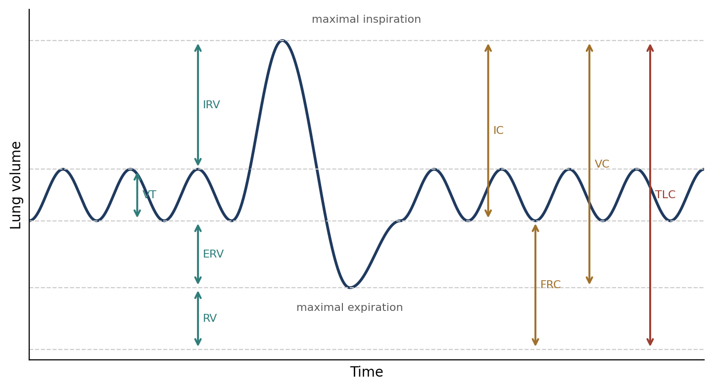
Figure 12.1. Lung volumes and capacities.
Table 12.2. Lung volumes and capacities.
| Term | Definition |
|---|---|
| Tidal volume (VT) | Volume inspired or expired in a single breath |
| Inspiratory reserve volume (IRV) | Volume that can be inspired above the normal tidal volume |
| Expiratory reserve volume (ERV) | Volume that can be expired by forceful expiration after a normal expiration |
| Residual volume (RV) | Air remaining in the lungs after the most forceful expiration |
| Minute ventilation (VE) | VT × respiratory frequency (f): the volume inspired or expired per minute |
| Inspiratory capacity (IC) | VT + IRV: the air that can be inhaled after a normal expiration, distending the lungs maximally |
| Functional residual capacity (FRC) | ERV + RV: the air in the lungs after a normal expiration |
| Vital capacity (VC) | IRV + VT + ERV: the maximum air that can be expelled after filling the lungs maximally |
| Total lung capacity (TLC) | IRV + VT + ERV + RV: the maximum volume of the lungs with the greatest inspiratory effort, or full inflation to 30 cm H₂O |
12.4.2 Respiration Basics
Gas transfer is the passing of gases across a membrane. Breathing depends on a pressure gradient between the alveoli and the outside atmosphere. During inspiration, active muscular effort expands the chest wall and contracts the diaphragm; expiration is normally passive, as the chest wall returns to its resting position. The exception is the horse, which uses abdominal muscle contraction during expiration (a biphasic exhalation).
Air flow depends on the diameter of the airways: the smaller the diameter, the greater the resistance. Part of each breath reaches only the upper airway and the tracheobronchial tree, where no gas exchange occurs. This is the anatomical dead space (VD anat). It is alveolar air that matters, because gas exchange with the blood happens only in the alveoli, by pressure gradients that allow oxygen to be absorbed and carbon dioxide eliminated.
Table 12.3. Terms describing blood gases and oxygenation.
| Term | Meaning |
|---|---|
| Hypercapnia | Elevated PaCO₂, causing respiratory acidosis |
| Hypocapnia | Lowered PaCO₂, causing respiratory alkalosis |
| Eucapnia | Normal PaCO₂ |
| Hypoxia | Abnormally low oxygen in the lungs, blood, and/or tissues, causing abnormal organ function or cell damage |
| Hypoxemia | Insufficient oxygenation of the blood to meet metabolic requirements |
| Apneic threshold | The PaCO₂ at which ventilation becomes zero and spontaneous breathing effort stops |
Under general anesthesia, the nasal and pharyngeal muscles relax and the cough reflex is abolished, which may allow airway obstruction, especially in brachycephalic dogs. This is one reason intubation is so valuable (Chapter 5).
Respiratory Rate and Pattern
Normal respiratory rates vary between species, but a change in breathing rate is a sensitive indicator of a change in the patient's status. Bradypnea is a sign of deep anesthesia or hypothermia. Tachypnea has many possible causes: anesthesia that is too light or too deep, hypoxemia, hypercapnia, hyperthermia, hypotension, atelectasis, the post-operative recovery phase or pain, and drugs (such as opioids).
Arrhythmic breathing patterns usually reflect a problem with respiratory control in the medulla, but some patterns are normal in certain species. A Cheyne-Stokes pattern (cycling between hyperventilation and hypoventilation) is normal in horses, but may indicate congestive heart failure or other heart or brain disorders in most species. Apneustic breathing may be seen in healthy cats and dogs, and in most species anesthetized with ketamine.
Respiratory Volume
Respiratory volume may be estimated visually, from reservoir bag movement, or with a ventilator or ventilometer. Normal tidal volume is 10–20 mL/kg per breath, and normal total minute ventilation in dogs is 150–250 mL/kg/min. Alveolar minute volume (the part that reaches the alveoli) may range from 20% to 70% of total minute ventilation.
Arterial Blood Gases
Arterial CO₂ and O₂ can be measured by drawing arterial blood and analyzing it with a blood-gas analyzer. Venous blood is not a good measure of respiratory function, because it has passed through a tissue bed where gas exchange has already taken place. Samples should be run immediately or stored in ice water, because blood cells continue to consume oxygen and produce CO₂ in the syringe.
PaCO₂ (the partial pressure of CO₂ in arterial blood) measures the patient's ventilatory status. The normal range is 35–45 mmHg (eucapnia). Venous CO₂ is usually 3–6 mmHg higher than arterial, and PaCO₂ can be estimated by measuring end-tidal CO₂.
PaCO₂ above 60 mmHg indicates excessive respiratory acidosis and may warrant mechanical ventilation.
PaCO₂ below 20 mmHg may indicate severe respiratory alkalosis and decreased cerebral blood flow.
Hypocapnia is most often caused by hyperventilation.
Hypercapnia may be caused by excessive anesthetic depth; intracranial or cervical disease; airway obstruction; thoracic or abdominal restrictive disease; pleural space filling disorders (air or fluid); terminal pulmonary parenchymal disease; improper ventilator settings; hyperthermia; or recent bicarbonate therapy.
PaO₂ (the partial pressure of O₂ in arterial blood) measures the oxygenating efficiency of the lungs. It measures oxygen dissolved in the blood, which is related to, but not the same as, hemoglobin saturation (SaO₂). PaO₂ is usually 80–110 mmHg breathing room air (21% oxygen). Animals breathing 100% oxygen have values around 500 mmHg (small animals) or over 200 mmHg (horses). Hypoxemia occurs at PaO₂ below 80 mmHg. Figure 12.3 shows how PaO₂ relates to hemoglobin saturation.
Table 12.4. Key respiratory monitoring values.
| Parameter | Normal / target | Significant values |
|---|---|---|
| PaCO₂ | 35–45 mmHg | > 60 mmHg: excessive respiratory acidosis, consider ventilation; < 20 mmHg: severe alkalosis, reduced cerebral blood flow |
| PaO₂ (room air) | 80–110 mmHg | < 80 mmHg: hypoxemia |
| PaO₂ (100% O₂) | ~500 mmHg (small animals); > 200 mmHg (horses) | — |
| SpO₂ | 90–95% breathing spontaneously; 95–100% ventilated on 100% O₂ | Falling values or lost waveform: check perfusion and probe |
| EtCO₂ | Usually somewhat lower than PaCO₂ | > 30–40 mmHg: animal will usually breathe on its own |
| Tidal volume | 10–20 mL/kg | — |
| Minute ventilation (dog) | 150–250 mL/kg/min | — |
End-Tidal CO₂ (EtCO₂)
A capnograph measures CO₂ in gas sampled from the breathing circuit, and EtCO₂ is the value at the end of exhalation, which approximates alveolar (and therefore arterial) CO₂. Its accuracy is affected by mechanical factors in the breathing circuit, such as volume, dead pockets, tubing diameter, and gas flow, and it is usually somewhat lower than PaCO₂. The shape of the waveform also carries information (Figure 12.2). A plateau that drops off to the right may indicate a leak in the circuit, because inspiratory pressure is not held.
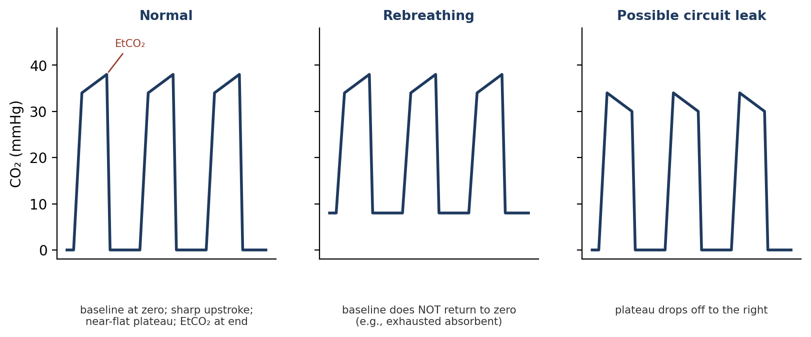
Figure 12.2. Capnogram waveforms: normal, rebreathing, and a pattern that may indicate a circuit leak (schematic).
EtCO₂ is useful when ventilating an animal. Animals with EtCO₂ over 30–40 mmHg will usually breathe on their own. Toward the end of a procedure, the animal should be weaned off the ventilator: gradually slow the rate and/or volume so that EtCO₂ rises above 30–40 mmHg, which stimulates spontaneous breathing. During open-skull procedures (craniotomies), values of 18–20 mmHg are described as helping to prevent brain swelling.
⚠ Check current guidance
Recommended EtCO₂ targets for neurosurgery vary. Chapter 7 cites hyperventilation to 30–35 mmHg to limit volatile-anesthetic-induced increases in cerebral blood flow, while 18–20 mmHg is cited here for craniotomies; very low values can reduce cerebral blood flow excessively. Check current references. A rising baseline on the capnogram (rebreathing) is a common sign of exhausted CO₂ absorbent (Chapter 9).
Pulse Oximetry (SpO₂)
A pulse oximeter measures the percentage of hemoglobin that is oxygenated and the heart rate. Its sensor shines red and infrared light through tissue and records how much is absorbed, either by light passing through the tissue to a receiver on the other side (transmission) or by light reflected back to the sensor (reflectance). Oxygenated and deoxygenated hemoglobin absorb these wavelengths differently. Because other tissues also absorb light, the oximeter uses the pulsatile part of the signal, and various computations, to isolate arterial blood. SpO₂ is broadly accurate for SaO₂. Normally, SpO₂ is 90–95% in spontaneously breathing animals and 95–100% in ventilated animals on 100% oxygen.
SpO₂ readings can be falsely lowered by positional factors (the probe slipping, thick tissue, or pigment), vasoconstriction, drying of the contact surface, and confusion with respiratory artifact or movement. If an animal becomes hypotensive, the pulse waveform first becomes smaller and then is lost. Some arrhythmias can be detected by watching the waveform and listening to the tone (a regular heartbeat produces a regular tone), and on most monitors the pitch of the tone rises with oxygen saturation. Heart rate in anesthetized dogs is usually about 60–100 bpm, and in anesthetized cats about 100 bpm.
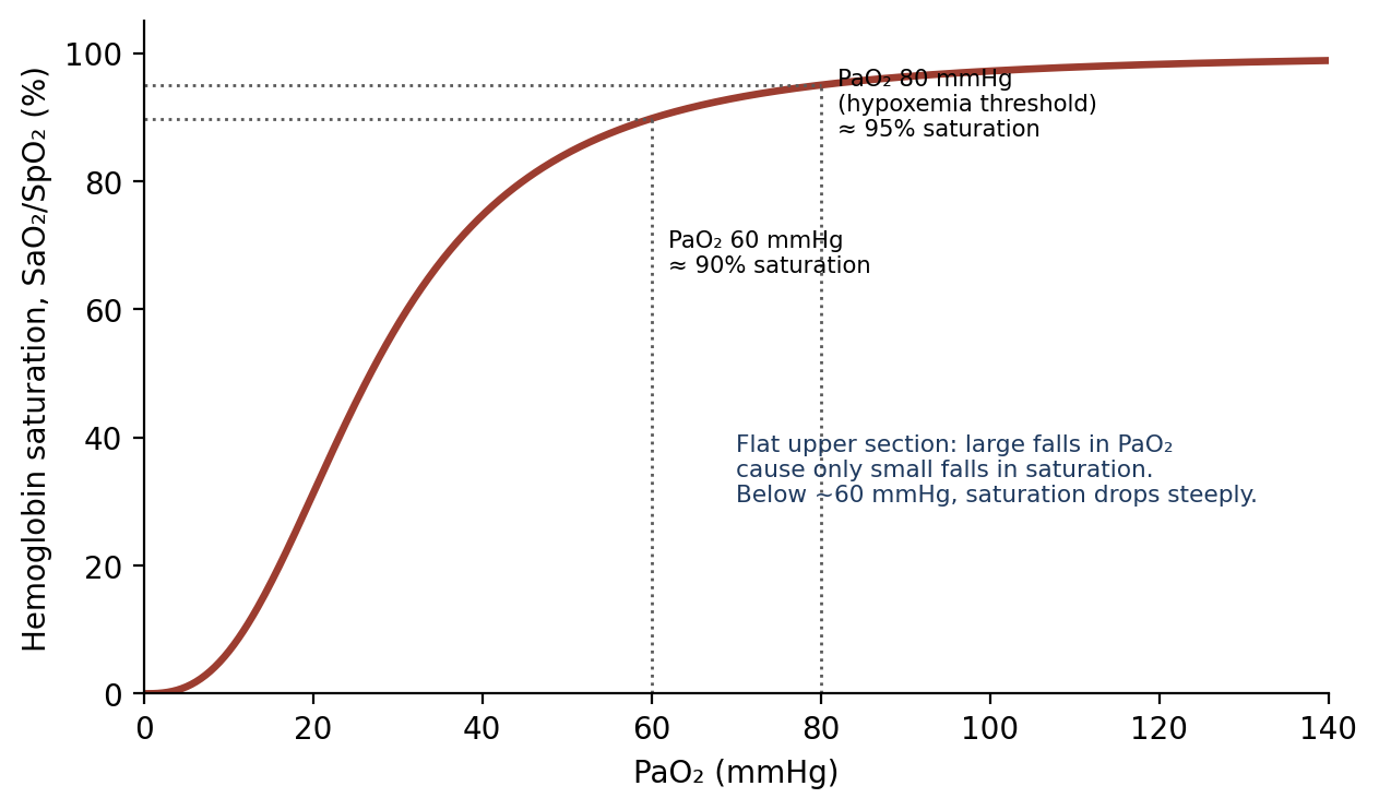
Figure 12.3. The oxygen–hemoglobin dissociation curve (representative). Saturation stays high until PaO₂ falls quite low, then drops steeply.
Why it matters
Because of the shape of the dissociation curve (Figure 12.3), SpO₂ is a late warning sign. An animal's PaO₂ can fall from 500 mmHg to 100 mmHg with almost no change in SpO₂. By the time SpO₂ drops noticeably, PaO₂ is already approaching the steep part of the curve, where oxygenation can deteriorate quickly.
Venous Admixture
Venous admixture is a collective term for all the ways blood can pass from the venous return to the arterial supply without being properly oxygenated, causing hypoxemia. Causes include equipment problems or decreased inspired oxygen supply, hypoventilation, bronchoconstriction, atelectasis, diffusion impairment (for example, from inhalation toxicity), and anatomic right-to-left shunts.
12.5 Cardiovascular Monitoring
12.5.1 Electrocardiogram (ECG)
The ECG monitors the electrical impulse as it is conducted through the heart (Figure 12.4). It shows electrical activity, not mechanical pumping, so a normal-looking ECG does not prove the heart is pumping effectively; it should be combined with pulse and blood pressure monitoring.
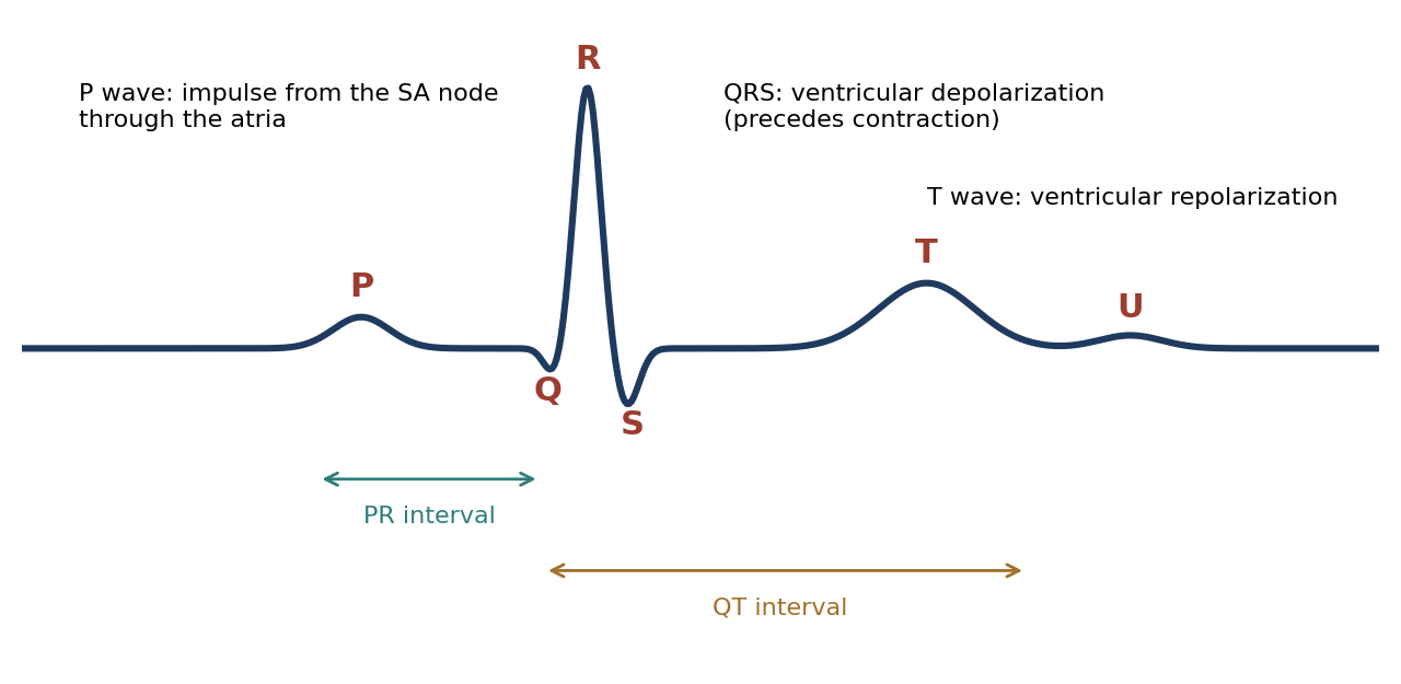
Figure 12.4. Components of the normal ECG.
Table 12.5. Components of the ECG.
| Component | Represents |
|---|---|
| P wave | Transmission of the impulse from the sinoatrial (SA) node through the atria |
| QRS complex | Ventricular depolarization, which precedes contraction |
| T wave | Ventricular repolarization |
| PR interval | Beginning of atrial excitation to the beginning of ventricular excitation |
| QT interval | Ventricular depolarization and repolarization |
| U wave | Uncertain origin; relatively uncommon; more often seen in larger species |
Rate abnormalities are defined relative to normal:
Tachycardia is an accelerated heart rate, typically more than 25% above normal; for example, rates above 160–180 bpm in cats and dogs.
Bradycardia is a slowed heart rate, typically more than 25% below normal; for example, rates below 60 bpm in dogs, 90 bpm in cats, and 20 bpm in horses.
Heart block indicates impaired electrical transmission through the atrioventricular (AV) node:
Table 12.6. Degrees of heart block.
| Degree | ECG finding |
|---|---|
| First-degree | Prolonged PR interval (every impulse conducted, but slowly) |
| Second-degree | Blocked P wave (some impulses not conducted; a P wave with no QRS) |
| Third-degree | Complete dissociation of P waves and QRS complexes |
Common Arrhythmias
Arrhythmias are abnormal depolarization, repolarization, and/or contraction of the heart, as seen on the ECG (Figure 12.5).
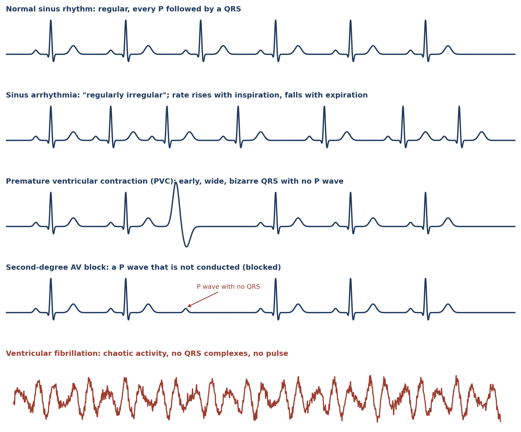
Figure 12.5. Representative ECG rhythm strips (schematic).
Sinus tachycardia shows the P wave appearing closer than normal after the preceding T wave (sometimes described as "T on P"). It is somewhat common and not typically dangerous, except in compromised patients or when it reflects inadequate coronary blood flow.
Sinus arrhythmia is a "regular irregularity": the heart rate increases with inspiration and slows with expiration. It is common in beagles, frequently seen in animals on a ventilator, and not a cause for concern.
Premature ventricular contractions (PVCs) are extra, abnormal beats originating in the ventricles, appearing as early, wide, bizarre QRS complexes. They are commonly caused by hypoxemia and/or hypercarbia and by traumatic myocarditis. They can also occur when a jugular catheter is placed too deep and irritates the heart: when placing an indwelling jugular catheter, watch the ECG for PVCs and back the catheter out if they appear. Single PVCs are not usually a problem, but a series may reduce cardiac output and coronary perfusion and may progress to ventricular fibrillation. Some species, such as pigs, are more susceptible to PVCs and should be pre-treated with anti-arrhythmic drugs for coronary procedures.
Ventricular fibrillation (VF) is a pulseless arrhythmia with irregular, chaotic electrical activity and ventricular contraction, in which the heart immediately loses its ability to pump; little or no blood leaves the heart. The sudden loss of cardiac output causes global tissue ischemia, putting the brain and myocardium most at risk. VF is the primary cause of sudden cardiac death. It can be treated with an electrical current (defibrillation), but defibrillation is not always effective and may cause burns and permanent damage to the heart. Before attempting resuscitation, consider whether it is a viable option, or whether the event automatically disqualifies the animal from the study protocol. Chapter 18 covers cardiopulmonary resuscitation.
Table 12.7. Summary of common arrhythmias.
| Arrhythmia | ECG features | Significance |
|---|---|---|
| Sinus tachycardia | Fast rate; P wave close after the T wave | Usually not dangerous, except in compromised patients or with poor coronary flow |
| Sinus arrhythmia | Rate rises with inspiration and falls with expiration | Normal; common in beagles and ventilated animals |
| PVCs | Early, wide, bizarre QRS without a preceding P wave | Single: usually not a problem. Series: reduced output; may lead to VF. Causes: hypoxemia, hypercarbia, myocarditis, deep jugular catheter |
| Heart block (1st–3rd degree) | Prolonged PR; blocked P waves; P–QRS dissociation | Impaired AV conduction |
| Ventricular fibrillation | Chaotic activity; no recognizable QRS | Cardiac arrest; defibrillation and CPR required |
12.5.2 Peripheral Perfusion Monitoring
Capillary refill time (CRT) is the time needed for a blanched mucous membrane to refill with blood after it is pressed. CRT should be 1–2 seconds, and non-pigmented gums should be pink. Other sites to observe color include the tongue, buccal mucous membrane, conjunctiva of the lower eyelid, and the mucous membranes of the prepuce or vulva. Pale membranes indicate poor perfusion, blood loss, or anemia; purple or blue membranes indicate cyanosis. When peripheral perfusion is low, pulse oximeters and indirect blood pressure monitors will most likely not work.
12.5.3 Central Venous Pressure (CVP)
CVP is the pressure inside the intrathoracic vena cava. It reflects the relationship between central blood volume (determined by venous return and cardiac output) and the capacity of the central veins to hold it. It is measured with a catheter placed through a jugular vein into the cranial (anterior) vena cava; the catheter may stimulate ectopic pacemaker activity (such as PVCs) if it touches the heart.
Table 12.8. Central venous pressure.
| Value / interpretation | |
|---|---|
| Normal, small animals | 0–10 cm H₂O |
| Normal, horses | 15–30 cm H₂O in lateral recumbency; 5–10 cm H₂O in dorsal recumbency |
| Below range | Hypovolemia; a rapid fluid bolus is suggested |
| Above range | Hypervolemia; fluid therapy should be stopped |
CVP is an important measurement when heart failure is a concern, but it does not predict or indicate cardiac output or stroke volume.
12.5.4 Arterial Blood Pressure
Arterial blood pressure is the primary indicator used to assess cerebral and coronary perfusion. It depends on blood volume and the capacity of the vessels to hold it, and is determined by cardiac output and systemic vascular resistance:
Systolic pressure is determined by stroke volume and arterial compliance.
Diastolic pressure is determined by systemic vascular resistance and heart rate.
Mean arterial pressure (MAP) is the average pressure and the most important, because it is the mean driving pressure for organ perfusion. It can be estimated as MAP ≈ diastolic + (systolic − diastolic) / 3 (Figure 12.6).
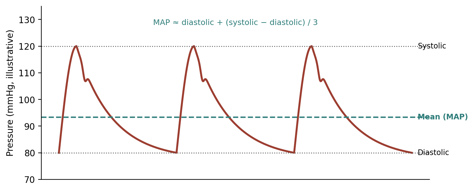
Figure 12.6. Arterial pressure waveform, showing systolic, diastolic, and mean pressure.
The strength of a peripheral pulse felt by palpation may not closely match central arterial pressure, so pressure should be measured. Methods are compared in Table 12.9.
Indirect Blood Pressure
Sphygmomanometry uses an occlusion cuff around an appendage over an artery, with a method to hear or detect blood flow. The cuff should be about 40% as wide as the circumference of the limb and placed snugly. If it is too tight, it partially occludes flow before inflation, giving an artificially low reading; if it is too loose, more pressure is needed to compress the artery, giving an artificially high reading. The cuff is inflated until it stops blood flow (cuff pressure above systolic). As cuff pressure is gradually released, blood begins to flow intermittently; at this point the manometer needle begins to oscillate, and the reading corresponds to systolic pressure. As pressure continues to fall, the point at which steady flow is detected and a pulse is palpated below the cuff corresponds to diastolic pressure.
Doppler ultrasound places a piezoelectric crystal over an artery. The crystal transmits ultrasound, and the movement of blood cells changes the frequency (pitch) of the reflected sound, so blood flow becomes audible. A pressure cuff is again used to occlude flow. As cuff pressure is released, the first audible pulse corresponds to systolic pressure. As pressure continues to fall, the sound changes character, which can be used to estimate diastolic pressure. Doppler readings correlate closely with direct arterial measurements.
⚠ Check current guidance
Doppler readings are sometimes described in reverse, with the first sound as diastolic pressure. The first pulse heard as the cuff deflates corresponds to systolic pressure; diastolic pressure is difficult to determine reliably with Doppler. Confirm against your exam references.
[ PHOTO PLACEHOLDER ] Suggested source: Wikimedia Commons (commons.wikimedia.org), search "Doppler blood pressure" or "veterinary blood pressure." Check the image's license and give the attribution it requires. |
|---|
Figure 12.7. Doppler blood pressure measurement: a cuff on the limb with the Doppler probe placed over the artery distal to it.
Oscillometry measures the fluctuation of pressure in the cuff as it slowly deflates, and provides a digital reading of systolic, diastolic, and mean pressure, and heart rate. It is the standard method in automatic blood pressure monitors.
Direct Blood Pressure
Direct measurement involves placing a catheter into an artery, surgically or percutaneously, connected to a transducer that converts the pressure wave into a digital signal. Fluid-filled catheters generally need to be flushed with heparinized saline (or a similar solution) to prevent clots at the tip. The catheter must be made of low-compliance material so that the pressure wave is transmitted without loss of amplitude. Solid-state catheters have no lumen but a pressure sensor at the tip that transmits the signal electronically, so no flushing is needed. Direct measurement gives a continuous reading and is the most accurate method.
Table 12.9. Methods of measuring arterial blood pressure.
| Method | How it works | Readings | Notes |
|---|---|---|---|
| Sphygmomanometry | Occlusion cuff and manometer | Systolic (needle oscillates); diastolic (steady flow) | Cuff ~40% of limb circumference; too tight reads low, too loose reads high |
| Doppler ultrasound | Cuff plus ultrasound crystal over artery | Systolic (first pulse heard) | Correlates closely with direct measurement |
| Oscillometry | Detects cuff pressure oscillations | Systolic, diastolic, mean, HR (digital) | Standard automatic monitor; may fail with poor perfusion |
| Direct (arterial catheter) | Catheter and transducer | Continuous systolic, diastolic, mean, waveform | Heparinized flush (unless solid-state); low-compliance tubing; most accurate |
12.5.5 Cardiac Output
Cardiac output (the volume of blood the heart pumps per minute) is more relevant to systemic perfusion (flow) than any pressure value, because a normal pressure can be maintained by vasoconstriction even when flow is low. Cardiac output is reduced by hypovolemia, ventricular restrictive disease, decreased contractility, bradycardia, tachycardia, arrhythmias, regurgitation (retrograde flow), and stenosis.
It is generally measured by thermodilution using a Swan-Ganz (pulmonary artery) catheter. A known volume of saline at a known, cold temperature is injected into the right side of the heart, and a temperature sensor near the catheter tip in the pulmonary artery measures how quickly the blood temperature changes. The faster the cold saline is carried past the sensor, the higher the cardiac output.
12.6 Temperature
Anesthetized animals lose the ability to thermoregulate normally (Chapter 5). Temperature falls because of clipping relatively large areas, evaporation of prep solutions, evaporation from and chilling of tissues within incisions, and vasodilation caused by anesthetic agents and adjuncts. Hypothermia prolongs recovery. Prevent it with heating blankets (water-circulating or warm-air), heated tables, warm IV and irrigation fluids, and covered extremities.
Hyperthermia is also possible and dangerous. It may be caused by overheating from heating pads and tables, or by anesthetic reactions such as malignant hyperthermia in swine (Chapter 18). Normothermic patients tolerate anesthesia better and recover faster, so body temperature should be monitored continuously to avoid both extremes.
Chapter Summary
Monitoring keeps anesthesia deep enough to prevent awareness, pain, and movement, but light enough to avoid cardiopulmonary depression. Requirements fall during an anesthetic, so the anesthetist should keep trying to reduce the dose. Depth is judged from dosing history, movement (unless paralyzed), reflexes, and trends in heart rate, blood pressure, and respiration; in paralyzed animals, rising heart rate and blood pressure suggest pain.
Respiratory monitoring uses breathing rate and pattern, tidal and minute volumes, arterial blood gases (PaCO₂ 35–45 mmHg; PaO₂ 80–110 mmHg on room air, hypoxemia below 80), EtCO₂ (slightly below PaCO₂), and pulse oximetry (a late warning because of the dissociation curve). Cardiovascular monitoring uses the ECG (P, QRS, T; tachycardia, bradycardia, heart blocks, sinus arrhythmia, PVCs, VF), CRT (1–2 seconds) and membrane color, CVP (0–10 cm H₂O in small animals), arterial pressure (MAP ≈ diastolic + one-third of pulse pressure, measured indirectly or directly), and cardiac output by thermodilution. Temperature must be monitored continuously to prevent hypothermia and hyperthermia.
Key Terms
Anatomical dead space: The part of each breath that fills the airways but does not reach the alveoli.
Apneic threshold: The PaCO₂ below which spontaneous breathing stops.
Capnograph: A monitor that measures and displays CO₂ in respired gas.
Capillary refill time (CRT): Time for a blanched mucous membrane to regain color; normally 1–2 seconds.
Central venous pressure (CVP): Pressure in the intrathoracic vena cava.
Cyanosis: Blue or purple discoloration of mucous membranes due to poorly oxygenated blood.
End-tidal CO₂ (EtCO₂): CO₂ concentration at the end of exhalation; approximates PaCO₂.
Heart block: Impaired electrical conduction through the AV node.
Hypercapnia / hypocapnia: Elevated / lowered PaCO₂.
Hypoxemia: Insufficient oxygenation of the blood (PaO₂ below 80 mmHg).
Mean arterial pressure (MAP): The average arterial pressure, which drives organ perfusion.
Oscillometry: Automatic indirect blood pressure measurement based on cuff pressure oscillations.
Premature ventricular contraction (PVC): An early beat originating in the ventricles.
Pulse oximetry (SpO₂): Non-invasive measurement of hemoglobin oxygen saturation.
Thermodilution: Measurement of cardiac output from the temperature change after injecting cold saline.
Venous admixture: Blood reaching the arterial circulation without being properly oxygenated.
Ventilation: Movement of gas into and out of the alveoli.
Ventricular fibrillation (VF): Chaotic ventricular electrical activity with no effective pumping.
Review Questions
1. Over the course of a single anesthetic, anesthetic requirements generally:
A. Increase steadily
B. Decrease overall
C. Stay exactly the same
D. Double every hour
2. In an animal that has received a neuromuscular blocking agent, which sign best indicates pain?
A. Movement
B. Increasing blood pressure and heart rate
C. Muscle tone
D. Palpebral reflex
3. Functional residual capacity (FRC) is:
A. Tidal volume plus inspiratory reserve volume
B. Expiratory reserve volume plus residual volume
C. The air moved in one breath
D. Total lung capacity minus tidal volume
4. The normal range for PaCO₂ is:
A. 20–30 mmHg
B. 35–45 mmHg
C. 60–80 mmHg
D. 80–110 mmHg
5. Hypoxemia is present when PaO₂ falls below:
A. 500 mmHg
B. 200 mmHg
C. 80 mmHg
D. 45 mmHg
6. If an animal becomes hypotensive, the pulse oximeter waveform will typically:
A. Become larger
B. First become smaller, then be lost
C. Show a higher saturation
D. Not change
7. On the ECG, the QRS complex represents:
A. Atrial depolarization
B. Ventricular depolarization
C. Ventricular repolarization
D. Conduction through the AV node
8. Third-degree heart block is characterized by:
A. A prolonged PR interval
B. An occasional blocked P wave
C. Complete dissociation of P waves and QRS complexes
D. Chaotic electrical activity with no QRS
9. A dog's blood pressure is 120/80 mmHg. Its approximate mean arterial pressure is:
A. 100 mmHg
B. 93 mmHg
C. 40 mmHg
D. 13 mmHg
10. An indirect blood pressure cuff that is applied too loosely will give a reading that is:
A. Artificially low
B. Artificially high
C. Accurate
D. Unreadable
Answer Key
1. B. Requirements trend downward because redistribution sites gradually fill, and they also vary with surgical stimulation and body temperature. Anesthetists should repeatedly try to reduce the amount given.
2. B. A paralyzed animal cannot move, but it can still feel pain. Rising blood pressure and heart rate are good indicators of pain in this situation.
3. B. FRC is the air remaining in the lungs after a normal expiration: ERV plus RV.
4. B. Normal PaCO₂ is 35–45 mmHg. Above 60 mmHg indicates excessive respiratory acidosis and may warrant ventilation.
5. C. PaO₂ is usually 80–110 mmHg breathing room air; hypoxemia occurs below 80 mmHg.
6. B. Poor peripheral perfusion reduces the pulsatile signal, so the waveform shrinks and then disappears.
7. B. The P wave is atrial depolarization, the QRS is ventricular depolarization (preceding contraction), and the T wave is ventricular repolarization.
8. C. First-degree block prolongs the PR interval, second-degree block has blocked P waves, and third-degree block shows complete dissociation of P waves and QRS complexes.
9. B. MAP ≈ diastolic + (systolic − diastolic)/3 = 80 + 40/3 ≈ 93 mmHg.
10. B. A loose cuff needs extra pressure to compress the artery, so the reading is artificially high. A cuff applied too tightly gives an artificially low reading.
Chapter 13: Pain Assessment & Management
Learning objectives
After studying this chapter, you should be able to:
Define the key terms used to describe pain, suffering, and distress, and distinguish types of pain.
Describe the physiology of pain, including nociceptor types, the four steps of nociception, and wind-up.
Explain pre-emptive and multimodal analgesia, and identify where each analgesic class acts.
Compare opioids, NSAIDs, local anesthetics, alpha-2 agonists, and miscellaneous analgesics.
Recognize behavioral and physiological signs of pain in common laboratory species and use a pain scoring system.
Preventing and relieving pain is both an ethical and a regulatory obligation (Chapter 2), and it is good science: pain alters behavior, immune function, metabolism, and cardiovascular physiology, all of which can confound research data. This chapter explains how pain is generated, how analgesic drugs interrupt it, and how to recognize it in animals that cannot tell us what they feel.
13.1 Definitions
Pain is an unpleasant (noxious) sensory and emotional experience associated with actual or potential tissue damage. Two related terms describe its emotional consequences. Suffering is an unpleasant emotional state that is internalized and not outwardly expressed, originating from either a physical or physiological source. Distress is the outward expression of suffering, through behavior or emotion, that an observer can see, such as anxiety, fear, aggression, or hyperactivity.
Pain has its own specialized vocabulary. The terms are grouped below by theme.
Table 13.1. General terms and drug-related terms.
| Term | Definition |
|---|---|
| Algology | The science and study of pain |
| Analgesia | Absence or decrease of pain in the presence of a stimulus that would normally be painful |
| Analgesic | A drug that induces analgesia |
| Anesthesia | Absence of all sensory modalities; may be local, regional, or general |
| Anesthetic | A drug that induces regional or general anesthesia |
Table 13.2. Terms describing abnormal pain sensitivity and sensation.
| Term | Definition |
|---|---|
| Allodynia | Pain caused by a stimulus that does not normally provoke pain |
| Hyperalgesia | Increased response to a stimulus that is normally painful |
| Hypoalgesia | Diminished sensitivity to noxious stimulation |
| Dysesthesia | An unpleasant spontaneous or evoked abnormal sensation, especially induced by touch |
| Paresthesia | A spontaneous or evoked abnormal sensation (tingling, tickling, pricking, or burning) with no apparent physical cause; not painful, as opposed to dysesthesia |
Table 13.3. Terms describing types and sources of pain.
| Term | Definition |
|---|---|
| Somatic | Describes input from body tissues other than viscera (skin, joints, muscles, and other deep tissues) |
| Somatic pain | General nociceptor-based pain originating from skin, joints, and muscles |
| Visceral pain | Pain originating from abdominal organs; more "internal," poorly localized, not evoked by burning or cutting, and not evoked from all organs (such as the liver and kidneys); may arise from nociceptors or receptors with other functions |
| Inflammatory pain | Spontaneous pain and hypersensitivity in response to tissue damage and inflammation |
| Neuropathic pain | Spontaneous pain and hypersensitivity associated with damage to the nervous system |
| Central pain | Pain associated with a lesion of the CNS |
| Chronic pain | Pain that persists longer than the expected time for healing, or pain associated with progressive disease |
| Deafferentation pain | Pain caused by loss of sensory input into the CNS (e.g., brachial plexus avulsion, other peripheral nerve lesions) or by CNS pathology |
| Causalgia | Prolonged burning pain, allodynia, and hyperpathia after a traumatic nerve lesion, often with vasomotor and sudomotor (sweating) dysfunction and later trophic changes |
Table 13.4. Terms describing nerves and nerve roots.
| Term | Definition |
|---|---|
| Neuralgia | Pain in the distribution of a nerve or nerves |
| Neuritis | Inflammation of a nerve or nerves |
| Neuropathy | Disturbance of function or pathological change in a nerve |
| Radiculalgia | Pain along the distribution of one or more sensory nerve roots |
| Radiculopathy | Disturbance of function or pathological change in one or more nerve roots |
| Radiculitis | Inflammation of one or more nerve roots |
Table 13.5. Terms describing nociception and responses to stimuli.
| Term | Definition |
|---|---|
| Nociception | Reception, conduction, and CNS processing of signals generated by stimulation of nociceptors; the physiological process that, when completed, results in conscious perception of pain |
| Nociceptor | A receptor preferentially sensitive to a noxious stimulus, or to a stimulus that would become noxious if prolonged |
| Nociceptor threshold | The minimum strength of stimulus that will cause a nociceptor to generate an impulse |
| Noxious stimulus | A stimulus that is actually or potentially damaging to tissue, or of a quality or intensity that triggers nociceptive reactions |
| Pain threshold | The least pain a subject can recognize; usually higher than the nociceptor threshold; relatively constant among species and individuals |
| Pain tolerance | The greatest pain a subject will tolerate; varies considerably between species and individuals and is influenced by experience, environment, stress, and drugs |
| Pain tolerance range | The difference between the pain detection threshold and the pain tolerance threshold |
| Reflex | An involuntary, purposeful, orderly reaction to a stimulus. The reflex arc consists of a receptor, a primary afferent nerve fiber, a region of integration in the spinal cord or brain stem, and a lower motor neuron leading to an effector (skeletal or smooth muscle, or glands) |
| Reaction | A combination of reflexes producing a widespread movement in response to a stimulus; mass reflexes not under voluntary control and not involving the cerebral cortex |
| Response | A willful movement of the body or its parts that requires involvement of the somatosensory cerebral cortex |
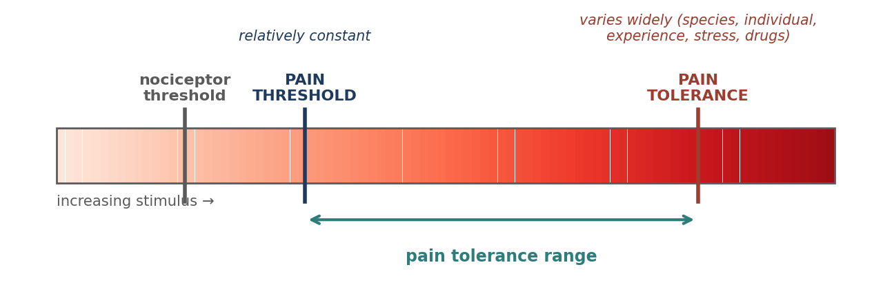
Figure 13.1. Nociceptor threshold, pain threshold, and pain tolerance.
Why it matters
The distinction between reflex, reaction, and response matters during anesthesia. A withdrawal reflex or a mass reaction can occur in an anesthetized animal without any conscious experience of pain, because neither requires the cerebral cortex. A response, by definition, does involve the cortex. This is why movement alone does not prove an animal is aware, and why the absence of movement does not prove it is not (Chapter 12).
Wind-Up (Central Sensitization)
Wind-up (hypersensitization) is a cascade of events resulting from ongoing stimulation of nociceptors and activation of N-methyl-D-aspartate (NMDA) receptors. It causes hyperalgesia and opioid tolerance, and it results from untreated pain.
Unlike many receptors, nociceptors do not fatigue with repeated stimulation. Instead, they become more sensitive, with a lowered threshold and a prolonged, enhanced response. After a barrage of afferent nociceptor impulses, the neurons of the spinal dorsal horn become hypersensitized by the release of excessive neurotransmitters, which lowers their firing threshold and increases their rate of discharge. The receptive field can even expand to include distant nerves. As a result, a stimulus causes more pain than it otherwise would (Figure 13.2), and more analgesics are needed to relieve it.
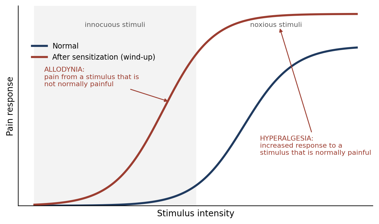
Figure 13.2. Sensitization shifts the pain response, producing allodynia and hyperalgesia (schematic).
Wind-up can be prevented with local pre-emptive analgesics and/or prompt or pre-emptive general analgesics. Once established, it is hard to reverse: peripheral nerve blocks are less effective, systemic opioids have little effect at normal therapeutic doses, and in severe cases anesthesia followed by epidural or intrathecal local anesthetics may be required.
13.2 Common Types of Pain
Table 13.6. Common types of pain.
| Type | Characteristics |
|---|---|
| Acute pain | Occurs immediately after a stimulus; severity varies; responds well to treatment; subsides once the stimulus is removed |
| Chronic pain | Persists well past the initial stimulus (3–6 months); severity varies; may or may not respond well to treatment and may require a multimodal approach; can result in allodynia, hyperalgesia, and opioid tolerance |
| Physiological pain | A protective mechanism that causes avoidance; little or no tissue injury; stops once the stimulus is removed |
| Pathological pain | Results from tissue injury, with inflammation, nerve damage, and release of neurotransmitters with ongoing nociceptor stimulation; can lead to hyperalgesia; persists after the stimulus is removed |
Physiological pain is useful: it teaches an animal to withdraw from a hot surface before it is burned. Pathological pain, including surgical pain, has lost that protective value and continues after the stimulus is gone, which is why it should be treated.
13.3 Physiology of Pain
When tissue is damaged, the damaged cells release substances that stimulate nociceptors and promote inflammation. Noxious stimuli activate nociceptors, which become sensitized with continued stimulation, lowering their threshold. Sensitized nociceptors release glutamate and neurokinins from their terminals in the spinal cord, activating NMDA receptors, which are implicated in hypersensitivity (wind-up). Neurons in the spinal cord relay the signal to multiple areas of the brain, resulting in the perception of pain. A "gate control" mechanism in the spinal cord provides early inhibition of nociception, allowing escape.
Pain also triggers a stress response. Stimulation of medullary centers causes hyperventilation, increased cardiac output and blood pressure, and increased secretion of catecholamines and other hormones. Cortisol and other hormones raise blood glucose and ketone levels and increase metabolic rate and oxygen consumption. The magnitude and duration of these effects parallel the degree of tissue damage. These same physiological responses occur in properly anesthetized or unconscious patients, but they do not produce the sensation of pain, because the cerebral cortex is not functioning. Pre-emptive analgesics decrease this response.
The body also has its own pain control. Descending neurons from the brain modulate pain by reducing sensation, releasing neurotransmitters such as glutamate, norepinephrine, serotonin, GABA, and endorphins. Analgesia can be induced by blocking the nociceptive process at one or more points (Section 13.4).
Uncontrolled pain has wide-ranging harmful effects:
Cortically mediated increases in blood viscosity, clotting time, fibrinolysis, and platelet aggregation.
Intense vasoconstriction, leading to ischemia, tissue hypoxia, and release of substances toxic to the myocardium.
Possible renal failure.
Muscle spasms, disuse of the injured area, muscle atrophy, hypoventilation, weight loss, and dehydration.
Anxiety and fear, which enhance the stress response.
Shock, in cases of severe post-traumatic or post-operative pain.
13.4 Nociception
Pain is not the same as nociception. Pain is the product of higher brain centers processing the signals they receive. Nociception refers to the peripheral and central nervous systems processing information generated when noxious stimuli stimulate nociceptors. Nociception can occur in the absence of pain, as in an anesthetized animal.
13.4.1 Types of Nociceptors
Table 13.7. Comparison of nociceptor fiber types.
| Property | A-delta (Aδ) fibers | C fibers |
|---|---|---|
| Myelination | Myelinated | Unmyelinated |
| Conduction | Rapid | Slow |
| Sensation | "First pain": sharp, pricking | Dull, burning, longer-lasting pain |
| Activation | Mechanical and thermal stimuli | Chemicals released in damaged or inflamed tissue; high threshold at rest; once inflammatory mediators lower their threshold, also activated by thermal and mechanical stimuli |
| Role in sensitization | — | Produce hyperalgesia |
If you stub a toe, the sharp, immediate pain is carried by A-delta fibers, and the slower, throbbing ache that follows is carried by C fibers. C fibers are relatively insensitive under normal conditions, but inflammation lowers their threshold dramatically, which is a major source of post-operative hyperalgesia.
13.4.2 Parts of the Nociceptive Process
Nociception involves four distinct processes, each of which can be modified by analgesics (Figure 13.3):
Transduction is the translation of a noxious stimulus into electrical nerve impulses at the peripheral nociceptor. It can be blocked by local anesthetics (injected at the site of injury or incision, or given IV), and decreased by NSAIDs or corticosteroids, which reduce prostaglandin production at the site of injury.
Transmission is the propagation of nerve impulses through the nervous system to the spinal cord. It is inhibited by local anesthetic blockade of peripheral nerves or plexuses, or by injection into the epidural or subarachnoid space, and prevented by alpha-2 agonists.
Modulation occurs once the signal reaches the spinal cord, where it stimulates the release of chemicals (such as endorphins) that inhibit the dorsal horn cells as the signal passes to the brain. It is affected by local anesthetics, alpha-2 agonists, NMDA antagonists, and anticonvulsants.
Perception is the final phase and occurs only in the conscious patient: the subjective, emotional experience of pain, which results in vocalization, withdrawal, and sometimes aggression. It is interrupted by anesthesia, and inhibited or altered by opioids, alpha-2 agonists, benzodiazepines, phenothiazines, and general anesthetics.
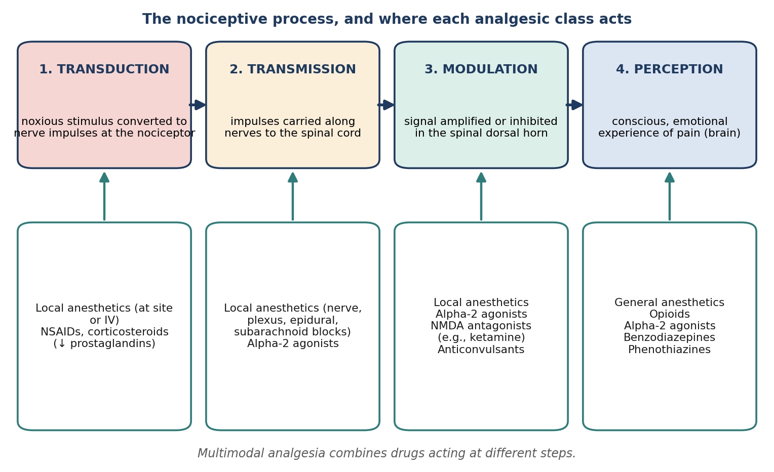
Figure 13.3. The four steps of nociception and the analgesic classes that act at each.
13.5 Response to Injury
Damaged cells in traumatized tissue release numerous chemicals that promote inflammation. Inflammation increases the permeability of capillary walls, allowing macrophages to infiltrate, and increases blood flow to the area, producing erythema and edema. Neurotransmitters are released that stimulate nociceptors. Initially this is protective: the animal guards the area to prevent further injury.
13.6 Control of Pain
Pre-emptive analgesia means giving analgesics before the noxious stimulus (surgery). Blocking or inhibiting the nociceptive process before it begins prevents hypersensitivity, and decreases the amount of anesthesia and post-operative analgesia needed.
Multimodal ("balanced") analgesia uses a combination of analgesics that act on more than one part of the nociceptive process (Figure 13.4). For example: buprenorphine and meloxicam before surgery, a lidocaine block before the incision, and a bupivacaine splash before closing the incision. Because each drug works at a different point, lower doses of each can be used, reducing side effects.
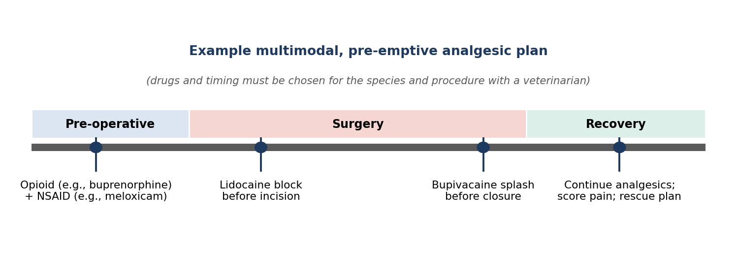
Figure 13.4. An example of a pre-emptive, multimodal analgesic plan.
Non-pharmaceutical methods may also be beneficial and should be considered when possible, such as ice packs during recovery, and splints or bandages that provide support and reduce swelling and inflammation. Good nursing care (warm, quiet, comfortable housing and easy access to food and water) also reduces pain and distress.
13.7 Analgesics
Analgesics are divided into five main classes based on their mode of action: opioids, NSAIDs, local anesthetics, alpha-2-adrenoceptor agonists, and miscellaneous drugs (Table 13.8). Chapters 6 and 11 cover many of these drugs in detail; this section focuses on their use for analgesia.
Table 13.8. Analgesic classes.
| Class | Examples | Main site of action | Key points |
|---|---|---|---|
| Opioids | Morphine, fentanyl, buprenorphine, butorphanol | Perception (CNS); also spinal | Drugs of choice for severe acute pain; controlled substances; reversible with naloxone |
| NSAIDs | Carprofen, meloxicam | Transduction (↓ prostaglandins) | Anti-inflammatory, analgesic, antipyretic; GI and renal risks |
| Local anesthetics | Lidocaine, bupivacaine | Transduction, transmission, modulation | Block Na⁺ channels; prevent central sensitization |
| Alpha-2 agonists | Xylazine, medetomidine, dexmedetomidine | Transmission, modulation, perception | Sedation, muscle relaxation, analgesia; reversible |
| Miscellaneous | Tramadol, ketamine, gabapentin | Various (e.g., NMDA antagonism) | Adjuncts, especially for wind-up and nerve pain |
13.7.1 Opioids
Opioids are also called "narcotic analgesics" because of their ability to induce narcosis. They were originally extracted from the opium poppy (as opium and laudanum), which contains about 20 active compounds called opiates, including morphine and codeine; the term opioid describes derivatives of these compounds. Opioids bind to specific receptors and mimic the body's own endogenous opioids (endorphins). There are mu (µ), delta (δ), and kappa (κ) receptors, distributed in the brain, spinal cord, and periphery; most activity, both analgesic and adverse, is at the mu receptor.
Opioids act by raising the pain threshold or decreasing the perception of pain in the CNS, and they alter the emotional component of pain to make it more tolerable. They are the drugs of choice for severe, acute pain. They are well absorbed after GI, IV, SC, IM, intrathecal, and (in special preparations) transdermal administration. They are controlled substances requiring special licenses and documentation.
Side effects vary by species:
CNS depression with miosis, hypothermia, bradycardia, and respiratory depression in primates, dogs, rats, and rabbits.
CNS stimulation with mydriasis, panting, tachycardia, and hyperkinesis in horses, cats, ruminants, and swine.
Depression of the respiratory and cough centers, and possible nausea and vomiting.
Mice and rats quickly develop tolerance.
Opioids are categorized as agonists, partial agonists, or mixed agonist-antagonists (Chapter 6, Figure 6.1):
Full agonists, such as morphine and fentanyl, are potent analgesics that decrease the amount of anesthesia required. They have more serious potential side effects than mixed agonist-antagonists, including respiratory depression, bradycardia, vomiting, and constipation. They can be combined with tranquilizers for neuroleptanalgesia, given IV, IM, transdermally, or epidurally (with or without local anesthetics), and reversed with naloxone.
Morphine is active at all three receptor types and is the most effective analgesic. It typically lasts 3–4 hours. Its poor lipid solubility means that, given epidurally or into the subarachnoid space, it stays there and produces long-lasting analgesia of 12–24 hours. It frequently causes vomiting, and IV administration can cause histamine release.
Fentanyl is described as 250 times more potent than morphine (see the note in Chapter 6), with a rapid onset and short duration (peak at 30 minutes), so it is commonly given as a continuous infusion during surgery. Its respiratory depression may persist for hours, and it causes vagally mediated bradycardia unless countered with atropine. It may be combined with a benzodiazepine to induce anesthesia in dogs with cardiovascular instability, and is available as a transdermal patch.
Mixed agonist-antagonists, such as butorphanol, have agonist or partial agonist activity at one or more opioid receptors and can antagonize a full agonist at others. Butorphanol is a mu antagonist and kappa agonist. It causes less respiratory depression than full agonists and can be used post-operatively to reverse the narcosis of fentanyl while still providing some analgesia. It has a ceiling effect and is not routinely used for analgesia today because of its short duration (frequent dosing).
Partial agonists, such as buprenorphine, have both agonist and antagonist activity at the mu receptor. Buprenorphine can be used to reverse pure mu agonists, has a relatively prolonged duration, and also has a potential ceiling effect.
Table 13.9. Opioid categories.
| Category | Example | Receptor activity | Key points |
|---|---|---|---|
| Full agonist | Morphine, fentanyl | Mu (morphine: mu, delta, kappa) | Most potent analgesia; most side effects; reversed by naloxone |
| Mixed agonist-antagonist | Butorphanol | Mu antagonist, kappa agonist | Less respiratory depression; ceiling; short duration; can reverse fentanyl narcosis |
| Partial agonist | Buprenorphine | Partial mu agonist | Long duration; ceiling; can reverse pure mu agonists |
13.7.2 Non-Steroidal Anti-Inflammatory Drugs (NSAIDs)
NSAIDs are weak organic acids with anti-inflammatory, analgesic, and antipyretic (fever-reducing) properties. They inhibit prostaglandin production by inhibiting the cyclooxygenase (COX) enzymes. There are two main COX isoenzymes: COX-1, which is largely responsible for prostaglandins that protect the stomach lining and maintain kidney blood flow, and COX-2, which is largely induced by inflammation.
NSAIDs are either non-selective (inhibiting both COX isoenzymes) or selective for COX-2. Non-selective NSAIDs have more serious side effects, especially gastric ulceration and renal toxicity. Because renal blood flow decreases during anesthesia, the kidneys are more susceptible to NSAID toxicity at that time. Carprofen and meloxicam are COX-2-selective inhibitors with a reasonable margin of safety when used pre-operatively.
⚠ Check current guidance
Carprofen and meloxicam are often described more precisely as COX-2-preferential rather than strictly COX-2-selective. NSAID choice, dosing, and pre-operative use depend on species, hydration, blood pressure, and renal function, and NSAIDs should generally not be combined with corticosteroids or other NSAIDs. Check current references and consult a veterinarian.
13.7.3 Local Anesthetics
Local anesthetics decrease or prevent sodium (Na⁺) permeability of neuronal membranes, stopping signal transmission along peripheral nerves. Because the nociceptive signal is blocked before it reaches the spinal cord, they prevent central sensitization. They are classified by duration: lidocaine is short-acting with a rapid onset, and bupivacaine is long-acting with a slower onset. Duration can be extended by adding a vasoconstrictor such as epinephrine (Chapter 11). Uses include:
Topical: most are applied to mucous membranes, though some preparations are absorbed through skin. 0.5% proparacaine is recommended for examining eyes. Lidocaine and benzocaine sprays assist intubation (benzocaine has been implicated in methemoglobinemia and should be used sparingly and with caution). Xylocaine jelly lubricates endotracheal tubes and urinary catheters. EMLA cream (lidocaine and prilocaine) numbs skin.
"Splash block": applied to exposed tissues before closure, and to nerves before they are cut during amputations. Soaker catheters (wound or diffusion catheters) left subcutaneously can infuse local anesthetic after major surgery such as amputation; infusion rates must be calculated to avoid toxicity.
Infiltration: multiple intradermal or SC injections along the proposed incision line, which may contain epinephrine (1:200,000) to increase effect and duration.
Field block: intradermal or SC infiltration followed by injection deep enough to reach the nerves, used for large areas.
Regional block: injection into the connective tissue around a nerve, producing loss of sensation and/or paralysis in the region it supplies. It requires smaller volumes than a field block, reducing the risk of toxicity.
Epidural: given alone or with other analgesics such as opioids. Combining drugs allows smaller doses of each, reducing adverse effects. Higher doses can cause motor deficits.
13.7.4 Alpha-2-Adrenergic Agonists
Stimulation of alpha-2 adrenoceptors produces sedation, muscle relaxation, and analgesia. This class includes xylazine, medetomidine, and dexmedetomidine, which can be reversed with alpha-2 antagonists such as yohimbine and atipamezole (Chapter 6). Note that reversing an alpha-2 agonist also reverses its analgesia, so other analgesics must be in place.
13.7.5 Miscellaneous Analgesics
Tramadol is a synthetic opioid agonist that also inhibits serotonin and norepinephrine reuptake in the spinal cord. Its main metabolite has moderate opioid activity. It is available only as an oral formulation in the US, but as an injectable in the UK.
Ketamine is an NMDA antagonist. Given as a constant rate infusion (CRI) at sub-anesthetic doses during surgery, it reduces MAC and can help prevent hypersensitivity (wind-up). It is more effective for somatic than visceral pain, and can be given epidurally.
Gabapentin is an analogue of the neurotransmitter GABA, believed to increase GABA production as part of the body's endogenous inhibition of nociception. It is used to treat nerve (neuropathic) pain.
⚠ Check current guidance
Gabapentin's main mechanism is now generally attributed to binding the α2δ subunit of voltage-gated calcium channels rather than to increasing GABA. The efficacy of oral tramadol varies considerably between species. Check current references for both drugs.
13.8 Pain Recognition & Assessment
The structures involved in pain sensation are very similar in humans and animals. It is therefore reasonable to assume that if something is painful in people, is damaging or potentially damaging to tissue, and/or induces escape or adverse emotional responses in an animal (signs of distress, avoidance, vocalization, changes in posture), it should be considered painful to the animal. If a procedure is likely to cause pain, it is appropriate to give preventive analgesics. With accurate selection and dosing, pain can be relieved without severe side effects.
Clinical analgesia is not the complete absence of pain, but the reduction of pain intensity to a tolerable level. Pre-operative analgesia is highly effective because it prevents central sensitization (wind-up), suppresses the neuroendocrine response to pain, improves tissue healing and mobility, and is the most effective means of controlling post-operative pain.
Recognizing and Assessing Pain
To relieve pain, you must be able to recognize it, and to know whether your analgesic plan is working. This is difficult, because pain cannot be measured directly:
You must be familiar with the species and strain to distinguish normal from abnormal behavior. Signs of pain vary not only between species, but between strains and individuals.
Assessment requires careful observation and experience with the species and with the individual animal.
Many laboratory species will not show obvious signs of mild to moderate pain. Prey species in particular tend to hide signs of weakness.
No single sign always indicates a specific amount of pain.
Some signs used for pain assessment may also be seen in healthy animals. For example, healthy cats may hiss, scratch, be aggressive, and vocalize; rats frequently vocalize when handled; and swine normally vocalize when handled unless well acclimated.
Table 13.10. General signs of pain.
| Category | Signs |
|---|---|
| Behavior | Lethargy, listlessness; avoidance; hiding; aggression; vocalization; biting or licking the injured area; disinterest in surroundings; "inwardly" focused; failure to make a nest |
| Posture and movement | Hunched or abnormal posture; "writhing"; disuse of a limb |
| Face and coat | Grimacing; eye squinting; ruffled coat |
| Intake and output | Decreased food and water consumption; decreased elimination |
Table 13.11. Measurable physiological effects of pain.
| System | Effect |
|---|---|
| Cardiovascular | Vasoconstriction (pale mucous membranes); increased heart rate, blood pressure, and cardiac output |
| Respiratory | Rapid, shallow breathing (especially with abdominal and thoracic pain) |
| Eyes | Pupil dilation; porphyrin staining around the eyes in rats |
| Blood chemistry (increased) | Epinephrine, norepinephrine, cortisol, glucose, glucagon, lipids, ketones, amino acids |
| Blood chemistry (decreased) | Phosphorus, magnesium, testosterone, insulin |
| Local | Heat in the affected area |
Physical signs also depend on the procedure performed (Table 13.12).
Table 13.12. Signs of pain by procedure.
| Procedure | Typical signs |
|---|---|
| Orthopedic or limb surgery | Decreased weight bearing, from limping to disuse |
| Thoracotomy | Shallow respirations; decreased mobility |
| Spinal surgery | Decreased mobility; inability to rest |
| Abdominal surgery | Shallow respirations; hunched posture; tucked abdomen |
Species-Specific Signs of Pain
All species show avoidance of painful stimuli and, when avoidance is not possible, may show aggression. Species-specific signs of mild to moderate pain and of severe or chronic pain are summarized in Table 13.13.
Table 13.13. Species-specific signs of pain.
| Species | Mild to moderate pain | Severe or chronic pain |
|---|---|---|
| Mouse | Partially closed eyelids; rough coat; hunched posture; scratching; increased aggression and apprehension; vocalization when handled; self-mutilation; abdomen tucked up or pressed to cage floor; writhing or abdominal stretch; decreased nest building; isolation from cage mates | Weight loss; dehydration; soiled coat; sunken eyes; sunken or distended abdomen; hunched posture; ataxia; hypothermia; decreased vocalization; wasting of back muscles |
| Rat | Partially closed eyelids; rough coat; scratching; increased aggression and apprehension; vocalization when handled; porphyrin staining around eyes and nose; writhing or abdominal stretch; abdomen tucked up or pressed to cage floor; failure to explore a new environment | Closed eyes; weight loss; dehydration; soiled coat; sunken or distended abdomen; ataxia; hypothermia; decreased vocalization; wasting of back muscles; incontinence; recumbency with tucked head; self-mutilation |
| Guinea pig | Sunken, dull eyes; respiratory changes; increased timidity and sleepiness; arched back; increased vocalization when handled | Weight loss; scaly skin; dehydration; decreased timidity; unresponsiveness; excessive salivation; increased barbering; loss of righting reflex; decreased vocalization; hypothermia |
| Rabbit | Ocular discharge; constipation or diarrhea; depression; excessive grooming; stretched or tucked posture (depending on procedure); teeth grinding; dullness or aggression; lack of appetite; abnormal gait and posture | Teeth grinding; weight loss; dehydration; wasting of lower back muscles; fecal staining; decreased night feces production; general unresponsiveness |
| Nonhuman primate | Masks many signs; licking at site; disuse; decreased activity, food, and water intake; behavior changes; hunched posture; failure to interact with neighbors; staying on the cage floor | Hunched or crouching posture; clenching or grinding teeth; disuse of limb; anorexia; weight loss; decreased grooming and socialization; increased aggressive attention from other NHPs |
| Dog | Decreased alertness; stiff posture; limping; panting; licking; biting; increased aggression and vocalization; decreased appetite; isolation; inwardly focused; decreased activity; standing for long periods; listlessness | Crouched or hunched posture; disuse of limb; unwillingness to move; difficulty lying down; depression; increased aggression; vocalization when handled; restlessness; listlessness; eyes focused inwardly |
| Cat | Increased aggression; decreased food intake; excessive licking or grooming; limping; immobility; abnormal vocalization | Hunched, crouching, or stretched posture; increased aggression; anorexia; weight loss; vocalizing; wild escape behavior; unkempt appearance; stiff gait; disuse of limb |
| Pig | Changes in gait and posture; increased handling avoidance; increased vocalization; decreased activity and food intake | Depression; unwillingness to move; attempts to hide; anorexia; decreased socialization |
| Sheep and goat | Lying with legs extended; limping; stomping; mild ataxia; depression; restlessness; teeth grinding; increased aggression; separation from the herd; hanging head (sheep are more stoic and less likely to show pain) | Rolling; frequent looking at or kicking the abdomen; falling over; walking backward; rapid, shallow breathing; weight loss; teeth grinding; grunting; vocalization when handled; rigidity; unwillingness to move; disuse of limb |
Why it matters
Notice how many signs of severe pain in rodents (weight loss, dehydration, hypothermia, hunched posture, soiled coat) are also signs of general illness. Weighing animals regularly after surgery is one of the most objective and useful ways to detect a problem, whatever its cause.
Pain Scoring Scales
A pain scoring system (analog scale) turns observations into a score that can be compared over time and between observers. To use one effectively:
Establish the parameters to score, decide what score indicates insufficient analgesia, and have a rescue plan (additional analgesia, veterinary consultation, or humane endpoint) ready.
Make the scale species-specific.
In socialized species, the scale can be used together with physiological parameters such as heart rate, respiratory rate, and/or blood pressure.
First observe the animal from outside the cage or pen, without interaction, then open the cage and observe how it reacts to your presence. You need to know what the normal reactions for that species are.
Table 13.14 shows an example scoring sheet. Each criterion is scored from 0 (normal) to 3, and the scores are totaled. The wording should be adapted to the species (for example, rabbits "hop" while dogs walk), and the intervention threshold set in advance.
Table 13.14. Example pain scoring sheet (adapt wording and thresholds for the species).
| Criterion | 0 | 1 | 2 | 3 |
|---|---|---|---|---|
| Attitude and posture (from a distance, no handling) | Alert, ears up, eyes bright and open, relaxed; at the front of the cage or lying normally in a relaxed posture at the back | Notices and watches the technician; ears not fully up; less relaxed muscles; quieter; eyes open; comes to the front of the cage or relaxes in a normal position at the back | Decreased activity; squinted, dull eyes; ears down; tense posture at the back of the cage; does not lie normally; "inwardly" focused | No interest in the technician unless the cage is opened or the animal handled; hunched posture; dull appearance; eyes very squinted or bulging |
| Gait and movement (distance observation) | Normal movement around cage; fully weight bearing; hops or walks around cage; rises up normally | Moves slowly and carefully but freely; not hesitant to move but may move slowly or gingerly; at least touches limb to floor | Non-weight bearing; very stiff movement; sits or lies abnormally with legs placed abnormally; moves with effort and reluctantly; may need to be prompted to move | Very reluctant or unwilling to move; may fall asleep while sitting; may become frantic when approached |
| Appetite | Normal | May eat less than normal, but eating something, even if only treats | Has not eaten in past 24 hours | No interest in food, water, or enrichment for more than 24 hours |
| Elimination | Normal | Decreased output over 24 hours | Decreased urine; little or no feces over 24 hours | No urine or feces over 24 hours |
Several validated, species-specific scales have been published, such as grimace scales (which score facial features like orbital tightening, nose and cheek bulging, and ear and whisker position) for mice, rats, rabbits, and other species, and composite pain scales for dogs and cats. Using a validated scale improves consistency between observers.
[ PHOTO PLACEHOLDER ] Suggested source: The NC3Rs (nc3rs.org.uk) publishes grimace scale posters for several species; check its terms of use and request permission if needed. Alternatively, cite the original published scale and photograph your own animals. |
|---|
Figure 13.5. A grimace scale for a laboratory species (for example, the Mouse Grimace Scale), showing facial action units scored 0, 1, and 2.
⚠ Check current guidance
Validated pain assessment tools are being developed and refined for more species. Check current references and your institution's approved scoring systems before choosing a scale.
Chapter Summary
Pain is an unpleasant sensory and emotional experience; suffering is internal, and distress is its visible expression. Nociception (the nervous system's processing of noxious stimuli) can occur without pain. Fast, myelinated A-delta fibers carry sharp first pain, and slow, unmyelinated C fibers carry dull, burning pain and drive hyperalgesia. Nociception has four steps (transduction, transmission, modulation, perception), and untreated pain causes wind-up via NMDA receptors, producing allodynia, hyperalgesia, and opioid tolerance. Uncontrolled pain also triggers a damaging stress response.
Pre-emptive analgesia prevents sensitization, and multimodal analgesia combines drugs acting at different steps: opioids (full, partial, and mixed agonists), NSAIDs (COX inhibitors with GI and renal risks), local anesthetics (topical, splash, infiltration, field, regional, epidural), alpha-2 agonists, and miscellaneous drugs (tramadol, ketamine, gabapentin). Recognizing pain requires knowing each species' normal behavior and its specific signs, using general, physiological, and procedure-specific signs together, and applying a species-specific pain scoring scale with a predetermined intervention threshold and rescue plan.
Key Terms
A-delta fiber: A myelinated, fast-conducting nociceptor fiber carrying sharp first pain.
Allodynia: Pain from a stimulus that does not normally provoke pain.
C fiber: An unmyelinated, slow-conducting nociceptor fiber carrying dull, burning pain.
Cyclooxygenase (COX): The enzyme family inhibited by NSAIDs to reduce prostaglandin production.
Distress: The outward expression of suffering that an observer can see.
Grimace scale: A pain assessment tool that scores changes in facial expression.
Hyperalgesia: An increased response to a stimulus that is normally painful.
Multimodal analgesia: Using analgesics that act on more than one part of the nociceptive process.
NMDA receptor: A glutamate receptor in the spinal cord implicated in wind-up.
Nociception: Nervous system processing of signals generated by noxious stimuli.
Nociceptor: A receptor preferentially sensitive to noxious stimuli.
Pain: An unpleasant sensory and emotional experience associated with actual or potential tissue damage.
Pain tolerance: The greatest level of pain a subject will tolerate.
Pre-emptive analgesia: Giving analgesics before the noxious stimulus.
Splash block: Local anesthetic applied directly to exposed tissues before closure.
Suffering: An unpleasant emotional state that is internalized and not outwardly expressed.
Wind-up: Central sensitization of dorsal horn neurons caused by ongoing nociceptor stimulation.
Review Questions
1. Pain caused by a stimulus that does not normally provoke pain is called:
A. Hyperalgesia
B. Allodynia
C. Hypoalgesia
D. Paresthesia
2. Which statement about pain threshold and pain tolerance is correct?
A. Both vary widely between individuals
B. Pain threshold is relatively constant, while pain tolerance varies considerably
C. Pain tolerance is always lower than pain threshold
D. Neither is affected by drugs
3. Wind-up is best prevented by:
A. Waiting until the animal shows pain before treating
B. Pre-emptive local and/or systemic analgesia
C. Peripheral nerve blocks after severe pain is established
D. Withholding opioids
4. Which nociceptor fibers are unmyelinated, slow-conducting, and associated with dull, burning pain?
A. A-delta fibers
B. C fibers
C. A-beta fibers
D. Motor fibers
5. NSAIDs reduce pain primarily at which step of the nociceptive process?
A. Transduction
B. Transmission
C. Modulation
D. Perception
6. Which step of the nociceptive process occurs only in the conscious patient?
A. Transduction
B. Transmission
C. Modulation
D. Perception
7. Giving buprenorphine and meloxicam before surgery, a lidocaine block before incision, and a bupivacaine splash before closure is an example of:
A. Monotherapy
B. Multimodal (balanced) analgesia
C. Neuroleptanalgesia
D. Reversal
8. Butorphanol is best described as:
A. A full mu agonist
B. A mu antagonist and kappa agonist
C. An NSAID
D. An NMDA antagonist
9. Why are non-selective NSAIDs riskier than COX-2-selective NSAIDs?
A. They do not reduce pain
B. They are more likely to cause gastric ulceration and renal toxicity
C. They are opioids
D. They cause sedation
10. Porphyrin staining around the eyes and nose is a sign of pain or stress in which species?
A. Dogs
B. Rats
C. Sheep
D. Pigs
Answer Key
1. B. Allodynia is pain from a normally non-painful stimulus. Hyperalgesia is an increased response to a stimulus that is normally painful.
2. B. Pain threshold is relatively constant among species and individuals; pain tolerance varies widely and is influenced by experience, environment, stress, and drugs.
3. B. Wind-up results from untreated pain. It can be prevented by pre-emptive local and/or systemic analgesia; once established, peripheral blocks and normal systemic opioid doses are less effective.
4. B. C fibers are unmyelinated and slow. A-delta fibers are myelinated, fast, and carry sharp "first pain."
5. A. NSAIDs and corticosteroids reduce prostaglandin production at the site of injury, decreasing transduction.
6. D. Perception, the subjective emotional experience of pain, occurs only in the conscious patient and is interrupted by anesthesia.
7. B. Multimodal analgesia combines drugs that act on more than one part of the nociceptive process.
8. B. Butorphanol is a mixed agonist-antagonist: a mu antagonist and kappa agonist. It has a ceiling effect and short duration.
9. B. Inhibiting both COX isoenzymes increases the risk of gastric ulceration and renal toxicity, especially when renal blood flow is reduced during anesthesia.
10. B. Porphyrin (red) staining around the eyes and nose is a recognized sign of pain or stress in rats.
Chapter 14: Basic Anatomy & Physiology
Learning objectives
After studying this chapter, you should be able to:
Describe the structure and functions of skin, muscle, connective tissue, bones, and joints.
Compare smooth, cardiac, and skeletal muscle, and define the terms used to describe muscle attachments and movements.
Describe the organization of the central and peripheral nervous systems, including the sympathetic and parasympathetic divisions.
Trace blood flow through the heart and describe the conduction system, cardiac cycle, and major surgical arteries and veins.
Identify the main structures of the respiratory, digestive, excretory, lymphatic, reproductive, and endocrine systems.
Every surgical procedure, anesthetic decision, and monitoring technique in this book depends on anatomy and physiology. Knowing where a vessel runs, which membranes line a cavity, or how the heart conducts its impulses lets you understand why a technique works and what can go wrong. This chapter reviews the body systems most relevant to surgical research. Anatomy varies between species, so use species-specific references alongside it.
14.1 Skin
The skin covers the body and is continuous with the mucous membranes of the digestive, respiratory, and urogenital tracts, as well as the conjunctivae of the eyelids, the lacrimal duct, and the tympanic membrane. It acts as a physical barrier between the environment and underlying tissues, and helps regulate temperature and hydration and provide sensation.
Skin has three main layers (Figure 14.1): the epidermis, a stratified squamous epithelium; the dermis, a connective tissue bed containing blood vessels, lymphatics, muscles, and nerve endings; and a subcuticular layer of adipose tissue.
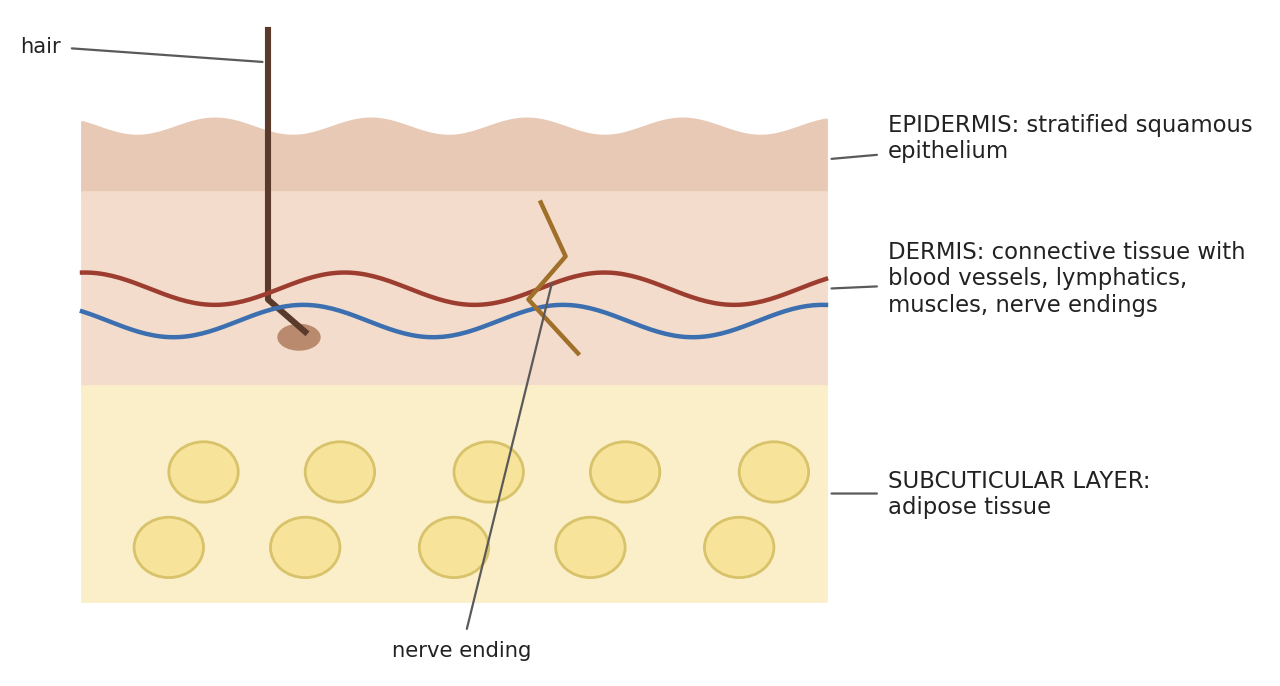
Figure 14.1. The layers of the skin.
Each layer has specialized cells and functions. The epidermis consists mainly of keratinocytes, which synthesize keratin, a protective, threadlike protein. It also contains melanocytes (pigment-producing cells), Merkel cells (slow-adapting touch receptors found where tactile sensitivity is high), and immune cells such as Langerhans (dendritic) cells, which capture antigens and migrate to lymph nodes to start immune responses. The dermis is made mostly of collagen, the stress-resistant protein also found in tendons, ligaments, and bone linings; it makes up most of the skin and gives it pliability, elasticity, and tensile strength, binds water, helps regulate temperature, and contains sensory receptors. The subcutaneous layer (panniculus) is made mostly of fat cells and connective tissue; it insulates and cushions underlying muscles and organs, stores energy, and acts as an endocrine organ (fat cells produce leptin, which regulates body weight, and convert some hormones).
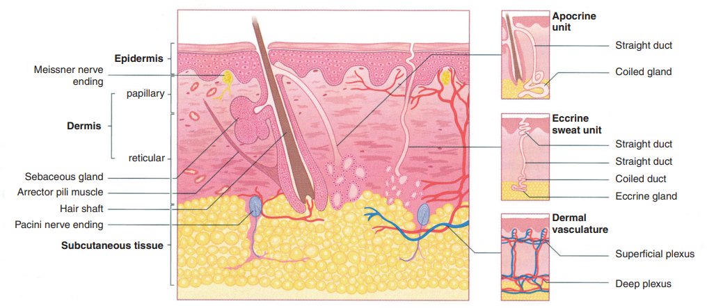
Figure 14.2. Detailed structure of the skin, showing epidermis, dermis, subcutaneous tissue, glands, hair follicles, and nerve endings (human skin shown).
Why it matters
Because the skin is the body's main barrier, every incision breaches it, which is why skin preparation and asepsis (Chapter 4) are so important. The dermis is the strong, vascular layer that holds sutures; the epidermis alone has little holding strength (Chapter 16).
14.2 Muscle
A muscle is made of hundreds to thousands of individual muscle cells called fibers. Each fiber is enclosed in connective tissue called endomysium. Fibers are gathered into bundles called fascicles, bound by perimysium, and the fascicles are bound together by epimysium to form the whole muscle (Figure 14.3). Muscles are divided into three types, defined by appearance and function: smooth, cardiac, and striated (skeletal) (Table 14.2).
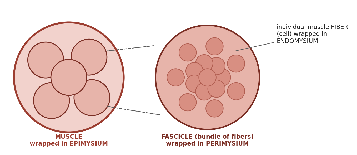
Figure 14.3. Organization of a skeletal muscle and its connective tissue layers.
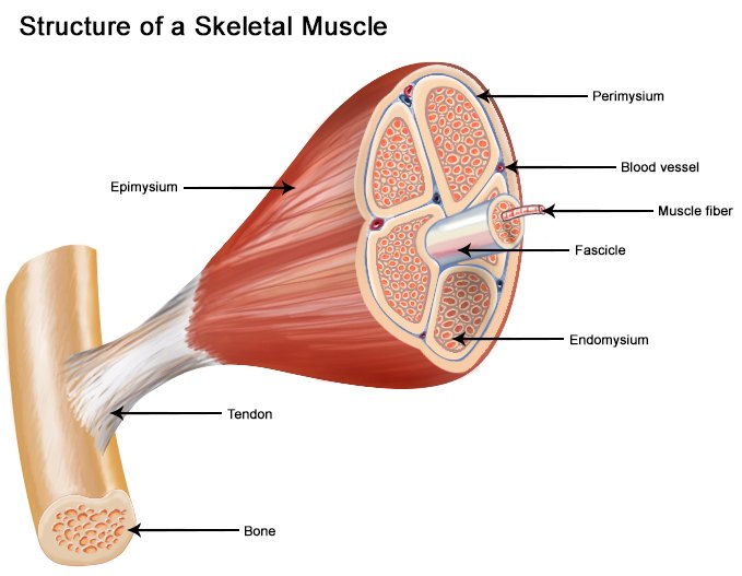
Figure 14.4. Structure of a skeletal muscle, from tendon and epimysium to fascicles and individual fibers.
14.2.1 Smooth Muscle
Smooth muscle is found in the walls of hollow organs and blood vessels and is controlled by the autonomic nervous system. Its cells are spindle-shaped with no striations, and it cannot be controlled voluntarily. It makes up almost all visceral muscle except cardiac muscle and forms part of the walls of blood vessels. It produces slow, steady contractions, moving food and waste through the GI tract and bladder, and producing vasoconstriction. Smooth muscle is organized into sheets of fibers surrounded by endomysium and connected by strands of collagen and elastin.
Table 14.1. Functions of smooth muscle in different body systems.
| System | Function |
|---|---|
| Gastrointestinal | Propulsion of food (peristalsis) |
| Cardiovascular | Regulation of blood flow and pressure through vascular resistance |
| Renal | Regulation of urine flow |
| Genital | Uterine contractions; propulsion of sperm |
| Respiratory | Regulation of bronchiolar diameter |
| Integument | Raising hair (arrector pili muscles) |
| Eye | Pupil dilation and constriction; changing lens shape |
14.2.2 Cardiac Muscle
Cardiac muscle forms the bulk of the heart and is optimized for rhythmic contraction under the control of the autonomic nervous system. It is found only in the heart. It is striated (with shorter cells than skeletal muscle), short and branched, and involuntary. It works in a steady rhythm set by the heart's pacemaker. Its cells are surrounded by endomysium and connected by intercalated discs, which let the electrical impulse spread rapidly from cell to cell.
14.2.3 Striated (Skeletal) Muscle
Striated muscle forms bundles surrounded by connective tissue envelopes and is largely controlled by the somatic nervous system. Its cells are the longest of the three types. It is also called skeletal muscle, because it attaches to and moves bones, and it can be voluntarily controlled.
Each skeletal muscle has two attachments. The origin is the more fixed point, and the insertion is the more movable point. In the limbs, the insertion is always distal to the origin. Muscle movements are described with the terms in Table 14.3.
Table 14.2. Comparison of the three types of muscle.
| Feature | Smooth | Cardiac | Skeletal (striated) |
|---|---|---|---|
| Location | Walls of hollow organs and blood vessels | Heart only | Attached to bones |
| Striations | None | Yes (shorter cells) | Yes |
| Cell shape | Spindle-shaped | Short, branched | Long (longest cells) |
| Control | Involuntary (autonomic) | Involuntary (pacemaker; autonomic regulation) | Voluntary (somatic) |
| Contraction | Slow, steady | Steady, rhythmic | Rapid, voluntary |
| Connections | Collagen and elastin strands | Intercalated discs | Connective tissue envelopes |
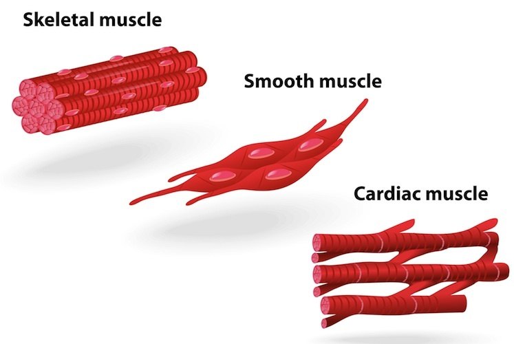
Figure 14.5. Skeletal, smooth, and cardiac muscle cells.
Table 14.3. Terms for muscle movements.
| Movement | Definition |
|---|---|
| Extension | Opens a joint or straightens bone alignment |
| Flexion | Closes a joint or angulates the bones |
| Adduction | Moves an extremity toward the center of the body |
| Abduction | Moves an extremity away from the center of the body |
| Circumduction | Circular movement of a limb |
| Rotation | Movement of a part around its long axis |
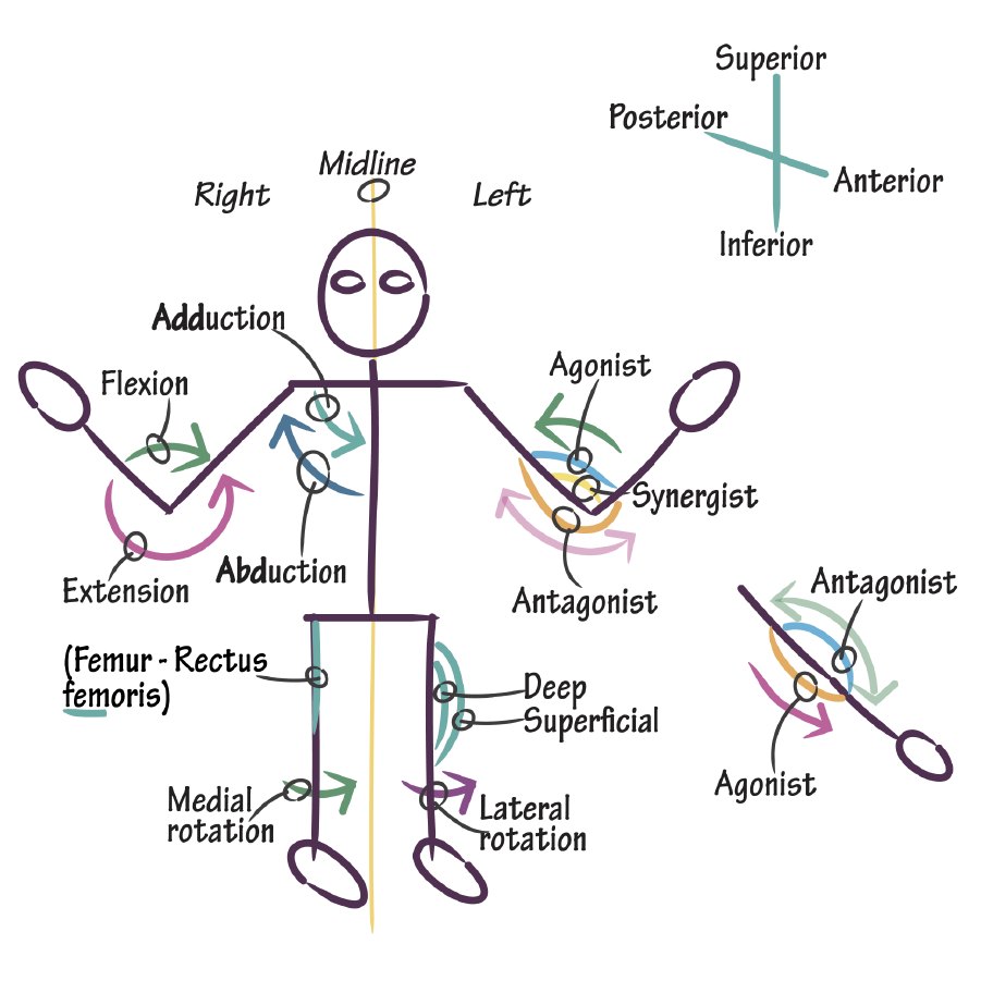
Figure 14.6. Anatomical directions and muscle actions: flexion and extension, abduction and adduction, rotation, and agonist and antagonist muscles (human figure).
Muscle names often describe the muscle itself, which makes them easier to learn (Table 14.4). For example, the biceps brachii has two heads (biceps) and lies in the arm (brachii).
Table 14.4. How muscles are named.
| Basis of name | Examples |
|---|---|
| Size | Vastus (huge), maximus (large), longus (long), minimus (small), brevis (short) |
| Shape | Deltoid (triangular), rhomboid (rhombus), latissimus (wide), teres (round), trapezius (trapezoid) |
| Direction of fibers | Rectus (straight), transverse (across), oblique (diagonal), orbicularis (circular) |
| Location | Pectoralis (chest), gluteus (buttock), brachii (arm); supra- (above), infra- (below), sub- (under), lateralis (lateral) |
| Number of origins (heads) | Biceps (two), triceps (three), quadriceps (four) |
| Origin and insertion | Sternocleidomastoideus (sternum and clavicle to mastoid process); brachioradialis (brachium to radius) |
| Action | Abductor, adductor, flexor, extensor, levator (lifts), masseter (chewer) |
14.3 Connective Tissue
Connective tissue binds structures together, forms a supporting framework for organs and the body, stores fat, transports substances, protects against disease, and helps repair damage. It is characterized by abundant intercellular matrix with relatively few cells, and usually (but not always) has a good blood supply. There are three broad types:
Loose connective tissue lies beneath the skin and around organs. A loose arrangement of collagen and elastic fibers in a gel-like matrix gives support, flexibility, and cushioning; it carries blood supply to overlying epithelium, responds quickly to damage or antigens, stores lipid (adipose tissue), and allows diffusion of nutrients, gases, and wastes.
Dense connective tissue is packed with fibers and fibroblasts and resists mechanical stress. In dense regular tissue (tendons and ligaments) collagen fibers run in parallel, giving strength along one axis; in dense irregular tissue (the dermis and organ capsules) fibers run in all directions, resisting stress from many directions. Elastic dense tissue in large arteries stores tension during each heartbeat.
Specialized connective tissue includes cartilage, blood, and bone.
Connective tissue envelops, separates, and connects muscles, nerves, and blood and lymphatic vessels, and provides routes for nerves and vessels to travel. It may blend with the periosteum of bone. Several named connective tissue coverings and membranes are important in surgery (Table 14.5).
Table 14.5. Connective tissue coverings and membranes.
| Structure | Description |
|---|---|
| Periosteum | Connective tissue covering bone |
| Perichondrium | Connective tissue covering cartilage |
| Peritoneum | Serous membrane, largely connective tissue, lining the abdomen |
| Parietal peritoneum | Covers (lines) the abdominal, pelvic, and scrotal cavities |
| Visceral peritoneum | Covers the abdominal, pelvic, and scrotal organs |
| Connecting peritoneum | Connects organs to other organs or to the parietal peritoneum |
| Common dorsal mesentery | Peritoneal fold that incorporates most of the abdominal organs |
| Pericardium | Fibrous sac enveloping the heart |
| Mediastinum | Sheet separating the two sides of the thoracic cavity, incorporating the heart, thymus, vena cava, and aorta; may be complete (two separate sides) or incomplete, leaving an opening between the sides (as in pigs) |
| Pleurae | Serous membranes lining the thoracic cavity and covering the lungs |
| Meninges | Protective membranes covering the brain and spinal cord |
| Dura mater | Thick, fibrous, most superficial meninx |
| Arachnoid membrane | Delicate meninx lining the deep surface of the dura mater |
| Pia mater | Delicate meninx coating the surface of the brain, spinal cord, nerve roots, and optic nerve |
Why it matters
Whether the mediastinum is complete matters in thoracic surgery and trauma. If it is incomplete, air or fluid entering one side of the chest (a pneumothorax or pleural effusion) can spread to the other side and affect both lungs. The completeness of the mediastinum varies between species and individuals; check species-specific references.
14.4 Skeleton
The skeleton is "the framework the body hangs on." It supports and protects the body, provides attachment points for muscles, and acts as a system of levers for motion. It is made of individual bones connected by ligaments and muscles.
14.4.1 Bones
Bone is a storage site for calcium and phosphorus, and the bone marrow produces red blood cells and several types of white blood cells. The skull is made of the incisive, nasal, maxilla, zygomatic, lacrimal, frontal, palatine, pterygoid, sphenoid, parietal, occipital, and temporal bones; the occipital bone bears the sagittal and nuchal crests. The radius and tibia are the main weight-bearing bones of the limbs.
There are two types of bone tissue (Figure 14.7):
Compact (cortical) bone is dense tissue that forms the outer shell of all bones.
Spongy (cancellous) bone is filled with pockets and lies between compact bone surfaces. It contains red bone marrow. Long bones also have a medullary cavity containing yellow bone marrow. The medullary cavity and the inner surface of the compact bone are lined with a fibrous tissue called endosteum.
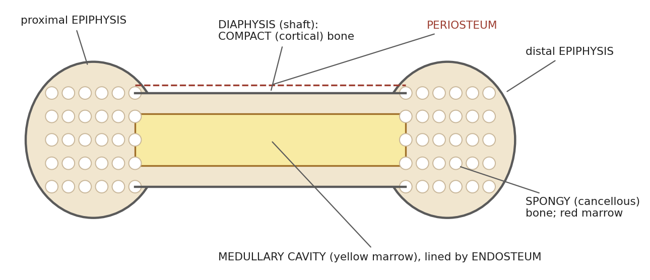
Figure 14.7. Structure of a long bone.
Compact bone makes up about 80% of the skeleton and cancellous bone about 20%. Cancellous bone is a three-dimensional lattice of bony struts (trabeculae) aligned along lines of stress, with marrow and blood vessels filling the spaces. Subchondral bone is the smooth bone at the joint ends, beneath the articular cartilage. Microscopically, compact bone is built of cylindrical units (osteons) around central (Haversian) canals that carry blood vessels and nerves, connected by perforating (Volkmann) canals (Figure 14.8).
Bone marrow is either red or yellow. Red marrow forms all of the blood cells: red cells, platelets, and white cells (lymphocytes are produced in the marrow but mature in the lymphoid organs). Along with the liver and spleen, it also helps destroy old red cells. Yellow marrow is mainly a fat store, but it can convert back to red marrow when needed, such as after severe blood loss (Figure 14.9).
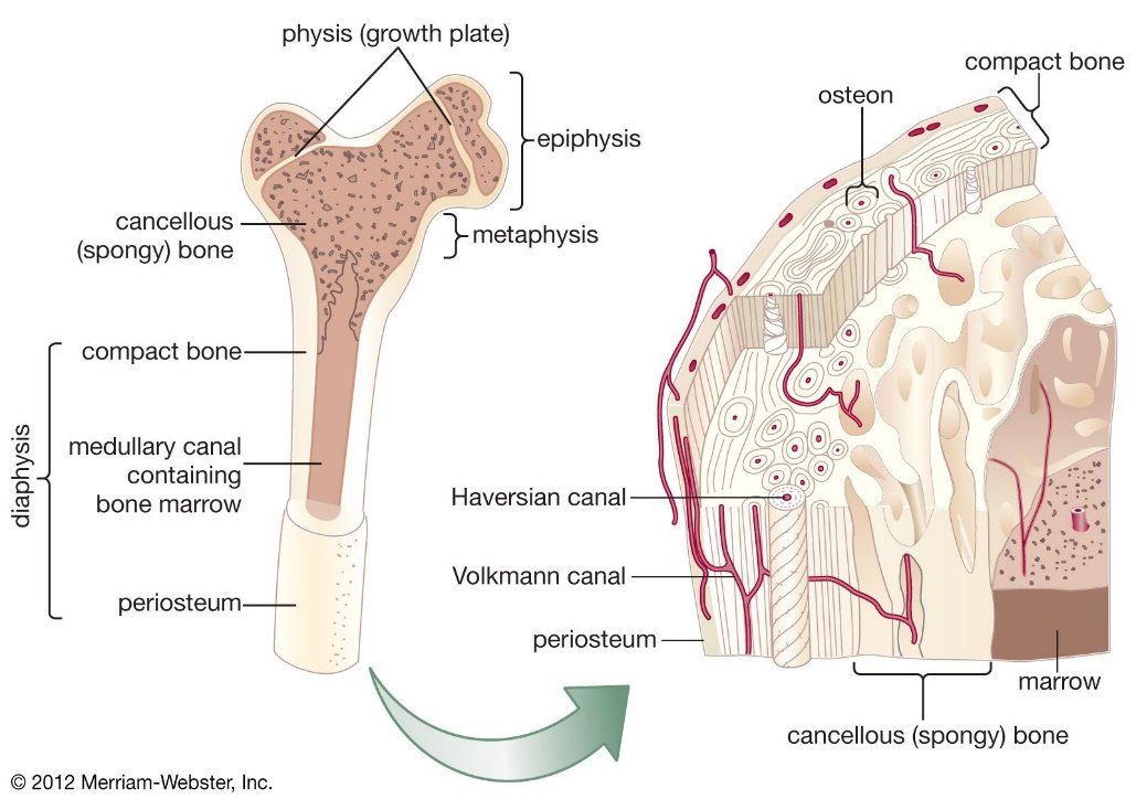
Figure 14.8. Long bone anatomy (left) and the microscopic structure of compact and cancellous bone (right).
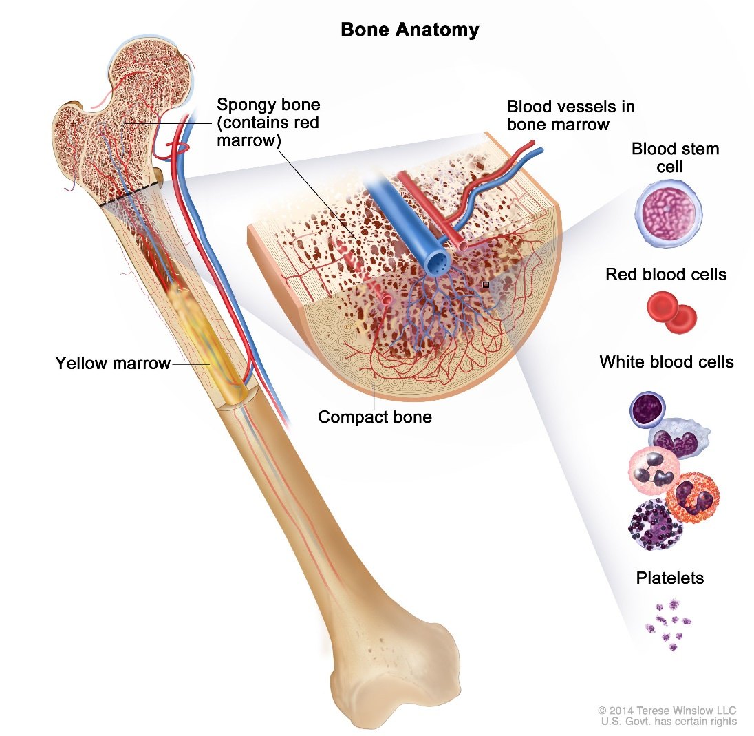
Figure 14.9. Bone marrow and the blood cells it produces.
⚠ Check current guidance
The terms for bone tissue are sometimes confused, with "cortical bone" used for the inner, pocketed bone. In standard terminology, cortical bone is the same as compact bone (the dense outer shell), and the inner, pocketed bone is called spongy, cancellous, or trabecular bone. Check how your exam references use these terms.
Table 14.6. Types of bones.
| Type | Structure | Examples |
|---|---|---|
| Long | A shaft (diaphysis) with proximal and distal ends (epiphyses) | All limb bones except the patella and the bones of the wrist and ankle |
| Short | Roughly cube-like; mainly spongy bone with a thin shell of compact bone | Bones of the wrist (carpus) and ankle (tarsus) |
| Sesamoid | A short bone embedded in a tendon or joint capsule | Patella |
| Flat | Thin; spongy bone (diploe) between two layers of compact bone; marrow but no marrow cavity | Sternum, ribs, most skull bones |
| Irregular | Same layered structure as flat bone | Vertebrae, pelvis, some skull bones |
14.4.2 Joints
Joints are areas where bones are joined by fibrous, elastic, and/or cartilaginous tissue. A ligament is fibrous tissue connecting bone to bone; a tendon is fibrous tissue connecting muscle to bone. Joints may be classified by function or by structure (Table 14.7).
Table 14.7. Classification of joints.
| By function | Movement | By structure | Description | Examples |
|---|---|---|---|---|
| Synarthrosis | None | Fibrous | Bones joined by fibrous tissue; no joint cavity | Skull sutures, teeth |
| Amphiarthrosis | Slight | Cartilaginous | Bones united by cartilage; no joint cavity; permits compression or stretching | Epiphyseal disc, intervertebral disc |
| Diarthrosis | Free | Synovial | Bones separated by a joint cavity | Limb joints |
Synovial joints may be plane, ball-and-socket, ellipsoidal, hinge, condylar, trochoid (pivot), or saddle joints. They have five main components (Figure 14.10):
Articular cartilage covering the bone ends, which has limited ability to regenerate.
An articular capsule enclosing the joint, with an outer fibrous capsule and an inner synovial membrane. (Hip and knee joints also have a fat pad between the fibrous capsule and the synovial membrane or bone.)
A joint cavity: the potential space within the capsule.
Synovial fluid, which fills the cavity, lubricates the joint, and transports nutrients and wastes. It is made of hyaluronic acid thinned by interstitial fluid from blood plasma.
Reinforcing ligaments (intrinsic, extracapsular, and intracapsular), which strengthen the joint.
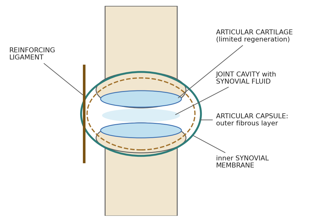
Figure 14.10. Structure of a synovial joint.
Many joints also have accessory structures. Bursae are fluid-filled sacs between bones, tendons, ligaments, or other structures that cushion friction. A meniscus is a crescent-shaped pad of cartilage within some joints (such as the stifle, or knee) that improves fit and absorbs shock (Figure 14.11). Tendons on each side of a joint attach the muscles that move it.
Figure 14.11. Side view of a knee (stifle) joint, showing bone, cartilage, synovial membrane and fluid, meniscus, bursa, ligament, tendon, and muscle (human knee shown).
Table 14.8. Types of freely movable (synovial) joints.
| Type | Movement allowed | Examples |
|---|---|---|
| Ball-and-socket | Forward, backward, sideways, and rotating movements | Shoulder, hip |
| Hinge | Bending and straightening only | Elbow, stifle (knee), digits |
| Pivot | Limited rotation | Atlas–axis (neck) |
| Ellipsoidal | All movements except pivoting | Carpus (wrist) |
| Plane, condylar, and saddle | Gliding and combined movements | Various |
Why it matters
Because articular cartilage regenerates poorly, joint surgery must handle it gently, and contamination of a joint is especially serious: the joint cavity is a closed, poorly vascular space where infection can quickly destroy cartilage.
14.5 Nervous System
The nervous system is divided into the central nervous system (CNS), the brain and spinal cord, and the peripheral nervous system (PNS), the nerves that connect the CNS to the rest of the body (Figure 14.12).
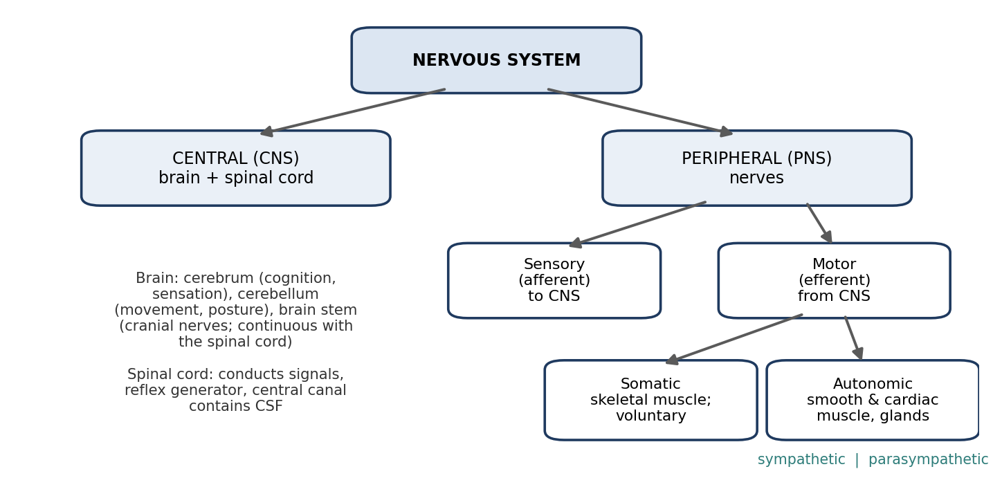
Figure 14.12. Organization of the nervous system.
14.5.1 Central Nervous System
The brain comprises the cerebrum, the primary site of cognitive and sensory functions; the cerebellum, which coordinates movement and posture; and the brain stem, which is continuous with the spinal cord and is the source of all cranial nerves except the olfactory nerves. The brain processes information received through peripheral nerves and sends commands. It floats in cerebrospinal fluid (CSF), which cushions it. CSF contains inorganic ions, protein, sugar, and a few cells; it is derived from the blood and returns to the bloodstream through the arachnoid villi and lymphatics.
The spinal cord extends from the brain stem through the vertebral canal. It conducts signals to and from the brain, acts as a reflex generator, and processes information and discharges commands. It has a central canal that carries CSF.
Most of the CNS is made of gray matter and white matter (Figure 14.13). Gray matter consists mainly of neuron cell bodies and processes and interprets information; white matter consists mainly of myelinated axons that carry information between regions. In the brain, gray matter forms the outer cerebral cortex; in the spinal cord, the arrangement is reversed, with a butterfly-shaped core of gray matter surrounded by white matter.
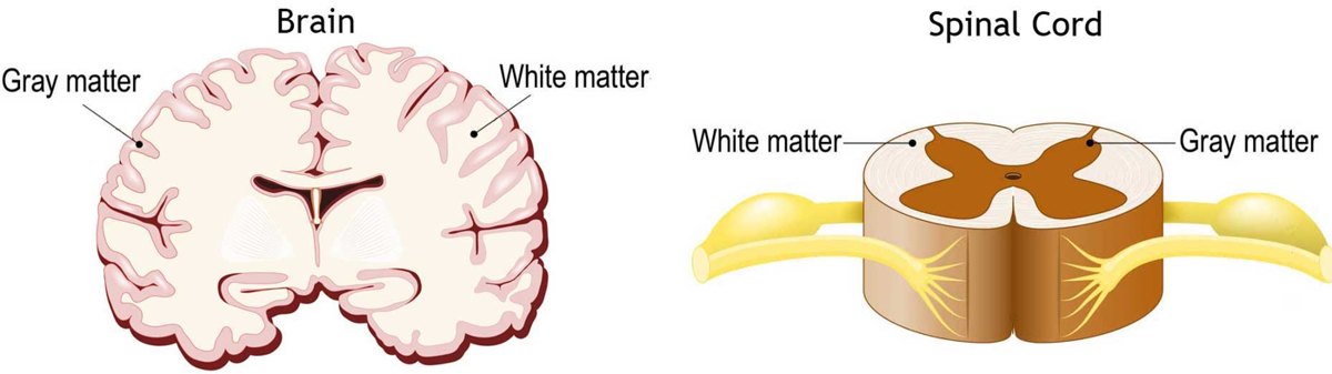
Figure 14.13. Gray and white matter in the brain and spinal cord (cross-sections).
The brain stem consists of the midbrain, pons, and medulla oblongata, and regulates vital functions such as breathing, heart rate, and balance. The medulla, where the brain meets the spinal cord, controls heart rhythm, breathing, and blood flow, which is why it is the last region depressed by anesthetics (Chapter 7). The cerebrum is divided into lobes: the frontal lobe (personality, decision-making, voluntary movement, smell), parietal lobe (touch, pain, and spatial relationships), occipital lobe (vision), and temporal lobe (hearing, memory, and some smell). Other important structures include:
The hypothalamus, which regulates body temperature, hunger, thirst, and sleep, and controls the pituitary gland.
The pituitary gland, the "master gland," which regulates many other endocrine glands (Section 14.13).
The pineal gland, which secretes melatonin in response to light and dark, regulating daily (circadian) rhythms.
The amygdala (emotion, memory, and the "fight or flight" response) and hippocampus (memory, learning, and spatial navigation).
The ventricles, fluid-filled spaces deep in the brain that produce CSF.
The meninges (Figure 14.14) protect the brain and spinal cord. The dura mater is thick and tough; around the brain it has two layers, an outer (periosteal) layer lining the skull and an inner (meningeal) layer. The arachnoid is a thin, web-like layer without nerves or blood vessels, and CSF circulates in the subarachnoid space beneath it, cushioning the CNS and removing waste. The pia mater is a thin, highly vascular membrane on the surface of the brain and spinal cord.
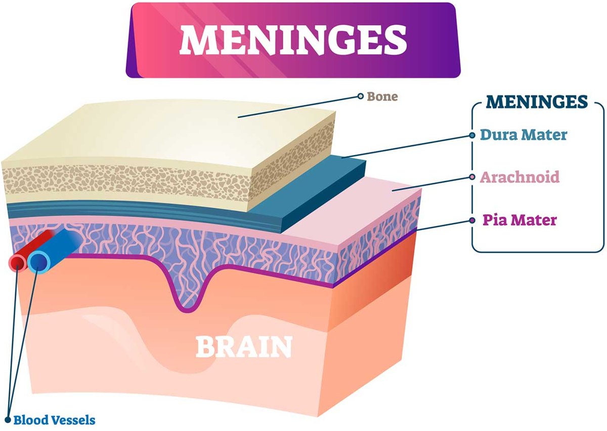
Figure 14.14. The meninges: dura mater, arachnoid, and pia mater.
Twelve pairs of cranial nerves arise from the brain (Table 14.9). The vagus nerve (X) is especially important in anesthesia: it carries parasympathetic signals that slow the heart, which is why vagal stimulation (for example, during intubation or traction on viscera) can cause bradycardia (Chapter 6).
Table 14.9. The cranial nerves.
| Number and name | Main function |
|---|---|
| I Olfactory | Smell |
| II Optic | Vision |
| III Oculomotor | Most eye movements; pupil constriction |
| IV Trochlear | Eye movement (one muscle) |
| V Trigeminal | Sensation from the face, teeth, jaw, and mouth; chewing muscles |
| VI Abducens | Eye movement (lateral) |
| VII Facial | Facial expression, taste, tear and salivary glands |
| VIII Vestibulocochlear | Hearing and balance |
| IX Glossopharyngeal | Taste, swallowing, salivation |
| X Vagus | Parasympathetic supply to the heart, lungs, and digestive tract; throat and larynx |
| XI Accessory | Muscles of the neck and shoulder |
| XII Hypoglossal | Tongue movement |
14.5.2 Peripheral Nervous System
The sensory (afferent) division conveys impulses from sensory organs to the CNS. The motor (efferent) division transmits impulses from the CNS to muscles and glands. The efferent division is divided into the somatic nervous system, which conducts impulses to skeletal muscle and allows voluntary control, and the autonomic nervous system (ANS), which regulates smooth muscle, cardiac muscle, and glands.
The ANS has two divisions that regulate each other (Table 14.10).
Table 14.10. Divisions of the autonomic nervous system.
| Sympathetic | Parasympathetic | |
|---|---|---|
| Origin | Spinal cord between T1 and L2 (thoracolumbar) | Brain stem and sacral spinal cord (craniosacral) |
| Heart rate | Accelerates | Decreases |
| Blood vessels | Constricts | Dilates |
| Blood pressure | Raises | Lowers |
| Common description | "Fight or flight" | "Rest and digest" |
This is the physiological basis of several drug effects described in Chapter 6. For example, anticholinergics such as atropine block parasympathetic (vagal) effects, which is why they increase heart rate.
Neurons have a cell body (soma) containing the nucleus and cytoplasm. Dendrites are small branches from the soma that receive signals and conduct them toward the soma. The axon carries impulses away from the soma to the axon terminals, which transmit signals to muscles, glands, and other nerves.
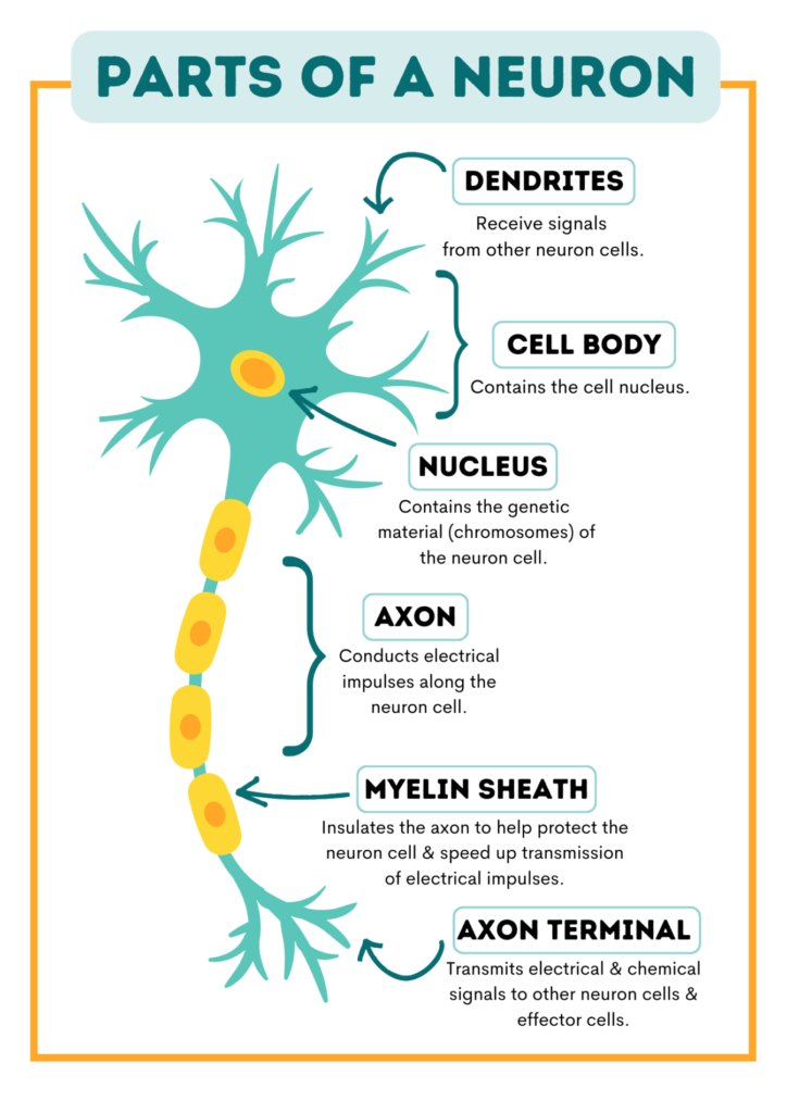
Figure 14.15. Parts of a neuron.
Nerves are bundles of nerve fibers. Fibers are bound into fascicles by perineurium, and fascicles and blood vessels are bound into a nerve by fibrous epineurium. In the CNS, bundles of nerve fibers are called tracts. Nerves are highly variable in structure.
Figure 14.16. Structure of a peripheral nerve: axons within endoneurium, fascicles wrapped in perineurium, and the whole nerve in epineurium, with its blood supply.
14.6 Cardiovascular System
14.6.1 Heart
The heart is the primary pump of the cardiovascular system and has four chambers. Blood flows through it as follows (Figure 14.17):
Deoxygenated blood enters the right atrium from the vena cava.
It passes through the right atrioventricular (AV) valve, called the tricuspid valve (although in many animals it consists basically of two cusps), into the right ventricle.
It is pumped through the pulmonary valve into the pulmonary artery and to the lungs for oxygenation.
Oxygenated blood returns through the pulmonary veins to the left atrium.
It passes through the left AV valve (bicuspid or mitral valve) into the left ventricle.
It is pumped through the aortic valve into the aorta and throughout the body.
Figure 14.17. Blood flow through the heart, and the cardiac conduction system.
Figure 14.18. Anatomy of the heart, showing chambers, valves, great vessels, and the layers of the heart wall and pericardium (human heart shown).
The heart muscle itself is supplied by the right and left coronary arteries, which divide into branches such as the right marginal, circumflex, and left marginal branches.
The Conduction System
Contraction is coordinated by a specialized conduction system. Contraction does not require outside stimulation, but its rate can be regulated by the autonomic nervous system.
The sinoatrial (SA) node is the pacemaker. It depolarizes spontaneously in a sinus rhythm, starting contraction of the atria, and the signal spreads through the atria to the AV node.
The atrioventricular (AV) node delays the impulse, allowing the atria to finish contracting and fill the ventricles, then passes it on.
The AV bundle (bundle of His) carries the impulse into the ventricular septum, where it divides into the right and left bundle branches.
The Purkinje fibers spread the signal evenly throughout the ventricles, producing an even contraction that wrings blood from the ventricles toward the outflow valves.
These events produce the waves of the ECG described in Chapter 12: the P wave (atrial depolarization), the QRS complex (ventricular depolarization), and the T wave (ventricular repolarization).
The Cardiac Cycle
Systole is the contraction phase of the heart, and diastole the resting phase. In mid to late diastole, pressure in the heart is low, the AV valves are open, and blood flows passively through the atria into the ventricles. In atrial systole, the atria contract, topping up the ventricles. Ventricular systole begins as the atria enter diastole: the ventricles contract, the AV valves close, and ventricular pressure rises sharply, ejecting blood. In early diastole, the ventricles begin to relax.
Figure 14.19. The cardiac cycle: diastole (filling) and systole (ejection), and the timing of the "lub" and "dub" heart sounds.
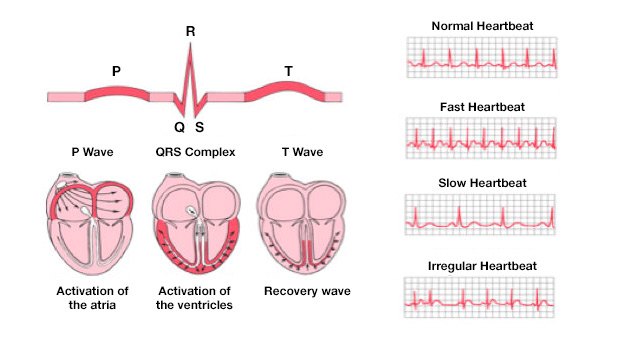
Figure 14.20. How the ECG waves correspond to electrical activation of the heart: the P wave (atria), QRS complex (ventricles), and T wave (recovery).
Cardiac output is the amount of blood pumped by each ventricle in one minute: heart rate × stroke volume, where stroke volume is the volume pumped by a ventricle with each contraction. Stroke volume is the difference between the volume in the ventricle at the end of filling and at the end of ejection: SV = end-diastolic volume (EDV) − end-systolic volume (ESV). The atria contract a fraction of a second before the ventricles, so they can empty fully into the ventricles before ventricular contraction begins.
Heart sounds come from the valves closing. The "lub" is closure of the AV valves, and the "dub" is closure of the semilunar (aortic and pulmonary) valves. Unusual sounds are called murmurs.
14.6.2 Vascular System
Most of the blood (about 70%) is in the venous system, with only about 10% in the arterial system. This large venous reservoir is why venous tone and positioning strongly affect how much blood returns to the heart.
Blood leaves the heart through the aorta (and pulmonary artery), and the coronary arteries supply the heart itself. Arteries carry blood to smaller arterioles, which carry it to capillaries, where oxygen is supplied to the surrounding tissues. Capillaries return deoxygenated blood to venules, which carry it to veins, which drain into the vena cava and back to the heart.
Arteries and veins have three layers (Figure 14.21): the tunica intima, a single layer of endothelium on a basement membrane; the tunica media, made of smooth muscle and elastic fibers, which is much thicker and stronger in arteries; and the tunica adventitia, made of collagen fibers. Capillaries are made only of endothelial cells and a sparse basement membrane, and they are the primary site of oxygen exchange between the blood and the tissues.
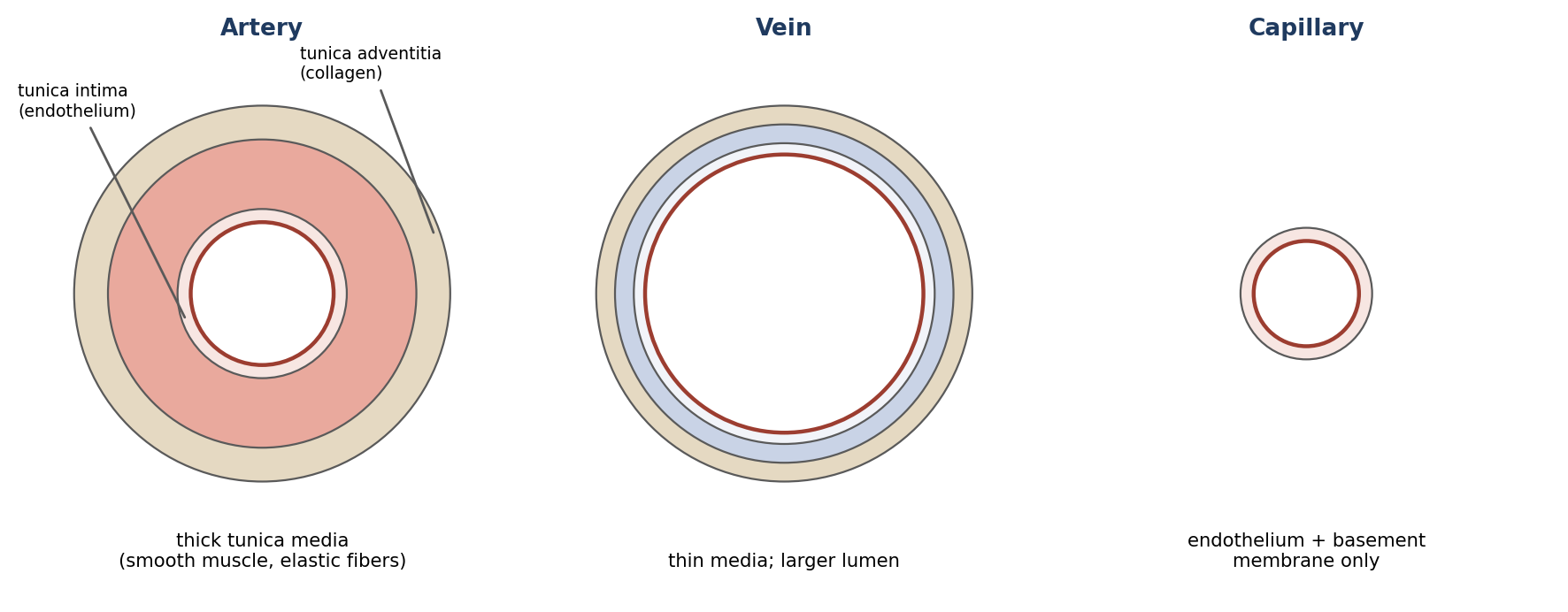
Figure 14.21. Wall structure of an artery, a vein, and a capillary (not to scale).
Table 14.11. Comparison of arteries and veins.
| Arteries | Veins | |
|---|---|---|
| Direction of flow | Away from the heart | Toward the heart |
| Blood carried (systemic) | Oxygenated | Deoxygenated |
| Pressure | High | Low |
| Wall | Thicker, more muscular and elastic; expands with each heartbeat | Thinner, less elastic |
| Valves | None (except at the heart) | Valves prevent backflow |
| Return aids | — | Skeletal muscle contraction squeezes veins and helps return blood against gravity |
The aorta carries all oxygenated blood from the heart. It extends cranially, then makes a 180° turn and proceeds caudally. It is arbitrarily divided into the ascending aorta, the aortic arch, and the descending aorta (thoracic and abdominal). The main arteries of surgical importance branch from it (Figure 14.22 and Table 14.12).
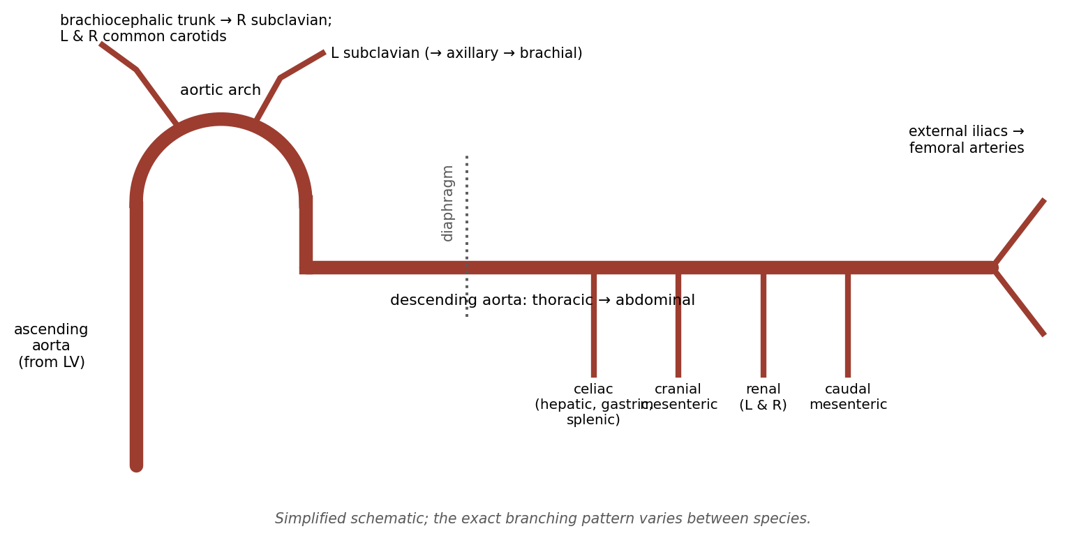
Figure 14.22. Major branches of the aorta.
Table 14.12. Major arteries of surgical importance.
| Artery | Origin | Supplies / continues as |
|---|---|---|
| Brachiocephalic trunk | Aortic arch | Both common carotid arteries and the right subclavian artery |
| Common carotid (L and R) | Brachiocephalic trunk | Branch into internal and external carotid arteries near C1–C2 |
| Left subclavian | Aortic arch | Continues as the axillary artery |
| Right subclavian | Brachiocephalic trunk | Continues as the axillary artery |
| Axillary | Subclavian | Continues as the brachial artery, supplying the forelimb |
| Celiac | Abdominal aorta | Hepatic, gastric, and splenic arteries |
| Cranial and caudal mesenteric | Abdominal aorta | Intestines |
| Renal (L and R) | Abdominal aorta | Kidneys |
| External iliac (L and R) | Termination of abdominal aorta | Continue as the femoral arteries (hind limbs) |
Table 14.13. Major veins of surgical importance.
| Vein | Course |
|---|---|
| Femoral (L and R) | Drain the hind limbs into the external iliac veins |
| External iliac (L and R) | Continue the femoral veins; join the internal iliac veins to form the common iliac veins |
| Common iliac | Join to form the caudal vena cava |
| Portal | Collects blood from the pancreas, spleen, and entire GI tract (except the anal canal) and carries it to the liver; the liver drains via the hepatic veins into the caudal vena cava |
| External jugular (L and R) | Continue the maxillary veins; drain into the brachiocephalic veins |
| Cephalic | Drains the forelimb along its lateral surface into the brachiocephalic vein; a common venipuncture site |
| Brachial | Drains the forelimb along its medial surface into the brachiocephalic vein |
| Subclavian (L and R) | Drain into the cranial vena cava |
| Vena cava | Collects venous blood; divided at the diaphragm into cranial and caudal vena cava, which drain into the right atrium |
Why it matters
The portal vein explains why oral drugs can be largely destroyed before they reach the general circulation, and why part of an intraperitoneal injection can be broken down by the liver (Chapter 8, pentobarbital): blood from the GI tract and much of the peritoneal cavity passes through the liver first.
14.7 Respiratory System
Besides exchanging oxygen and carbon dioxide, the respiratory system warms and humidifies inspired air, protects the body from inhaled particles, allows the sense of smell, and helps regulate acid–base balance: carbon dioxide forms carbonic acid in the blood, so rising CO₂ lowers blood pH (respiratory acidosis), and breathing out more CO₂ raises it (Chapters 12 and 18).
The respiratory system transports oxygen from the environment to the bloodstream (and removes carbon dioxide). Its main parts are:
The pharynx, the main passageway from the nose and mouth.
The larynx, a musculocartilaginous organ that protects the entrance to the trachea.
The trachea, a flexible tube with a smooth muscle wall supported by C-shaped rings of hyaline cartilage, carrying air to the left and right principal bronchi.
The bronchi, which divide into lobar bronchi, then segmental bronchi, then progressively smaller bronchioles ending in the alveoli.
The lungs, which contain the bronchi, bronchioles, and alveoli.
The diaphragm, the muscular wall separating the thoracic and abdominal cavities, which creates negative pressure in the thoracic cavity to inflate the lungs.
The lungs are passive organs: they inflate because the diaphragm and chest wall expand the thorax. Each lung lies in a pleural cavity that normally contains only a thin film of moistening fluid; this is a potential space that becomes a true cavity when air enters it, as in a pneumothorax. The left lung is slightly smaller than the right because of the heart's position. Lung tissue is highly vascular and filled with alveoli, the site of oxygen transfer to the blood, which can be visualized as millions of tiny bubbles surrounded by elastic connective tissue (stroma).
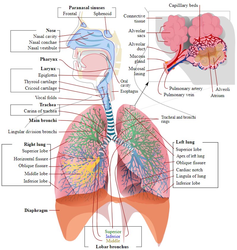
Figure 14.23. The respiratory tract, from the nasal cavity, pharynx, and larynx to the trachea, bronchi, lungs, and alveoli (human anatomy shown).
Why it matters
Because lung inflation depends on negative pressure in a sealed chest, opening the thorax (thoracotomy) or puncturing the pleura causes the lungs to collapse. This is why thoracic surgery requires intubation and positive pressure ventilation (Chapters 5 and 9).
14.8 Digestive System
The gastrointestinal tract runs from the mouth to the anus (Figure 14.24):
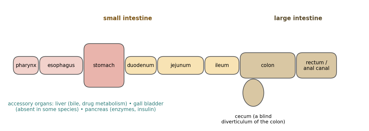
Figure 14.24. Simplified layout of the gastrointestinal tract (not to scale).
Pharynx: passageway from the mouth to the esophagus.
Esophagus: passageway from the pharynx to the stomach.
Stomach: stores and mixes food and adds digestive enzymes.
Small intestine: the duodenum (first and most fixed part, leaving the stomach), the jejunum (middle part), and the ileum (last part, connecting to the ascending colon).
Large intestine: the cecum (a diverticulum of the colon that does not itself connect the ileum and large intestine), the colon (connecting to the rectum), and the rectum (connecting to the anal canal, which exits the body).
Accessory digestive organs include:
The liver, the largest gland in the body, which produces bile (stored in the gallbladder in most species) and releases substances into the bloodstream that help metabolize fats and sugars. Bile neutralizes stomach acid entering the small intestine and emulsifies fats into small droplets that enzymes can digest. The number and arrangement of liver lobes varies between species.
The gallbladder, which stores and concentrates bile. Several mammals, including horses, deer, rats, and camelids (such as llamas), as well as several species of birds, lampreys, and all invertebrates, lack a gallbladder.
The pancreas, which has two lobes, secretes pancreatic juice for digestion, and produces insulin in its islet cells.
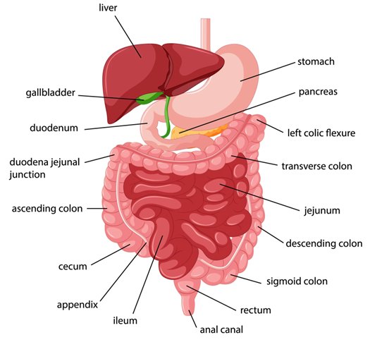
Figure 14.25. The digestive system (human anatomy shown); the arrangement and relative size of organs, especially the cecum and colon, differ considerably between species.
14.9 Filtration & Excretory System
The filtration and excretory organs eliminate waste products from the body. The liver produces the enzymes that metabolize the majority of administered drugs (Chapter 7). The kidneys filter blood to remove urea and produce urine, which is carried by the ureters to the bladder, where it is stored until it is eliminated through the urethra.
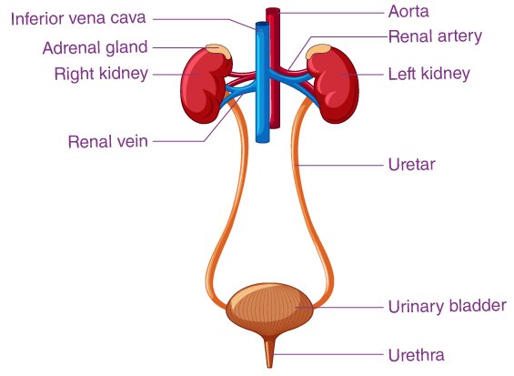
Figure 14.26. The urinary system: kidneys, renal arteries and veins, ureters, urinary bladder, and urethra (human anatomy shown).
14.10 Lymphatic System
The lymphatic system returns fluid and protein lost from the blood in the capillary beds to the circulation. The lymph nodes, spleen, thymus, and tonsils are phagocytic and remove foreign material, and the system produces and circulates the cells responsible for the body's immune response.
The thymus has two identical lobes, located cranioventral to the heart. It is where T cells mature.
The spleen is the largest organ of the lymphatic system. It filters the blood, removes old or damaged cells, and stores red blood cells and platelets, releasing them when needed; it lies parallel to the greater curvature of the stomach.
Lymph nodes are bean-shaped organs that filter and cleanse lymph as it passes through them, removing foreign material and damaged or cancerous cells; some are connected in groups called chains.
Bone marrow produces white blood cells, red blood cells, and platelets.
14.11 Special Senses
The eye is responsible for vision, the ear for hearing (and balance), and the nose for smell.
14.12 Reproductive System
Table 14.14. Reproductive organs.
| Sex | Organ | Description |
|---|---|---|
| Male | Scrotum | Skin pouch enclosing the testes |
| Male | Testes | Male gonads that produce spermatozoa |
| Male | Prostate gland | Accessory sex gland that contributes fluid to semen |
| Male | Penis | Composed of the roots, body, and glans |
| Male | Os penis | Bone in the glans penis of many mammals |
| Female | Ovaries | Paired organs that produce ova |
| Female | Oviducts (fallopian tubes) | Transport ova to the uterus |
| Female | Uterus | Consists of a neck (cervix), body, and two horns; allows passage of sperm to the oviducts |
| Female | Vagina | Dilatable canal from the uterus to the vulva |
14.13 Endocrine System
The endocrine system is composed of glands that secrete hormones directly into the circulation, where they affect other body systems. The major endocrine glands include the pineal gland, pituitary gland, hypothalamus, thyroid gland, parathyroid glands, adrenal glands, pancreas, ovaries, testes, liver, and gastrointestinal tract. The stress response to pain and surgery (Chapter 13), with its release of cortisol and catecholamines from the adrenal glands, is one example of endocrine activity that directly affects anesthesia and research data.
Table 14.15. Major endocrine glands and their hormones.
| Gland | Main hormones | Key actions |
|---|---|---|
| Hypothalamus | Thyrotropin-releasing hormone (TRH), gonadotropin-releasing hormone (GnRH), growth hormone-releasing hormone (GHRH), corticotropin-releasing hormone (CRH), somatostatin, dopamine | Controls the pituitary; regulates temperature, hunger, thirst, and sleep |
| Pituitary | Adrenocorticotropic hormone (ACTH), thyroid-stimulating hormone (TSH), luteinizing hormone (LH), follicle-stimulating hormone (FSH), prolactin, growth hormone (GH), melanocyte-stimulating hormone (MSH) | "Master gland": regulates the adrenal, thyroid, and reproductive glands, growth, and milk production |
| Pineal | Melatonin | Circadian (sleep–wake) rhythm |
| Thyroid | Thyroxine (T4), triiodothyronine (T3), calcitonin | Metabolic rate; calcitonin lowers blood calcium |
| Parathyroid | Parathyroid hormone (PTH) | Regulates calcium, phosphorus, and magnesium in blood and bone |
| Adrenal | Cortisol and aldosterone (cortex); epinephrine and norepinephrine (medulla) | Stress response, glucose and fluid balance, "fight or flight" |
| Pancreas (islets) | Insulin (and glucagon) | Regulates blood glucose |
| Liver | Insulin-like growth factor 1 (somatomedin), angiotensinogen, thrombopoietin, hepcidin | Growth, blood pressure regulation, platelet production, iron balance |
| Gastrointestinal tract | Gastrin, cholecystokinin (CCK), secretin, somatostatin, ghrelin, bombesin / gastrin-releasing peptide | Digestion, gut motility, mucosal growth, appetite |
| Ovaries | Estrogens, progesterone | Reproductive (estrous) cycle and pregnancy |
| Testes | Androgens (testosterone) | Male reproductive function and characteristics |
[ PHOTO PLACEHOLDER ] Suggested source: Search Wikimedia Commons (commons.wikimedia.org) for labeled rat or mouse anatomy, or use images from your institution's training materials. Check the image's license and give the attribution it requires. |
|---|
Figure 14.27. A labeled dissection or radiograph of a common research species (for example, a rat), showing the major thoracic and abdominal organs.
⚠ Check current guidance
Image credits: Figures showing photographic or illustrated anatomy (skin detail, muscle structure, bone, knee, nervous system, heart, cardiac cycle, ECG, respiratory, digestive, and urinary systems) were supplied from compiled study materials and appear to come from published sources (for example, Merriam-Webster and Encyclopaedia Britannica). Most depict human anatomy; the basic structures are similar in research species, but proportions and details differ. Before this textbook is distributed or published, confirm each image's source, obtain permission where required, and add a credit line.
Chapter Summary
The skin (epidermis, dermis, subcuticular fat) is the body's barrier. Muscle is organized as fibers (endomysium) in fascicles (perimysium) within the muscle (epimysium) and is smooth, cardiac, or skeletal; skeletal muscles run from origin to insertion and produce extension, flexion, adduction, abduction, circumduction, and rotation. Connective tissue forms the periosteum, peritoneum, pleurae, pericardium, mediastinum, and meninges (dura, arachnoid, pia). Bones (long, short, sesamoid, flat, irregular) have compact outer and spongy inner bone; joints are fibrous, cartilaginous, or synovial, and synovial joints have cartilage, a capsule, a cavity, synovial fluid, and ligaments.
The nervous system has a CNS (brain, spinal cord) and a PNS with sensory and motor divisions; the autonomic system's sympathetic division speeds the heart and constricts vessels, and the parasympathetic division does the reverse. Blood flows right atrium → tricuspid → right ventricle → pulmonary artery → lungs → pulmonary veins → left atrium → mitral → left ventricle → aorta, driven by the SA node → AV node → bundle of His → bundle branches → Purkinje fibers. Most blood is venous; vessels have intima, media, and adventitia layers. The respiratory, digestive, excretory, lymphatic, reproductive, and endocrine systems complete the picture, with species differences (such as the absent gallbladder in rats and horses) that matter in research.
Key Terms
Abduction / adduction: Movement of an extremity away from / toward the center of the body.
Atrioventricular (AV) node: Conduction tissue that delays the impulse between atria and ventricles.
Cardiac output: Heart rate × stroke volume.
Diaphysis / epiphysis: The shaft / end of a long bone.
Endomysium, perimysium, epimysium: Connective tissue around a muscle fiber, a fascicle, and a whole muscle.
Insertion / origin: The more movable / more fixed attachment of a muscle.
Intercalated disc: Junction between cardiac muscle cells that allows rapid impulse spread.
Ligament: Fibrous tissue connecting bone to bone.
Mediastinum: The partition between the two sides of the thoracic cavity.
Meninges: The dura mater, arachnoid membrane, and pia mater covering the brain and spinal cord.
Parasympathetic: The craniosacral division of the ANS; slows the heart and dilates vessels.
Periosteum: Connective tissue covering bone.
Portal vein: Vein carrying blood from the GI tract, spleen, and pancreas to the liver.
Sinoatrial (SA) node: The heart's pacemaker.
Sympathetic: The thoracolumbar division of the ANS; accelerates the heart and constricts vessels.
Synovial joint: A freely movable joint with a fluid-filled cavity.
Tendon: Fibrous tissue connecting muscle to bone.
Tunica intima, media, adventitia: The inner, middle, and outer layers of a blood vessel wall.
Review Questions
1. Which connective tissue layer surrounds each individual muscle fiber?
A. Epimysium
B. Perimysium
C. Endomysium
D. Periosteum
2. Which type of muscle is striated, branched, involuntary, and connected by intercalated discs?
A. Smooth muscle
B. Cardiac muscle
C. Skeletal muscle
D. Visceral smooth muscle
3. A muscle that moves a limb away from the center of the body is an:
A. Adductor
B. Abductor
C. Extensor
D. Flexor
4. Which meninx is the thick, fibrous, outermost layer?
A. Pia mater
B. Arachnoid membrane
C. Dura mater
D. Endosteum
5. The patella is an example of a:
A. Long bone
B. Flat bone
C. Sesamoid bone
D. Irregular bone
6. A ligament connects:
A. Muscle to bone
B. Bone to bone
C. Nerve to muscle
D. Skin to muscle
7. The sympathetic nervous system:
A. Slows the heart and dilates blood vessels
B. Accelerates the heart, constricts blood vessels, and raises blood pressure
C. Controls voluntary skeletal muscle
D. Originates only from the brain stem
8. Which is the correct order of the cardiac conduction system?
A. AV node → SA node → Purkinje fibers → bundle of His
B. SA node → AV node → bundle of His → bundle branches → Purkinje fibers
C. Purkinje fibers → bundle branches → AV node → SA node
D. SA node → bundle branches → AV node → Purkinje fibers
9. The "lub" heart sound is caused by:
A. Closure of the semilunar valves
B. Closure of the atrioventricular valves
C. Atrial contraction
D. Blood entering the aorta
10. Which species lacks a gallbladder?
A. Dog
B. Rat
C. Pig
D. Cat
Answer Key
1. C. Individual fibers are wrapped in endomysium, fascicles in perimysium, and the whole muscle in epimysium.
2. B. Cardiac muscle is striated (shorter than skeletal), short and branched, involuntary, and joined by intercalated discs.
3. B. Abduction moves an extremity away from the center of the body; adduction moves it toward the center.
4. C. The dura mater is thick, fibrous, and most superficial; the arachnoid lines its deep surface; the pia mater coats the brain and spinal cord.
5. C. A sesamoid bone is a short bone embedded in a tendon or joint capsule, such as the patella.
6. B. Ligaments connect bone to bone; tendons connect muscle to bone.
7. B. The sympathetic system (thoracolumbar origin) accelerates the heart, constricts vessels, and raises blood pressure; the parasympathetic system does the opposite.
8. B. The SA node initiates the impulse, the AV node delays it, and the bundle of His, bundle branches, and Purkinje fibers spread it through the ventricles.
9. B. "Lub" is AV valve closure at the start of ventricular systole; "dub" is semilunar valve closure.
10. B. Several mammals, including horses, deer, rats, and camelids, lack a gallbladder.
Chapter 15: Surgical Instruments
Learning objectives
After studying this chapter, you should be able to:
Explain how surgical instruments are named and identify the basic parts of a ringed instrument.
Distinguish thumb forceps, hand forceps, hemostats, and vascular clamps by their tips and jaw patterns, and select the least traumatic instrument for a task.
Identify common towel clamps, scissors, needle holders, retractors, and suction tips, and describe their uses.
Identify common cutting, cautery, bone, and biopsy instruments and surgical equipment.
Describe basic principles of instrument selection, use, and care.
A surgical instrument is a specially designed tool or device used to perform specific actions during surgery and the surgical manipulation of tissue. Each instrument is designed with a specific purpose, and choosing the right instrument for a tissue is one of the simplest ways to apply the principle of gentle tissue handling (Chapter 16).
Instrument names follow certain patterns. Some describe the action the instrument performs (for example, scalpel or hemostat); some carry the last name of the inventor (for example, Kocher forceps or Metzenbaum scissors); and some use a compound scientific name related to a type of surgery. Many instruments come in several sizes and in straight or curved versions, and the same name (such as Doyen or DeBakey) may be attached to several different instruments.
This chapter presents some of the more common instruments, grouped by function. Each instrument is shown with a photograph and its key features. Learning the parts of a ringed instrument (Figure 15.1) makes the descriptions easier to follow.
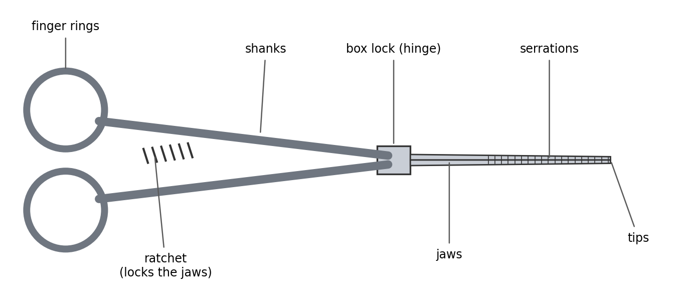
Figure 15.1. Parts of a ringed instrument (hemostat).
Why it matters
Most instrument identification questions come down to a few features: the tips (smooth, serrated, toothed, or fenestrated), the jaw pattern (full or partial, transverse or longitudinal serrations), whether there is a ratchet, and the size and curvature. Train yourself to look at those features first.
15.1 Thumb Forceps
Thumb forceps (also called tissue forceps or pick-ups) are spring-tensioned, tweezer-like instruments held between the thumb and fingers like a pencil. They have no lock, so they hold tissue only while pressure is applied. The design of the tips determines how traumatic they are: serrated tips are the least traumatic, toothed tips grip dense tissue such as skin securely, and fenestrated tips spread pressure over a ring (Figure 15.2).
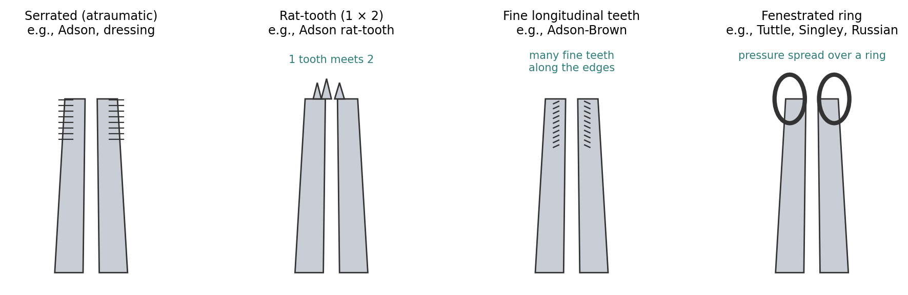
Figure 15.2. Common thumb forceps tip designs (schematic).
Figure 15.3. Adson Forceps |
Adson Forceps
|
|---|---|
Figure 15.4. Adson-Brown Forceps |
Adson-Brown Forceps
|
Figure 15.5. Mayo (Russian) Forceps |
Mayo (Russian) Forceps
|
Figure 15.6. Rat-Tooth Forceps |
Rat-Tooth Forceps
|
Figure 15.7. Tuttle Thoracic Tissue Forceps |
Tuttle Thoracic Tissue Forceps
|
Figure 15.8. DeBakey Thoracic Forceps |
DeBakey Thoracic Forceps
|
Figure 15.9. Singley Tissue Forceps |
Singley Tissue Forceps
|
Figure 15.10. Cushing Tissue Forceps (1 of 2)
Figure 15.11. Cushing Tissue Forceps (2 of 2) |
Cushing Tissue Forceps
|
Figure 15.12. Dressing Forceps |
Dressing Forceps
|
Figure 15.13. Dumont Forceps |
Dumont Forceps
|
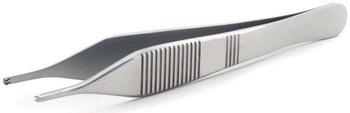
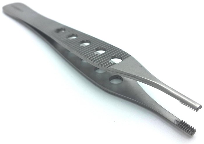
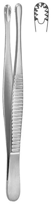
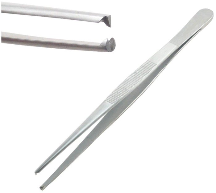
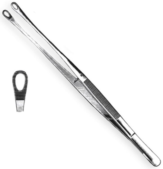
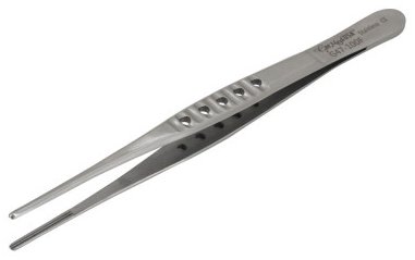
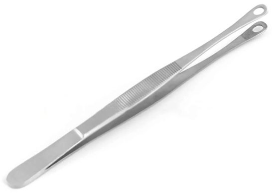



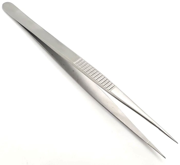
15.2 Hand Forceps (Ratchet Mechanism)
Ringed forceps with a ratchet can be locked onto tissue, freeing the surgeon's hands. Because a locked jaw applies constant pressure, these instruments are chosen carefully according to how much crushing the tissue can tolerate (Figure 15.1 shows the parts of a ringed instrument).
Figure 15.14. Allis Tissue Forceps |
Allis Tissue Forceps
|
|---|---|
Figure 15.15. Doyen Forceps |
Doyen Forceps
|
Figure 15.16. Babcock Intestinal & Tissue Forceps |
Babcock Intestinal & Tissue Forceps
|
Figure 15.17. Mixter (Thoracic) Forceps |
Mixter (Thoracic) Forceps
|
Figure 15.18. Mixter (Gallbladder) Forceps |
Mixter (Gallbladder) Forceps
|
Figure 15.19. Collin Gallbladder Forceps (Judd-DeMartel) |
Collin Gallbladder Forceps (Judd-DeMartel)
|
Figure 15.20. Duval Lung Forceps |
Duval Lung Forceps
|
Figure 15.21. Vulsellum Forceps |
Vulsellum Forceps
|
Figure 15.22. Alligator Forceps |
Alligator Forceps
|
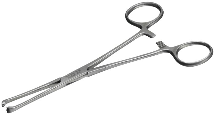
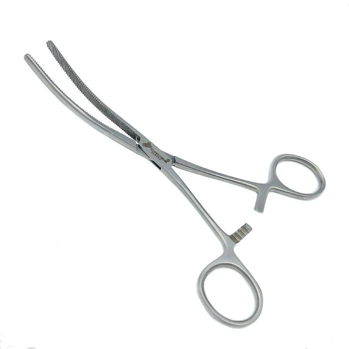
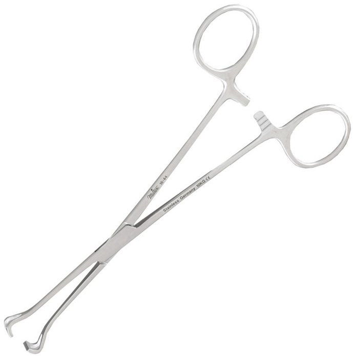
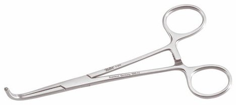
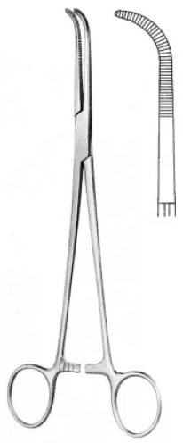
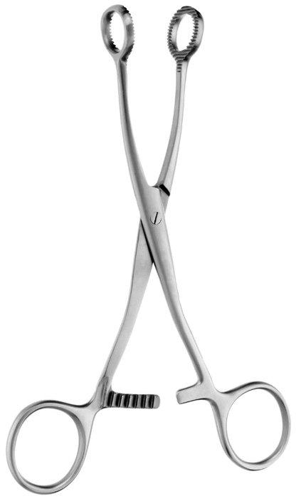
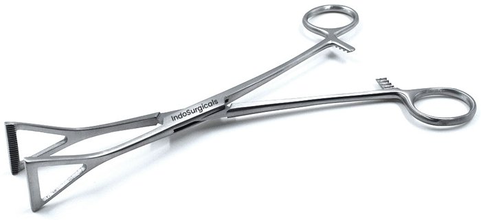
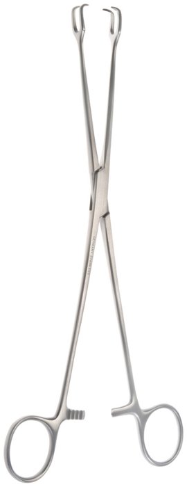
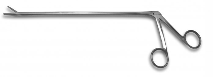
15.3 Hemostatic & Vascular Forceps (Ratchet Mechanism)
Hemostats clamp blood vessels so they can be ligated or cauterized. They are distinguished mainly by size and by the pattern of serrations on their jaws (Figure 15.23), which determines how well they resist slipping. Their proper use is clamping vessels for ligation and hemostasis, not grasping tissue (Chapter 16).
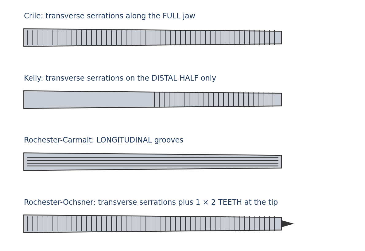
Figure 15.23. Jaw patterns of common hemostatic forceps (schematic).
Figure 15.24. Hartman Mosquito Forceps |
Hartman Mosquito Forceps
|
|---|---|
Figure 15.25. Halsted Mosquito Forceps |
Halsted Mosquito Forceps
|
Figure 15.26. Crile Hemostats |
Crile Hemostats
|
Figure 15.27. Jacobsen Hemostats |
Jacobsen Hemostats
|
Figure 15.28. Kelly Hemostats |
Kelly Hemostats
|
Figure 15.29. Rochester-Pean Hemostats |
Rochester-Pean Hemostats
|
Figure 15.30. Rochester-Carmalt Hemostats |
Rochester-Carmalt Hemostats
|
Figure 15.31. Rochester-Ochsner Hemostats |
Rochester-Ochsner Hemostats
|
Figure 15.32. Ferguson Angiotribe |
Ferguson Angiotribe
|
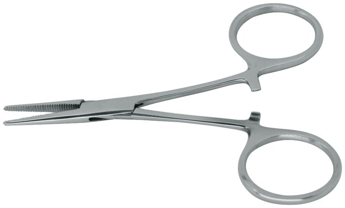
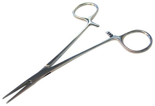
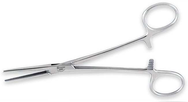
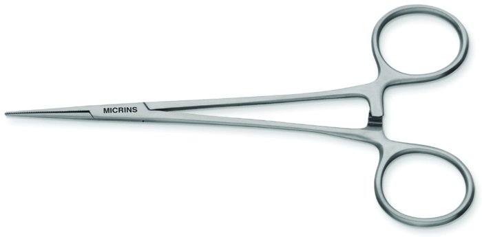
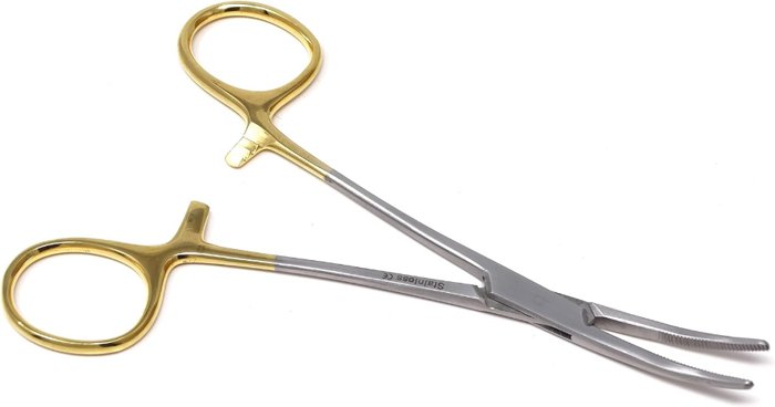
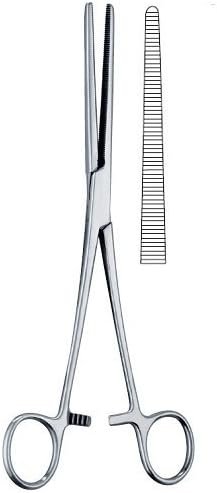
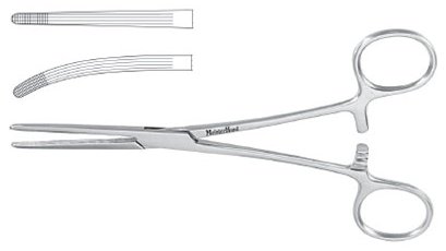
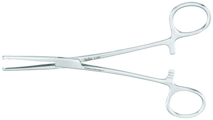
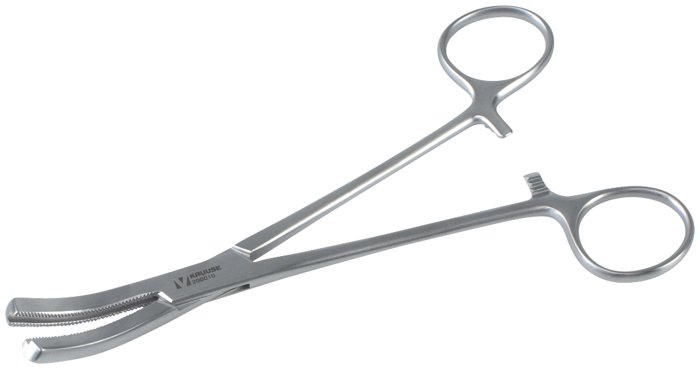
15.4 Sponge Forceps (Ratchet Mechanism)
Sponge forceps hold gauze sponges for skin preparation or for blotting fluid in deep areas where fingers cannot reach.
Figure 15.33. Chester and Ballenger Forceps |
Chester and Ballenger Forceps
|
|---|---|
Figure 15.34. Foerster Forceps |
Foerster Forceps
|
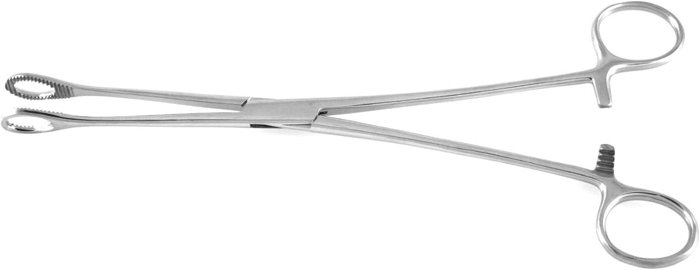
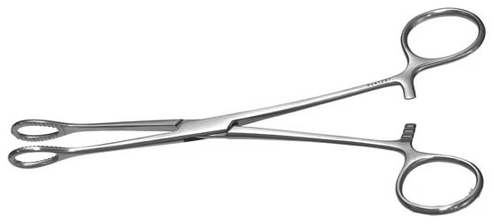
15.5 Vascular Clamps
Vascular clamps occlude blood flow through a vessel, fully or partially, with minimal damage to its wall. Partial (side-biting) clamping isolates a section of the vessel wall while blood continues to flow around it, which allows a vessel to be opened or joined (anastomosis) without stopping circulation entirely. Bulldog clamps are small, spring-loaded clamps without handles, with varying degrees of tension.
Figure 15.35. Cooley Cardiovascular Clamp |
Cooley Cardiovascular Clamp
|
|---|---|
Figure 15.36. DeBakey Abdominal Clamp |
DeBakey Abdominal Clamp
|
Figure 15.37. Satinsky Clamp |
Satinsky Clamp
|
Figure 15.38. Renal Clamp |
Renal Clamp
|
Figure 15.39. Serrefine (Bulldog) Clamp |
Serrefine (Bulldog) Clamp
|
Figure 15.40. Senning Bulldog Clamp |
Senning Bulldog Clamp
|
Figure 15.41. Diefenbach Bulldog Clamp |
Diefenbach Bulldog Clamp
|
Figure 15.42. Blalock Bulldog Clamp |
Blalock Bulldog Clamp
|
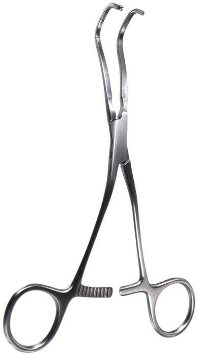
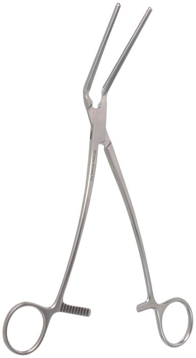
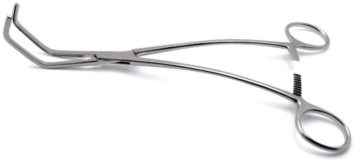
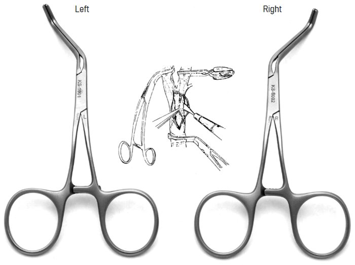
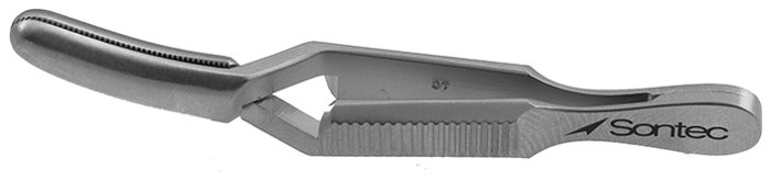
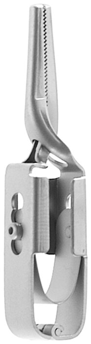
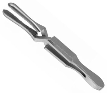
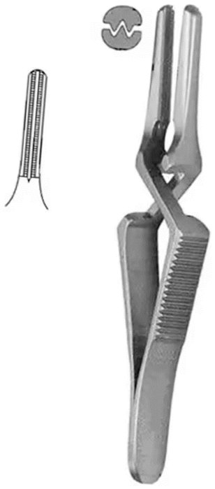
15.6 Towel Clamps
Towel clamps hold drapes in place to create and maintain the sterile field (Chapter 4). Penetrating clamps pierce the drape; non-penetrating clamps preserve the drape's barrier.
Figure 15.43. Backhaus Towel Clamp |
Backhaus Towel Clamp
|
|---|---|
Figure 15.44. Roeder Towel Clamp |
Roeder Towel Clamp
|
Figure 15.45. Edna Towel Clamp |
Edna Towel Clamp
|
Figure 15.46. Jones Towel Clamp |
Jones Towel Clamp
|
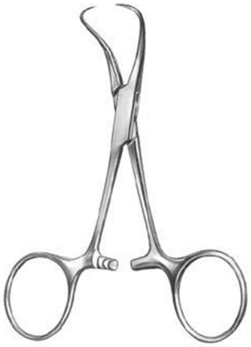
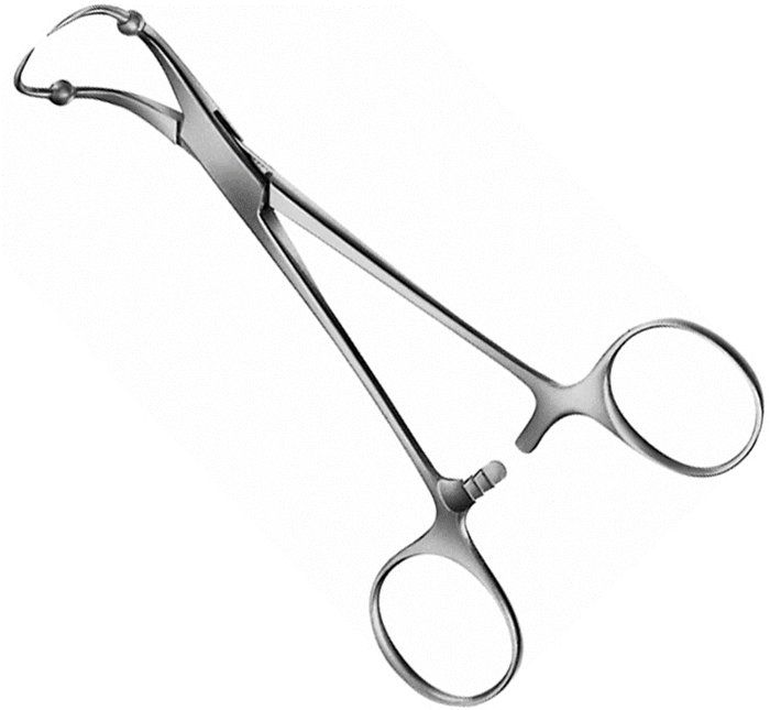
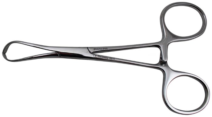
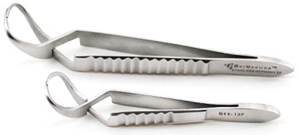
15.7 Scissors
Scissors are chosen by the tissue they will cut. Tissue scissors should never be used to cut suture, drapes, or wire, which dulls them; separate operating, suture, wire, and bandage scissors exist for those tasks. Tips may be sharp or blunt, and blades straight or curved (Figure 15.47). Cutting with scissors crushes as well as cuts, so scissors are more traumatic than a scalpel (Chapter 16).

Figure 15.47. Scissor tip and blade configurations (schematic).
Figure 15.48. Operating Scissors |
Operating Scissors
|
|---|---|
Figure 15.49. Mayo Scissors |
Mayo Scissors
|
Figure 15.50. Metzenbaum Scissors |
Metzenbaum Scissors
|
Figure 15.51. Enterotomy Scissors |
Enterotomy Scissors
|
Figure 15.52. Iris Scissors |
Iris Scissors
|
Figure 15.53. Strabismus Scissors |
Strabismus Scissors
|
Figure 15.54. Ragnell Scissors |
Ragnell Scissors
|
Figure 15.55. Stevens Tenotomy Scissors |
Stevens Tenotomy Scissors
|
Figure 15.56. Vannas (Micro-Vannas) Scissors |
Vannas (Micro-Vannas) Scissors
|
Figure 15.57. Wire Scissors |
Wire Scissors
|
Figure 15.58. Lister Bandage Scissors |
Lister Bandage Scissors
|
Figure 15.59. Doyen Scissors |
Doyen Scissors
|
Figure 15.60. Spencer Suture Scissors |
Spencer Suture Scissors
|
Figure 15.61. Littauer Suture Scissors |
Littauer Suture Scissors
|
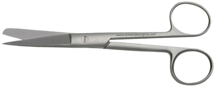
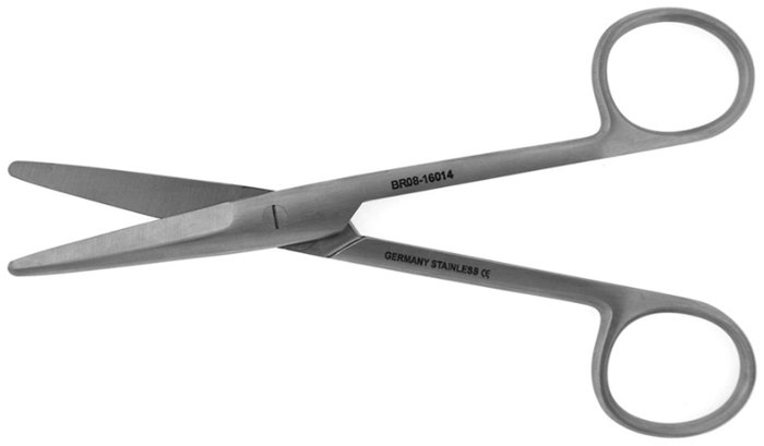
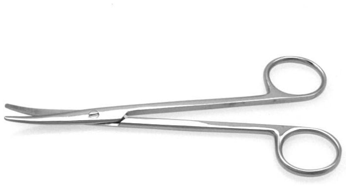
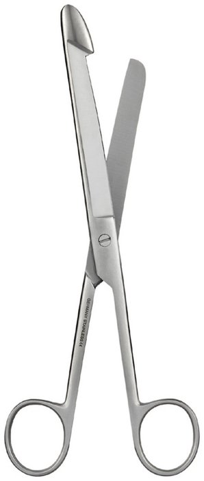
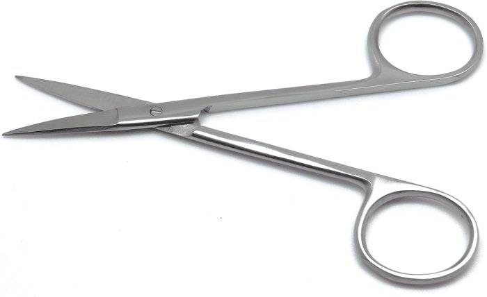
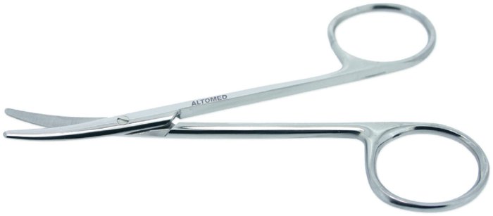

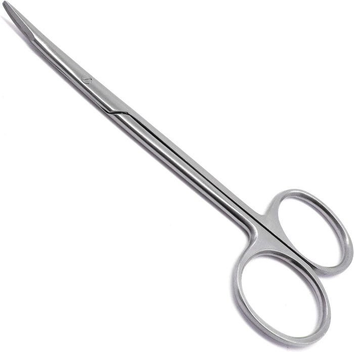
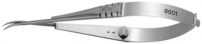
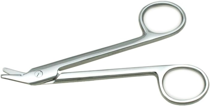
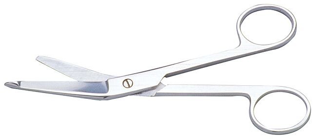
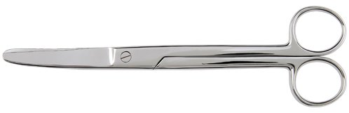
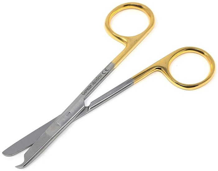
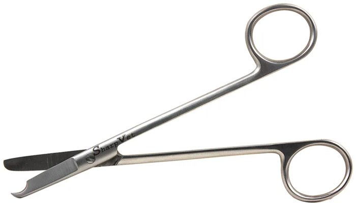
15.8 Needle Holders
Needle holders grip a curved suture needle firmly so it can be driven through tissue. The needle is usually grasped about one-third to one-half of the way back from the point (never at the swaged end, where the suture attaches) with the tip of the jaws. The size of the needle holder should match the size of the needle: small, delicate needle holders bend or lose their grip on large needles, while large ones can damage small needles.
Figure 15.62. Mayo-Hegar Needle Holder |
Mayo-Hegar Needle Holder
|
|---|---|
Figure 15.63. Olsen-Hegar Needle Holder |
Olsen-Hegar Needle Holder
|
Figure 15.64. Finochietto and DeBakey Thoracic Needle Holders (1 of 2)
Figure 15.65. Finochietto and DeBakey Thoracic Needle Holders (2 of 2) |
Finochietto and DeBakey Thoracic Needle Holders
|
Figure 15.66. Halsey Needle Holder |
Halsey Needle Holder
|
Figure 15.67. Castroviejo Needle Holder |
Castroviejo Needle Holder
|
Figure 15.68. Mathieu Needle Holder |
Mathieu Needle Holder
|
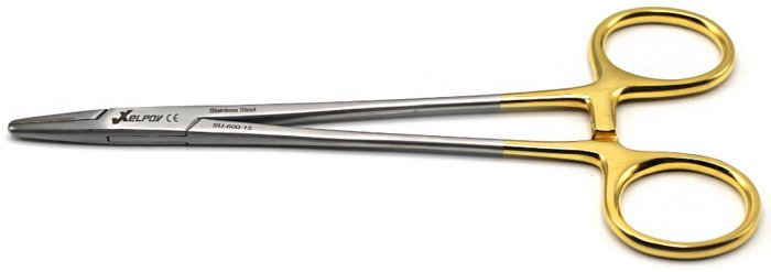
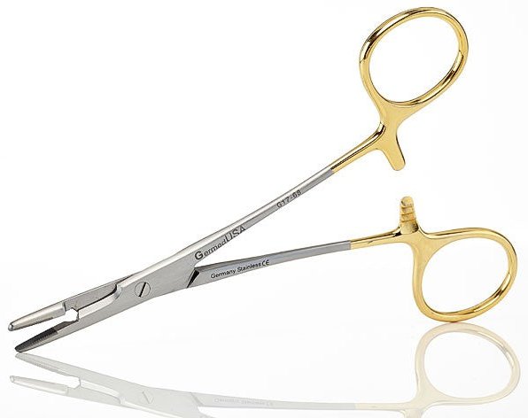
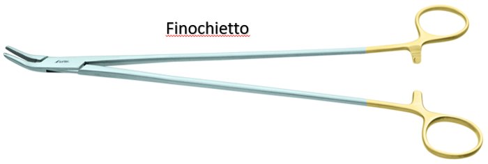
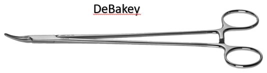
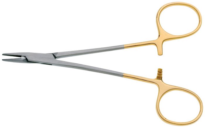
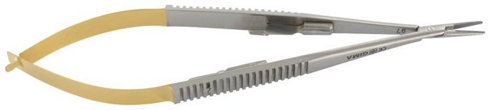
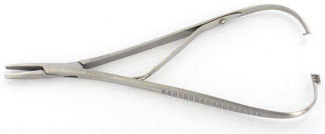
15.9 Self-Retaining Retractors
Self-retaining retractors hold a wound open by themselves, using a ratchet, rack, or screw, which frees the surgeon's and assistant's hands. Because they apply continuous pressure, they should be opened only as far as needed and released periodically during long procedures to avoid ischemic injury to the retracted tissue.
Figure 15.69. Balfour Retractor |
Balfour Retractor
|
|---|---|
Figure 15.70. Finochietto Retractor (Rib Spreader) |
Finochietto Retractor (Rib Spreader)
|
Figure 15.71. Wilson Rib Retractor |
Wilson Rib Retractor
|
Figure 15.72. DeBakey Retractor |
DeBakey Retractor
|
Figure 15.73. O'Sullivan-O'Connor and Wexler Abdominal Ring Retractors |
O'Sullivan-O'Connor and Wexler Abdominal Ring Retractors
|
Figure 15.74. Alm Retractor |
Alm Retractor
|
Figure 15.75. Colibri Retractor (Eye Speculum) |
Colibri Retractor (Eye Speculum)
|
Figure 15.76. Gelpi Retractor |
Gelpi Retractor
|
Figure 15.77. Weitlaner Retractor |
Weitlaner Retractor
|
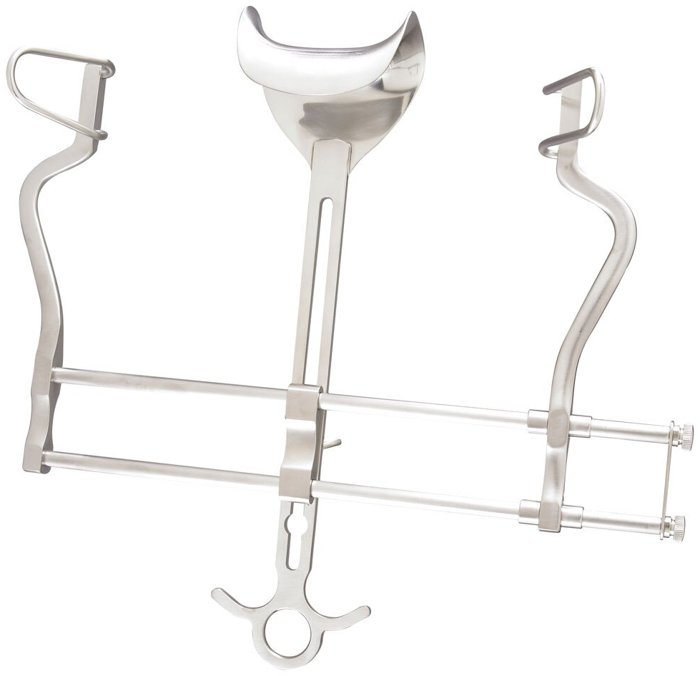
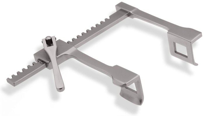
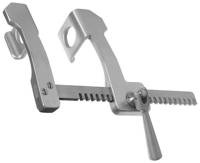
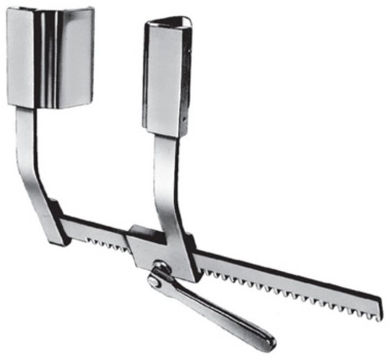
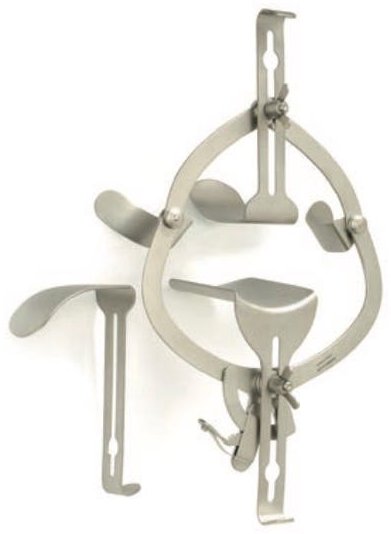
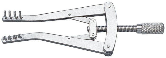
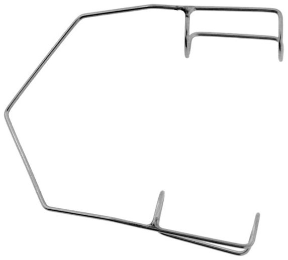
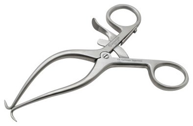
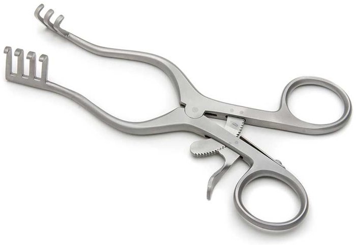
15.10 Manual Retractors
Manual (hand-held) retractors are held by an assistant, who can adjust the angle and force of retraction continuously. Sharp-pronged or toothed retractors grip fascia and skin; blunt blades and rakes are gentler on soft tissue.
Figure 15.78. Senn Retractor |
Senn Retractor
|
|---|---|
Figure 15.79. Army-Navy Retractor |
Army-Navy Retractor
|
Figure 15.80. Ochsner Ribbon (Malleable) Retractor |
Ochsner Ribbon (Malleable) Retractor
|
Figure 15.81. Parker Retractor |
Parker Retractor
|
Figure 15.82. Deaver Retractor |
Deaver Retractor
|
Figure 15.83. Mayo Abdominal Retractor |
Mayo Abdominal Retractor
|
Figure 15.84. Langenbeck Retractor |
Langenbeck Retractor
|
Figure 15.85. Sauerbruch Retractor |
Sauerbruch Retractor
|
Figure 15.86. Doyen Retractor |
Doyen Retractor
|
Figure 15.87. Volkmann Rake Retractor |
Volkmann Rake Retractor
|
Figure 15.88. Bernay Finger Rake Retractor |
Bernay Finger Rake Retractor
|
Figure 15.89. Love Nerve Retractor |
Love Nerve Retractor
|
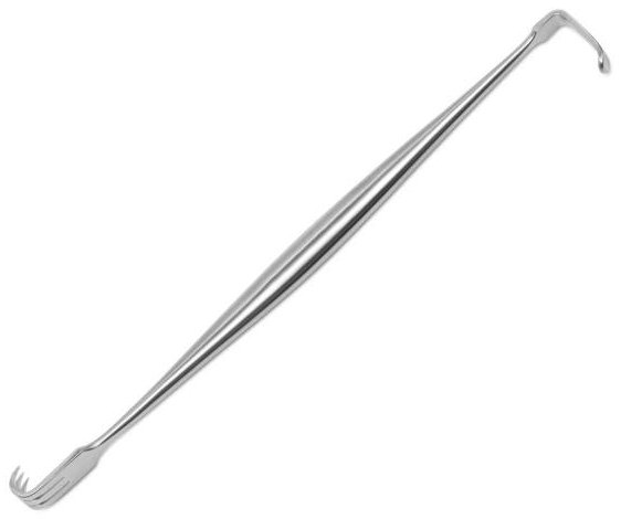
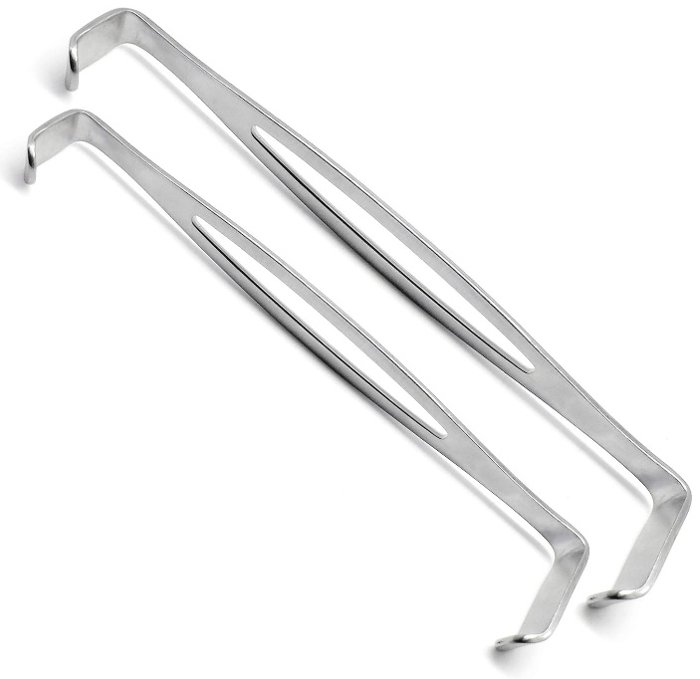

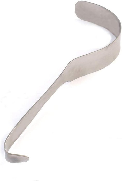
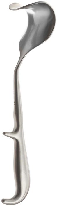
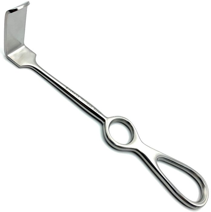
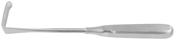
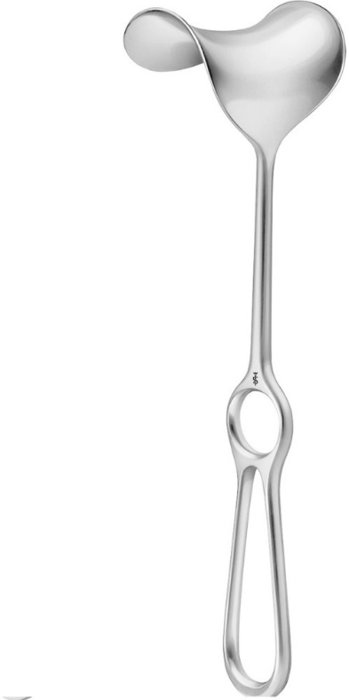
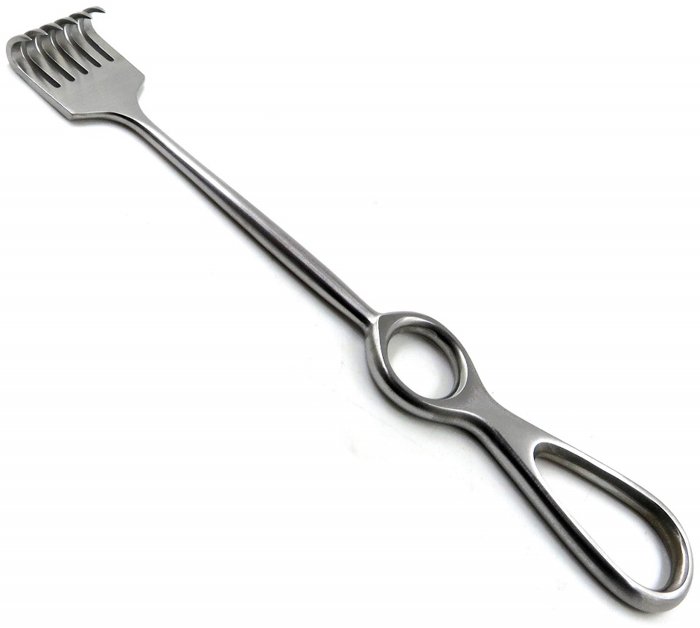

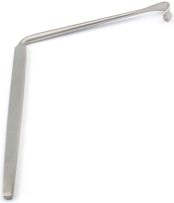
15.11 Suction Tips
Suction tips remove blood, irrigation fluid, and debris from the surgical field so the surgeon can see. Their design balances flow against the risk of damaging or plugging with tissue.
Figure 15.90. Yankauer Suction |
Yankauer Suction
|
|---|---|
Figure 15.91. DeBakey Suction |
DeBakey Suction
|
Figure 15.92. Frazier Suction |
Frazier Suction
|
Figure 15.93. Poole Suction |
Poole Suction
|
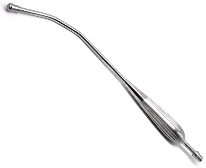

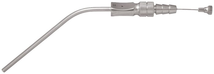
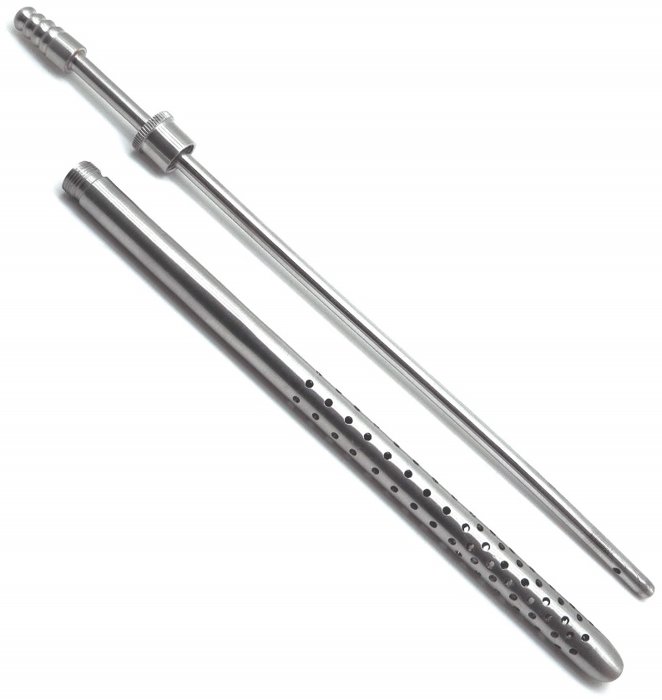
15.12 Elevators
Figure 15.94. Periosteal Elevator |
Periosteal Elevator
|
|---|---|
Figure 15.95. Matson Rib Elevator & Stripper |
Matson Rib Elevator & Stripper
|

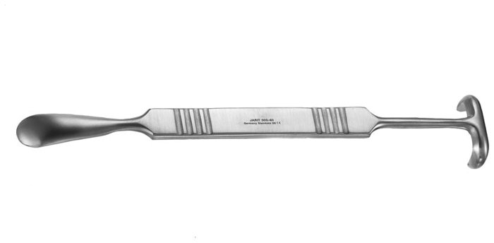
15.13 Tissue Cutting & Cautery
Figure 15.96. Scalpel |
Scalpel
|
|---|---|
Figure 15.97. Harmonic Scalpel |
Harmonic Scalpel
|
Figure 15.98. Cautery & Cautery Pen |
Cautery & Cautery Pen
|
Figure 15.99. CO₂ Laser |
CO₂ Laser
|
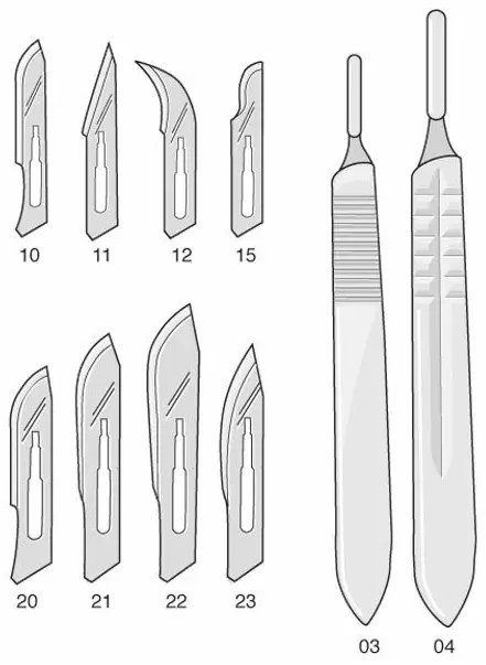
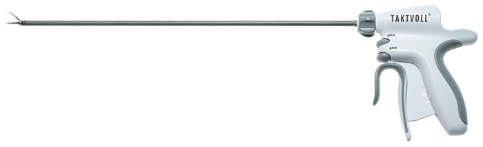
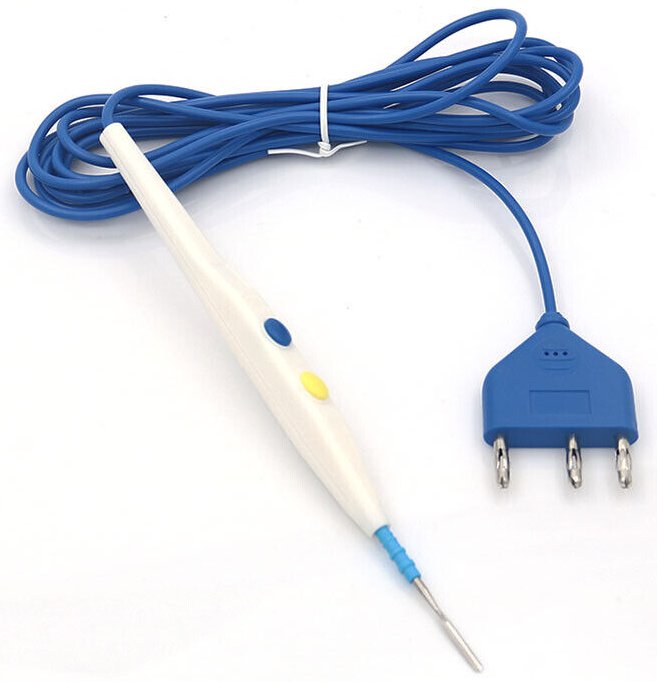
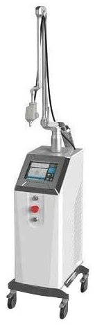
15.14 Bone Instruments
Orthopedic instruments cut, shape, hold, and fix bone. Bone instruments are heavy and powerful, so they must be used with care to avoid damaging the soft tissues, nerves, and vessels around the bone.
Other rongeurs commonly encountered include the Pilling-Ruskin, Beyer, and Zaufel-Jansen rongeurs.
Figure 15.100. Bailey Rib Approximator |
Bailey Rib Approximator
|
|---|---|
Figure 15.101. Bruns Bone Curettes |
Bruns Bone Curettes
|
Figure 15.102. Spratt Bone Curettes |
Spratt Bone Curettes
|
Figure 15.103. Galt Cranial Trephine |
Galt Cranial Trephine
|
Figure 15.104. Michele Trephine |
Michele Trephine
|
Figure 15.105. Lowman Bone Clamp |
Lowman Bone Clamp
|
Figure 15.106. Sherman Bone Plate |
Sherman Bone Plate
|
Figure 15.107. Kern Bone-Holding Forceps |
Kern Bone-Holding Forceps
|
Figure 15.108. Lebsche Sternum Knife |
Lebsche Sternum Knife
|
Figure 15.109. Stille-Liston (Liston) Bone-Cutting Forceps |
Stille-Liston (Liston) Bone-Cutting Forceps
|
Figure 15.110. Gigli Wire |
Gigli Wire
|
Figure 15.111. Stryker Bone and Cast Saw |
Stryker Bone and Cast Saw
|
Figure 15.112. Bone Chisel |
Bone Chisel
|
Figure 15.113. Osteotome |
Osteotome
|
Figure 15.114. Lempert Rongeur |
Lempert Rongeur
|
Figure 15.115. Ruskin Rongeur |
Ruskin Rongeur
|
Figure 15.116. Kerrison Rongeur |
Kerrison Rongeur
|
Figure 15.117. Jacobs Chuck & Key |
Jacobs Chuck & Key
|
Figure 15.118. Steinmann Pins |
Steinmann Pins
|
Figure 15.119. Kirschner Wire (K-Wire) |
Kirschner Wire (K-Wire)
|
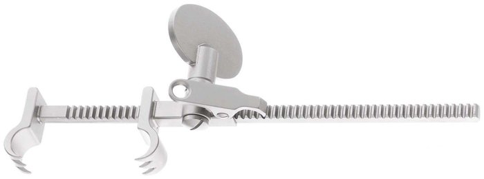
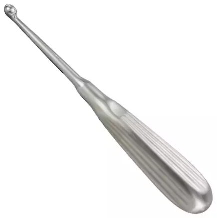

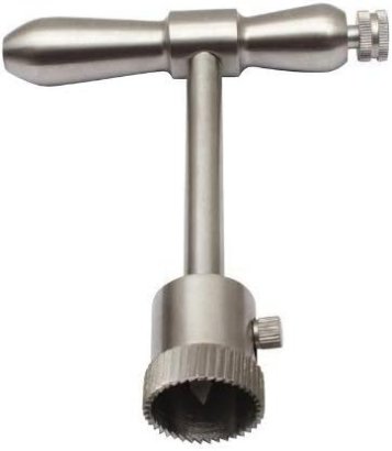
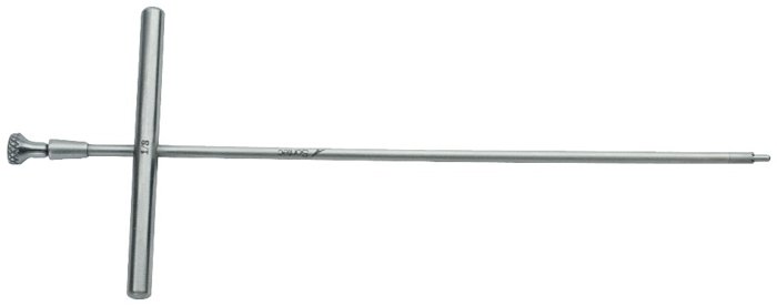
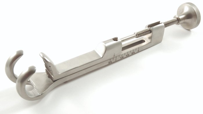
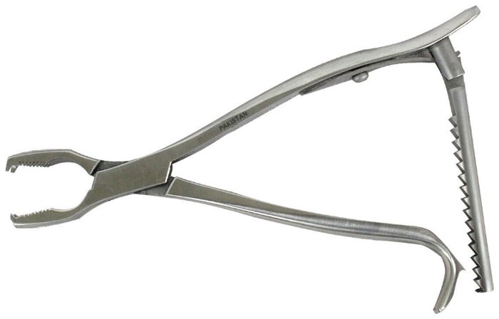
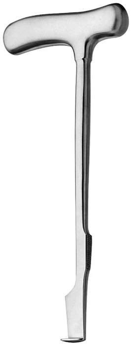
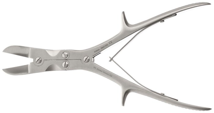
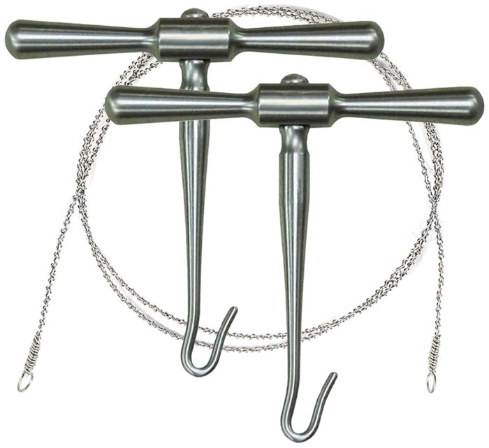
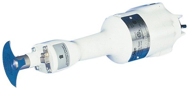
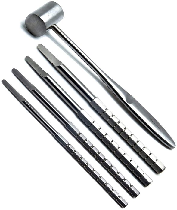

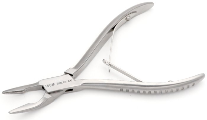
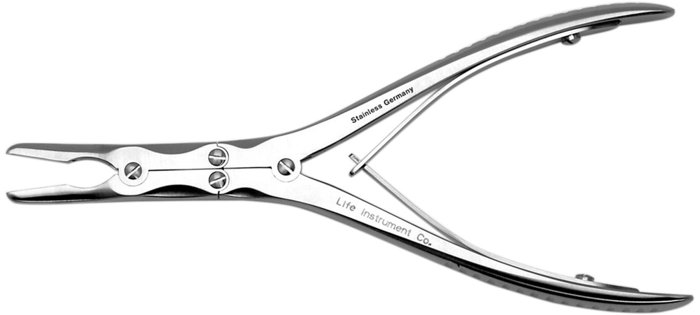
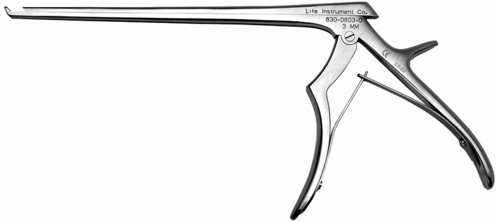

15.15 Soft Tissue Biopsy Instruments
Figure 15.120. Tru-Cut Biopsy Needle |
Tru-Cut Biopsy Needle
|
|---|---|
Figure 15.121. Tissue Biopsy Punch |
Tissue Biopsy Punch
|
Figure 15.122. Tissue Biopsy Cup |
Tissue Biopsy Cup
|
Figure 15.123. Tissue Curettes |
Tissue Curettes
|
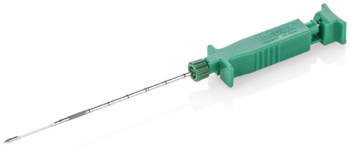

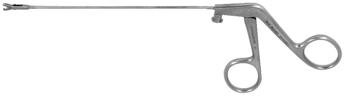
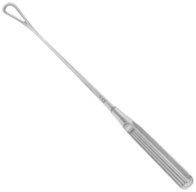
15.16 Basins & Bowls
Figure 15.124. Emesis Basin |
Emesis Basin
|
|---|---|
Figure 15.125. Sponge Bowl |
Sponge Bowl
|
Figure 15.126. Irrigation, Soaking, or Organ Bowl |
Irrigation, Soaking, or Organ Bowl
|
15.17 Miscellaneous Equipment & Instruments
Figure 15.127. Mayo Stand |
Mayo Stand
|
|---|---|
Figure 15.128. Surgical Trocar |
Surgical Trocar
|
Figure 15.129. Surgical Staplers |
Surgical Staplers
|
Figure 15.130. Stereotaxic Stand & Frames |
Stereotaxic Stand & Frames
|
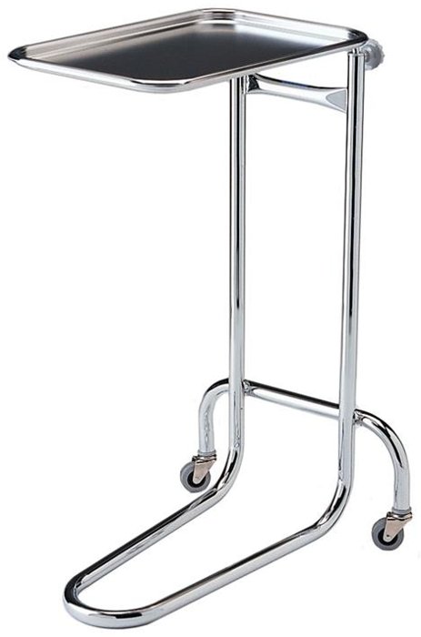
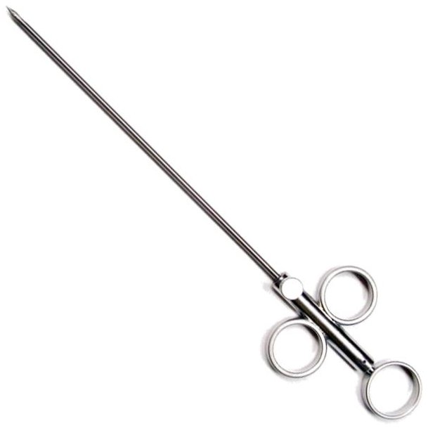
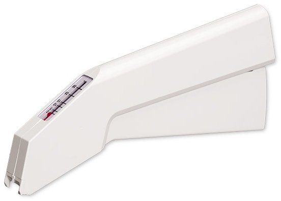
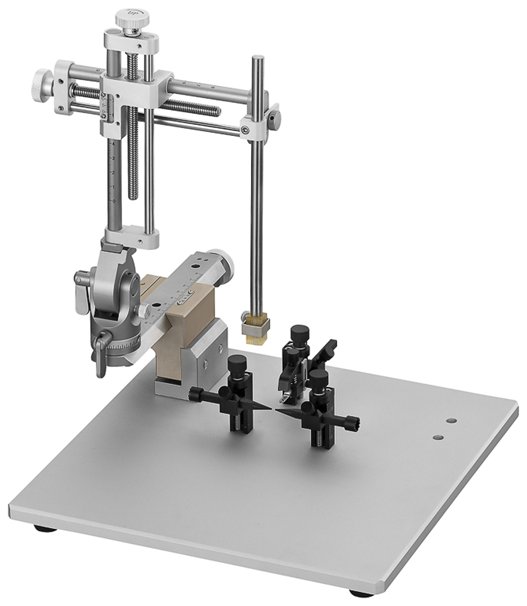
⚠ Check current guidance
Instrument names, spellings, and catalog descriptions vary between manufacturers, and the same name may refer to different instruments. Instrument descriptions here combine several sources; confirm specific features with your facility's instrument sets and current catalogs.
Instrument Use and Care
A few principles apply to all instruments:
Use each instrument only for its intended purpose. Tissue scissors used on suture or drapes become dull; hemostats used to grasp tissue crush it; needle holders used as pliers lose their grip.
Choose the least traumatic instrument that will do the job: smooth or finely serrated tips for delicate tissue, toothed tips only for dense tissue such as skin and fascia.
Inspect instruments before use: jaws should meet evenly, ratchets should hold, tips should be aligned, and scissors should cut cleanly along their full length.
Clean instruments promptly after use, before blood and tissue dry, with box locks and hinges open; then dry, lubricate if appropriate, and sterilize them in the open position (Chapter 3).
Protect delicate tips (such as microsurgical instruments) with tip guards during cleaning, storage, and sterilization.
⚠ Check current guidance
Image credits: The instrument photographs in this chapter were supplied from a compiled instrument study document. Several appear to be manufacturer or vendor catalog images (for example, from GerMedUSA, Sontec, and Scatter Instruments). Before this textbook is printed for distribution, shared, or published, confirm the source of each photograph, obtain permission where required, and add a credit line to each caption.
Chapter Summary
Instruments are named for their action, their inventor, or the surgery they serve. Thumb forceps range from atraumatic serrated tips (Adson, DeBakey) to toothed tips for dense tissue (rat-tooth, Adson-Brown) and fenestrated ring tips (Tuttle, Singley, Russian). Ratcheted hand forceps include the traumatic but secure Allis and the gentler Babcock and Doyen. Hemostats differ by size and serration pattern: mosquito, Crile (full transverse), Kelly (distal half), and the Rochester Pean, Carmalt (longitudinal), and Ochsner (with teeth). Vascular clamps (Cooley, DeBakey, Satinsky, renal, and bulldogs) occlude vessels fully or partially with minimal wall damage.
Towel clamps may penetrate (Backhaus, Roeder) or not (Edna). Scissors are matched to the tissue: Mayo for heavy tissue, Metzenbaum for delicate dissection, iris, tenotomy, and Vannas for fine and microsurgical work, and separate suture, wire, and bandage scissors. Needle holders range from the heavy Mayo-Hegar to the cutting Olsen-Hegar and the delicate Castroviejo. Retractors are self-retaining (Balfour, Finochietto, Gelpi, Weitlaner) or manual (Senn, Army-Navy, ribbon). Suction tips, elevators, cutting and cautery devices, bone and biopsy instruments, and basic equipment complete the surgical kit.
Key Terms
Box lock: The hinge joint of a ringed instrument.
Fenestrated: Having an opening (window), as in a ring-tipped forceps.
Hemostat: A ratcheted forceps used to clamp blood vessels.
Needle holder: A ratcheted instrument that grips a suture needle.
Partial (side-biting) clamping: Clamping part of a vessel wall while blood continues to flow past.
Ratchet: The interlocking teeth that lock a ringed instrument closed.
Retractor: An instrument that holds tissue aside to expose the surgical field.
Rongeur: A heavy, cupped-jaw instrument for removing bone in small pieces.
Self-retaining retractor: A retractor that holds itself open with a ratchet, rack, or screw.
Serrations: Ridges on the jaws of an instrument that improve grip.
Thumb forceps: Spring-tensioned, tweezer-like forceps without a lock.
Trephine: A cylindrical cutting instrument for removing a disc or core of bone.
Review Questions
1. Instruments named for the action they perform include:
A. Kelly and Allis
B. Scalpel and hemostat
C. Metzenbaum and Mayo
D. Backhaus and Balfour
2. Which thumb forceps have fine teeth running longitudinally along the edges of the tip, requiring less crushing pressure to grip?
A. Dressing forceps
B. Adson-Brown forceps
C. Rat-tooth forceps
D. Dumont forceps
3. Which forceps have broad, flared, smooth tips used to hold bowel and bladder atraumatically?
A. Allis
B. Babcock
C. Kocher
D. Backhaus
4. Kelly hemostats differ from Crile hemostats in that Kelly hemostats have:
A. Serrations along the full length of the jaws
B. Serrations only on the distal half of the jaws
C. Longitudinal grooves
D. 1 × 2 teeth at the tip
5. Which hemostat has long blades with transverse serrations and 1 × 2 teeth at the tip?
A. Rochester-Pean
B. Rochester-Carmalt
C. Rochester-Ochsner
D. Halsted mosquito
6. Which towel clamp must NOT be used on skin?
A. Backhaus
B. Roeder
C. Edna
D. Jones
7. Which scissors are designed for soft, delicate tissue dissection and have a longer handle-to-blade ratio than Mayo scissors?
A. Operating scissors
B. Metzenbaum scissors
C. Lister bandage scissors
D. Wire scissors
8. A needle holder that combines needle-holding jaws with scissor blades is the:
A. Mayo-Hegar
B. Olsen-Hegar
C. Castroviejo
D. Mathieu
9. Which blades fit a #3 scalpel handle?
A. 10, 11, 12, and 15
B. 20, 21, 22, and 23
C. Only 10
D. All blades fit all handles
10. When using a Kerrison rongeur in spinal surgery, upward traction is applied to:
A. Increase cutting speed
B. Prevent trauma to the soft tissue beneath the bone
C. Lock the jaws
D. Sharpen the blade
Answer Key
1. B. Instrument names follow patterns: the action performed (scalpel, hemostat), the inventor's name (Kelly, Allis, Metzenbaum), or a compound scientific name related to the type of surgery.
2. B. Adson-Brown forceps have 7 × 7 to 9 × 9 fine teeth along the edges, which grip with less crushing pressure.
3. B. Babcock forceps are more delicate than Allis forceps and are used to hold viscera atraumatically.
4. B. Crile hemostats are serrated along the entire jaw; Kelly hemostats only on the distal half.
5. C. The Rochester-Ochsner has transverse serrations plus 1 × 2 tip teeth for stronger grasping. The Carmalt has longitudinal grooves, and the Pean deep transverse serrations without teeth.
6. C. Edna towel clamps have flat pins that grip towels without puncturing them, but crush skin.
7. B. Metzenbaum scissors are for delicate tissue cutting and dissection. Mayo scissors cut heavy fascia and suture.
8. B. The Olsen-Hegar includes scissor blades so suture can be cut without changing instruments, with the risk of cutting suture accidentally.
9. A. Blades 10, 11, 12, and 15 fit the #3 handle; blades 20–23 fit the larger #4 handle.
10. B. Upward traction during placement and use protects the spinal cord and other soft tissue beneath the bone.
Chapter 16: Surgical Techniques
Learning objectives
After studying this chapter, you should be able to:
Apply the principles of gentle tissue handling, hemostasis, and elimination of dead space.
Describe the phases of wound healing, the factors that affect it, and the types of wound healing.
Classify surgical wounds and recognize common complications.
Compare suture materials by size, structure, absorption, and handling, and select appropriate sutures and needles.
Describe knot-tying principles and identify common interrupted and continuous suture patterns and their uses.
Surgical technique determines how well and how quickly a wound heals. Gentle tissue handling, careful hemostasis, appropriate suture and needle choice, and the right suture pattern all reduce pain, infection, and complications, which matters for both animal welfare and the quality of research data. This chapter brings together the principles of tissue handling, the biology of wound healing, and the materials and techniques of wound closure.
16.1 Tissue Handling
The goal of good tissue handling is to minimize tissue trauma, which leads to more rapid healing. The first priority is always aseptic technique (Chapter 4), because infection deters healing. Beyond that:
Plan the incision. Incisions heal side to side, not end to end, so there is little advantage to making an incision too small; a slightly longer incision that gives good exposure causes less trauma than forcing a small one. The best cosmetic results come from incisions parallel to the direction of the tissue fibers.
Cut cleanly. Make the skin incision in one stroke with even pressure on the scalpel, then use sharp dissection through the remaining tissues, preserving as many underlying nerves, vessels, and muscles as possible. Cutting with a sharp blade is minimally traumatic, with little damage to adjacent cells; cutting with scissors crushes and tears, causing more adjacent damage. Use blunt dissection to separate tissues along natural tissue planes.
Handle tissue gently and as little as possible. Use minimal tension, place retractors to avoid excessive tension, and provide gentle retraction with the proper instruments. Avoid impairing blood or lymph flow, which may change the local physiological state.
Do not crush tissue. Clamping tissue with hemostats or forceps crushes cells and is very traumatic, releasing vasoconstrictors and clotting factors. The proper use of hemostats is clamping vessels for ligation and hemostasis (Chapter 15).
Keep tissue moist. Dry tissue is dead tissue. Periodically irrigate the site, and lavage body cavities, with warm sterile fluid such as saline, LRS, or Tis-U-Sol.
Remove contamination. Lavage wounds with excessive debris thoroughly with sterile isotonic fluid ("the solution to pollution is dilution"), debride all devitalized tissue, and remove foreign material and anything nonessential.
Minimize exposure time.
Why it matters
The three "T's" of tissue handling summarize these principles: keep time to a minimum, remove existing trash (contamination) and prevent new contamination, and keep trauma to a minimum.
Hemostasis
Bleeding should be stopped whenever possible. It obscures the surgical field, and blood left in the wound forms hematomas that prevent wound apposition, increase dead space, and retard healing. Blood is also a natural food for microorganisms, and a large clot can protect them from the immune system. Achieving hemostasis before closure helps prevent post-operative hematomas and seromas.
Bleeding may be slowed or stopped by pressure, clamping, electrocautery or thermocautery, ligatures, and hemostatic agents, depending on the rate and volume of hemorrhage. Excessive pressure, like that applied by hemostats, and mass ligation of large areas of tissue may cause necrosis and prolong healing.
Why it matters
The three "P's" of hemostasis: Pressure (apply gentle, firm pressure), Patience (leave the gauze in place long enough for the type of bleeding present; repeatedly lifting it to look dislodges the forming clot), and Perseverance (continue until bleeding is controlled).
Dead Space
Dead space is an open area within closed tissue, created when tissue layers beneath the skin edges are not brought together, or when air or fluid is trapped between layers, especially in the poorly vascular fatty layer. It prevents tissue apposition and fills with serum or blood, which is an ideal medium for microorganisms; in many species, seromas form (Figure 16.1). Dead space is eliminated by closing tissue in layers, and post-operatively with a drain or pressure dressing if needed.
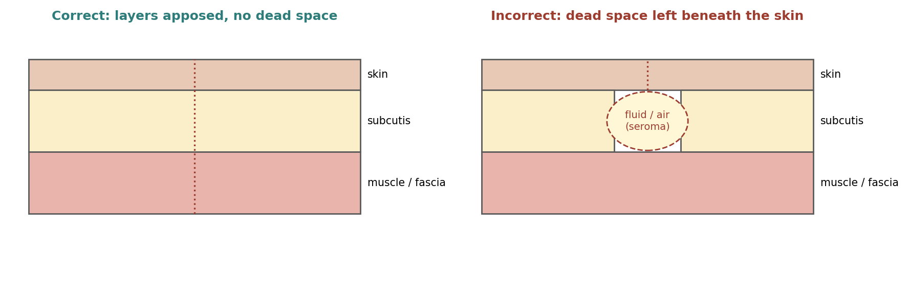
Figure 16.1. Layered closure that eliminates dead space, compared with a closure that leaves dead space beneath the skin (schematic).
16.2 Wound Healing
A wound is an injury to any of the body's tissues, especially one caused by physical means, that interrupts their continuity. Healing is the process of restoring health. A tissue is a collection of similar cells and the intercellular substances around them. The four basic tissue types are epithelium, connective tissue (including blood, bone, and cartilage), muscle, and nerve.
Wound healing is natural and spontaneous. When tissue is so disrupted that it cannot heal on its own, dead tissue and foreign bodies must be removed, infection treated, and the tissue held in apposition until healing gives the wound enough strength to withstand stress without support. Wounds can be closed with sutures, staples, clips, skin closure strips, or topical tissue adhesives.
Tissue Strength
Tensile strength is the load per unit of cross-sectional area at the point of rupture. It reflects the nature of the material rather than its thickness, and it affects a tissue's ability to withstand injury, but it is not related to how long the tissue takes to heal.
Breaking strength is the load required to break a wound regardless of its dimensions; it is the more clinically significant measurement.
Burst strength is the pressure needed to rupture a viscus (a large internal organ).
Tissues differ greatly. Skin and fascia are the strongest tissues but regain tensile strength slowly, while the stomach and small intestine are much weaker but heal rapidly. Strength can vary even within an organ: in the colon, the sigmoid region is about twice as strong as the cecum, yet both heal at the same rate. As collagen accumulates, strength increases rapidly, but it is many months before it plateaus.
Factors Affecting Wound Healing
Table 16.1. Patient factors that affect wound healing.
| Factor | Effect |
|---|---|
| Age | Skin and muscle lose elasticity; metabolism slows; circulation may be impaired; chronic disease, common with age, delays cellular response, collagen deposition, and gain in tensile strength |
| Weight (obesity) | Excess fat may prevent good closure; fat has a poor blood supply, making it the tissue most vulnerable to trauma and infection |
| Nutritional status | Malnutrition, or deficiencies of carbohydrate, protein, zinc, or vitamins A, B, and C, impair healing; nutrition supports cellular activity and collagen synthesis |
| Dehydration | Electrolyte imbalance affects cardiac function, cellular metabolism, blood oxygenation, and hormonal function |
| Blood supply | Oxygen is essential; skin heals fastest where blood supply is greatest; anything that compromises supply slows or arrests healing |
| Immune response | Immunodeficiency seriously compromises outcome; allergies can cause a heightened response |
| Chronic disease | Endocrine disorders, diabetes, malignancy, localized infection, or debilitating injury slow healing and increase complications; malignancy may alter tissue structure |
| Radiation therapy | Can considerably impair healing and cause wound complications |
Phases of Wound Healing
Wound healing proceeds through overlapping phases (Figures 16.2 and 16.3). Many references, including older ones, describe three phases (inflammatory, proliferative or migratory, and maturation), counting hemostasis as part of inflammation; others describe four, with hemostasis as a separate first phase. Both describe the same process.
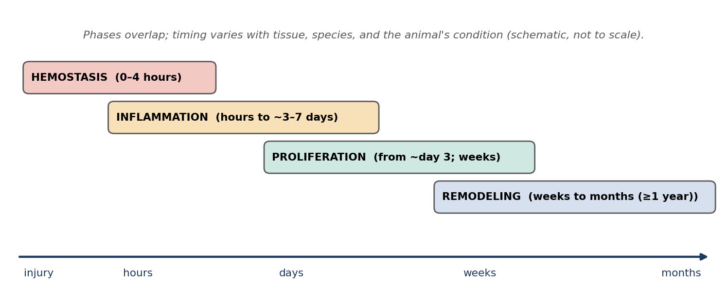
Figure 16.2. The phases of wound healing (schematic timeline).
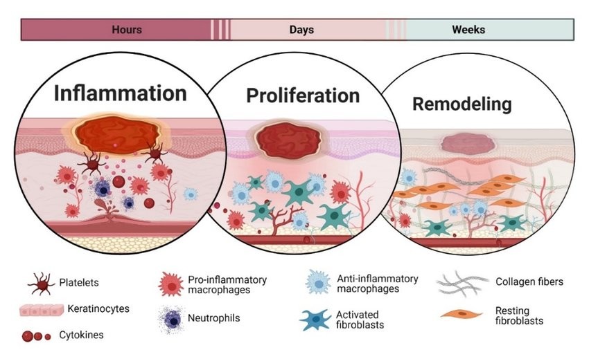
Figure 16.3. The inflammation, proliferation, and remodeling phases, and the key cells involved in each.
1. Hemostasis (0–4 hours). The key components are platelets and clotting factors. Immediately after injury, vessels spasm to limit blood loss; this vasoconstriction lasts 5–10 minutes and is followed by vasodilation. Histamine makes vessels porous, letting leukocytes enter the wound. When blood contacts collagen, platelets become sticky and form a platelet plug, and coagulation produces a fibrin mesh that serves as a scaffold for inflammatory cells. Platelets release substances that start repair, including platelet-derived growth factor (PDGF), transforming growth factor beta (TGF-β), platelet-activating factor, fibronectin, and serotonin. Injured cells release prostaglandins and cytokines that promote inflammation.
2. Inflammation (from hours to about 3–7 days). The key cells are neutrophils and macrophages. Clinically, inflammation produces swelling, redness, warmth, and pain, with an outpouring of tissue fluid and increased blood supply. Neutrophils are the first cells to arrive, attracted by fibronectin, growth factors, and kinins; they peak at 24–48 hours, phagocytose bacteria and debris, and secrete proteases that break down damaged tissue. Macrophages appear at about 72 hours and are the most important regulatory cells of this phase: they phagocytose bacteria and debris; recruit and activate fibroblasts, endothelial cells, and other inflammatory cells; promote angiogenesis (new vessel formation); and promote extracellular matrix synthesis. Epithelial cells may begin migrating across the wound as early as 48 hours. During the acute inflammatory phase the tissue gains no appreciable tensile strength; it depends entirely on the closure material to hold it together.
3. Proliferation (from about day 3, lasting weeks). Cells recruited by macrophage cytokines (fibroblasts, endothelial cells, and keratinocytes) rebuild the tissue in three overlapping processes:
Angiogenesis: endothelial cells proliferate to form a dense new capillary bed that supplies oxygen and nutrients, promoted by growth factors such as TNF-α, TGF-β, and vascular endothelial growth factor (VEGF). Lymphatics re-canalize.
Granulation: fibroblasts, attracted by PDGF and TGF-β, produce granulation tissue, a matrix of adhesive proteins, proteoglycans, glycosaminoglycan gel, collagen, and elastin. Collagen formation begins here, and tensile strength starts to increase rapidly. Some fibroblasts become myofibroblasts, whose actin filaments contract and pull the wound edges together. Wounds closed by primary intention have minimal contraction; skin grafting can be used to reduce contraction in undesirable locations.
Re-epithelialization: keratinocytes proliferate and migrate across the new matrix to cover the wound.
4. Remodeling (maturation; weeks to months). The key cells are fibroblasts and myofibroblasts. Remodeling begins when collagen production and breakdown are balanced (often cited as beginning around day 14). Disorganized collagen fibers are rearranged, cross-linked, and aligned along tension lines, so tensile strength increases even though collagen content stays about constant. Unneeded blood vessels are removed by apoptosis, so the scar grows paler, and contraction continues for weeks to months. Strength continues to improve for a year or more (Figure 16.4).
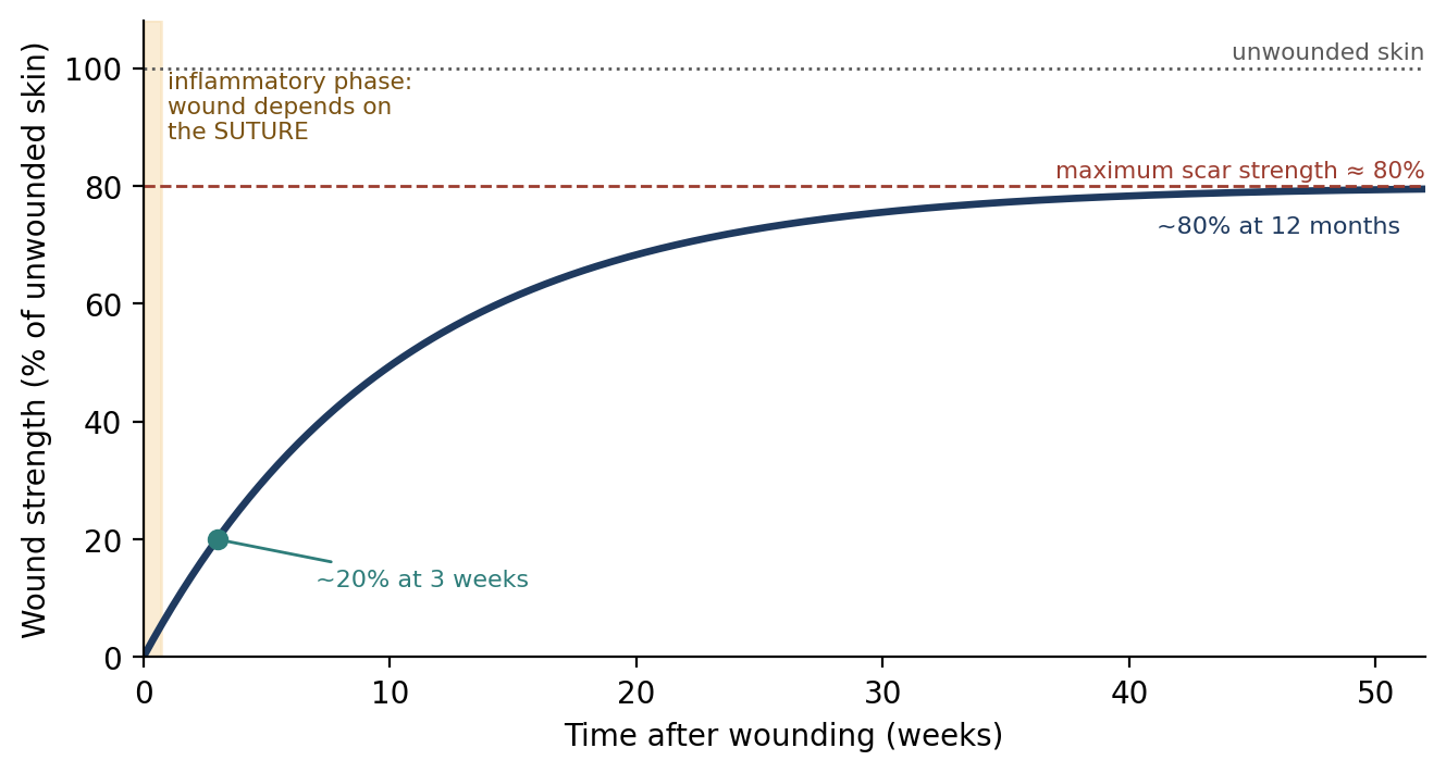
Figure 16.4. Gain in wound strength over time (schematic, based on the values cited in this chapter).
⚠ Check current guidance
Reported timings and strength values differ between references. Values cited here include: inflammation lasting 0–5 days (peaking within 24 hours and subsiding by day 3) or 3–7 days; collagen reaching about 20% of its strength at 3 weeks and 80% at 12 months; and healed skin regaining 70–90% (or a maximum of about 80%) of its original strength. Check which values your exam references use.
16.3 Types of Wound Healing
Table 16.2. Types of wound healing.
| Type | Description | Notes |
|---|---|---|
| First intention (primary union) | Wound edges are brought together during closure at the time of surgery | Fastest; least scar; minimal contraction |
| Second intention | Wound is left open and heals from the inside (bottom) up by granulation and contraction | Used when there is infection, excessive trauma, tissue loss, or imprecise apposition; slower; more granulation and scar tissue; excessive granulation tissue protruding above the surface may need treatment |
| Delayed primary closure (third intention) | Wound is debrided and left open (often packed with gauze changed twice daily) until infection is controlled and healthy red granulation tissue forms, then closed | Used for contaminated and dirty-infected wounds with tissue loss and high infection risk; closure possible in about 3–5 days if no infection; otherwise heals by second intention |
Infected tissue should not be closed, because it may dehisce (break open). Infection is resolved naturally or with topical and systemic treatment before closure.
16.4 Classification of Wounds
Surgical wounds are classified by their degree of contamination, which predicts infection risk (Table 16.3). Infection is generally defined as more than 10⁵ bacteria per gram of tissue. Inflammation is a normal part of healing and must be distinguished from infection, in which bacteria are present and causing damage.
Table 16.3. Classification of surgical wounds.
| Class | Definition | Examples and notes |
|---|---|---|
| Clean | Uninfected operative wound; no inflammation; respiratory, alimentary, genital, or uninfected urinary tracts not entered | Elective incisions under aseptic conditions (the majority of surgical wounds); closed by primary union, usually not drained |
| Clean-contaminated | Respiratory, alimentary, genital, or urinary tract entered under controlled conditions without unusual contamination; or a clean wound contaminated by entering a viscus with minimal spillage, treated at the time | Biliary tract, appendix, vagina, oropharynx; e.g., cholecystectomy, hysterectomy |
| Contaminated | Open traumatic wounds; gross spillage from the GI tract; GU or biliary procedures with infected urine or bile; a major break in aseptic technique | Lacerations, open fractures, penetrating wounds; can become infected within 6 hours |
| Dirty-infected | Heavily contaminated or clinically infected before the operation; an ongoing infection | Perforated viscera, abscesses, neglected traumatic wounds with devitalized tissue or foreign material; infection at surgery can increase infection rates about 4 times |
Antibiotic recommendations depend on wound class. Human surgical references commonly recommend: for clean and clean-contaminated wounds, an IV dose at induction or 30–60 minutes before incision, discontinued at closure or within 24 hours; for contaminated and dirty-infected wounds, an IV dose 30–60 minutes before incision, repeated every 1.5–2 hours during surgery, followed by therapeutic treatment for 2–3 days or longer, depending on reassessment after the third day.
⚠ Check current guidance
These antibiotic guidelines are drawn from human surgical practice. In research animals, antibiotic use must be decided with a veterinarian and approved in the IACUC protocol, because antibiotics may affect study results. Good aseptic technique, not routine antibiotics, is the primary means of preventing infection (Chapter 4).
Wound Complications
Infection. The source should always be determined. Before an infected wound is closed, it should be drained and debrided, and a small opening or drain left in place.
Dehiscence. Failure of the incision line, caused by too much tension on the tissue, improper suturing technique, or improper suture material.
16.5 Wound Closure
Proper closure restores the alignment of tissues and eliminates dead space (Figure 16.1). Several principles apply:
Closing tension: sutures must be tight enough to appose tissue and eliminate dead space, but loose enough to avoid discomfort, ischemia, and necrosis. Post-operative edema will increase tension on the sutures, so allow for it.
Cellular response: any implanted material, including suture, causes a tissue reaction, which can be complicated by infection, allergy, or trauma. Choose closure material that maximizes healing and minimizes infection risk.
Post-operative distraction forces: activity can stress a healing incision; abdominal fascia is strained by coughing, vomiting, urinating, and defecating, and tendons and extremities are under tension during healing. Retention sutures may be needed.
Immobilization: adequate immobilization of the closed wound is necessary for efficient healing and minimal scar formation.
When selecting sutures, consider the amount of tension on the wound, the number of layers of closure, the depth of suture placement, the anticipated edema, and the anticipated timing of suture removal.
16.6 Sutures
Definitions
Table 16.4. Suture terminology.
| Term | Definition |
|---|---|
| Suture | Any strand of material used to ligate blood vessels or approximate tissues |
| Knot tensile strength | The force (in pounds) a suture strand can withstand before breaking when knotted |
| Absorbable suture | Suture that degrades in tissue, losing its tensile strength within 60 days |
| Nonabsorbable suture | Suture that generally maintains its tensile strength for longer than 60 days |
| Ligature (tie) | A suture tied around a vessel to occlude its lumen |
| Free tie (freehand ligature) | A single strand used to ligate a vessel, duct, or other structure, often around a clamp |
| Stick tie (suture ligature, transfixion suture) | A strand attached to a needle, passed through the structure before ligating it, so it cannot slip off |
| Primary suture line | The sutures that hold wound edges in apposition during first-intention healing; may be continuous or interrupted |
| Deep sutures | Placed completely beneath the epidermis |
| Buried sutures | Placed so that the knot lies inside, beneath the layer being closed |
| Purse-string suture | A continuous suture placed around a lumen and tightened like a drawstring to close it |
| Subcuticular sutures | Placed in the dermis, beneath the epithelial layer |
| Secondary suture line | Retention, stay, or tension sutures that reinforce the primary line, eliminate dead space, and prevent fluid accumulation; also used to support second-intention healing or secondary closure after disruption |
| Memory | The tendency of a suture to return to the shape set by its manufacturing or packaging |
16.6.1 The Ideal Suture Material
The ideal suture would be usable in any procedure (varying only in size and strength); sterile; non-electrolytic, non-capillary, non-allergenic, and non-carcinogenic; non-ferromagnetic; easy to handle; minimally reactive and not predisposed to bacterial growth; able to hold a knot securely without fraying or cutting; resistant to shrinking; and absorbed with minimal reaction after serving its purpose. The ideal suture material does not exist. In practice, choose suture with high, uniform tensile strength (permitting finer sizes), good strength retention through the critical healing period followed by rapid absorption, a consistent diameter, sterility, pliability for handling and knot security, freedom from irritants, and predictable performance.
16.6.2 Suture Size
Size denotes the diameter of the strand and is stated in "ought" sizes (Figure 16.5). The more 0s, the smaller the diameter: 5-0 (00000) is smaller than 4-0, 2-0 is larger than 4-0, and 0 is larger than 2-0. Sutures and wires larger than 0 are numbered 1 and up, with larger numbers being larger. From smallest to largest: 7-0, 3-0, 0, 1, 3, 7.
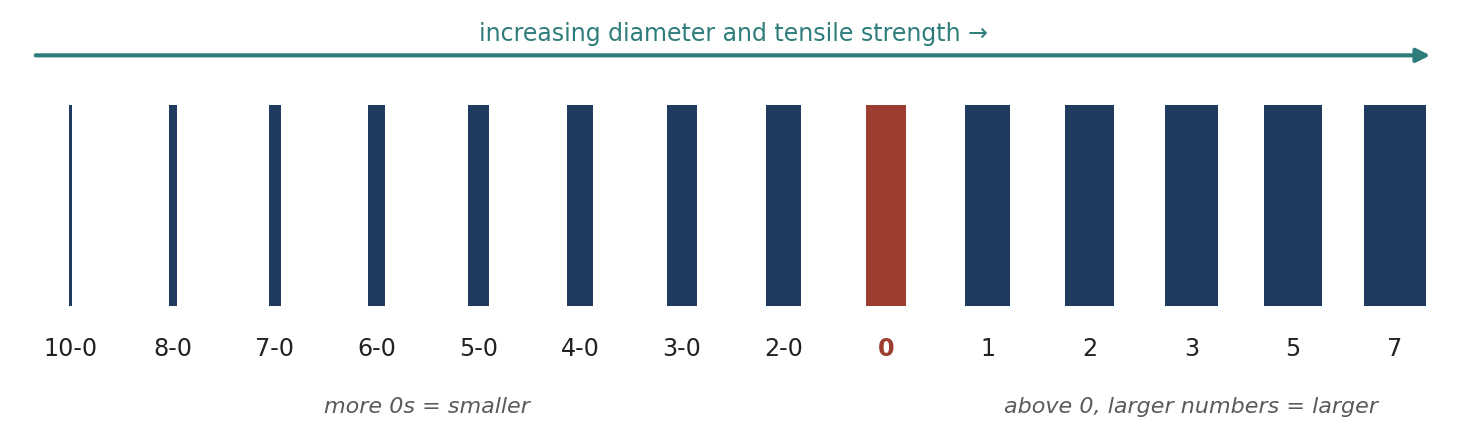
Figure 16.5. Relative suture sizes (schematic; not to scale).
Use the smallest diameter that will adequately hold the tissue: this minimizes trauma and the mass of foreign material left behind. Smaller sutures have less tensile strength, and the tensile strength of the tissue determines the size needed; a suture never needs to be stronger than the tissue it holds.
16.6.3 Monofilament and 16.6.4 Multifilament (Braided) Suture
Table 16.5. Monofilament versus multifilament suture.
| Monofilament | Multifilament (braided or twisted) | |
|---|---|---|
| Structure | A single strand | Several filaments twisted or braided together; may be coated |
| Passage through tissue | Less resistance; passes easily | More drag (less with coating) |
| Infection risk | Resists harboring microorganisms; good for skin (percutaneous) sutures | May harbor microorganisms and wick them along the strand; avoid for percutaneous sutures |
| Strength and handling | More "memory"; knots may slip; easily weakened by crushing or crimping with instruments | Greater tensile strength, pliability, flexibility, and knot security; easier to handle and tie |
| Typical uses | Skin, vascular surgery | Coated multifilaments are well suited for intestinal procedures |
16.6.5 Absorbable Suture
Absorbable sutures hold wound edges together temporarily, until the tissue can withstand normal stress. They are made from collagen from healthy mammals or from synthetic polymers, may be treated to lengthen absorption, and may be coated (for handling) or dyed (for visibility). Natural absorbables (surgical gut) are digested by body enzymes; synthetic absorbables are hydrolyzed, as water penetrates the filaments and breaks the polymer chains, which causes less tissue reaction.
Absorption occurs in two stages: first, tensile strength declines gradually and almost linearly over the first several weeks; second, the suture loses mass as leukocytes remove it. Loss of strength and loss of mass are independent processes. Limitations: fever, infection, or protein deficiency can accelerate absorption and the loss of strength; sutures that get wet before implantation may begin absorbing prematurely; and patients with impaired healing may not be good candidates.
Table 16.6. Absorbable sutures.
| Generic name (trade name) | Structure | Strength retention | Complete absorption | Notes |
|---|---|---|---|---|
| Plain surgical gut | Virtually monofilament collagen (sheep submucosa or beef serosa) | 7–10 days | ~70 days | For rapidly healing tissue needing little support (superficial vessels, subcutaneous fat); heat-treated (fast-absorbing) gut is for 5–7 day epidermal sutures only and not for internal use |
| Chromic gut | As above, treated with chromium salts | 10–14 days (some to 21 days) | > 90 days | Resists enzymes; less early reaction than plain gut; digested by proteolytic enzymes |
| Polyglactin 910 (Coated Vicryl) | Braided; coated | 75% at 2 wk; 50% at 3 wk; 25% at 4 wk; 0% by 5 wk | 56–70 days | Mild reaction; dyed violet or undyed |
| Polyglactin 910 (Vicryl Rapide) | Braided; lower molecular weight | 50% at 5 days; 0% at 10–14 days | 42 days | Fastest-absorbing synthetic; superficial skin and mucosa only; falls off in 7–10 days; not for ligation or ophthalmic, cardiovascular, or neurologic use |
| Polyglactin 910 (Vicryl Plus) | Braided; triclosan-coated | As Coated Vicryl | 56–70 days | Antibacterial coating protects against colonization (e.g., S. aureus, MRSA) |
| Polyglycolic acid (Dexon) | Braided | — | 60–90 days | Homopolymer of glycolide |
| Poliglecaprone 25 (Monocryl) | Monofilament | Undyed: 50–60% at 7 d, 20–30% at 14 d, 0% by 21 d. Dyed: 60–70% at 7 d, 30–40% at 14 d, 0% by 28 d | 91–119 days | Very pliable; virtually inert; subcuticular closure and soft tissue; not for neural, cardiovascular, ophthalmic, or microsurgery |
| Polydioxanone (PDS II) | Monofilament | 70% at 2 wk; 50% at 4 wk; 25% at 6 wk | ~6 months (minimal until day 90) | Extended support; slight reaction; clear or violet |
| Polyglyconate (Maxon) | Monofilament | 81% at 2 wk; 59% at 4 wk; 30% at 6 wk | ~180 days | High initial strength |
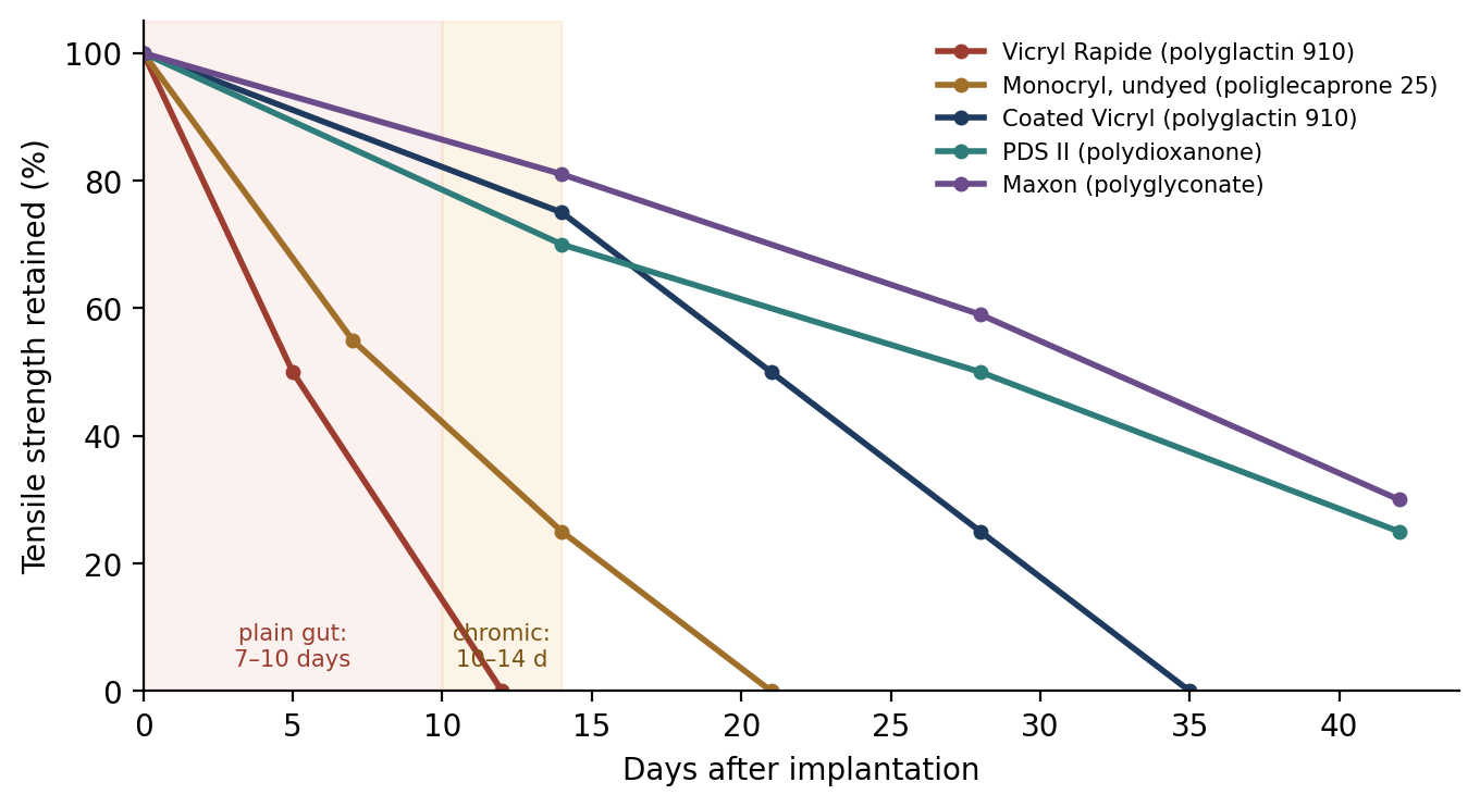
Figure 16.6. Tensile strength retention of common absorbable sutures (plotted from the values in Table 16.6).
⚠ Check current guidance
Strength-retention and absorption figures vary between manufacturers, product versions, sizes, and references, and some sources list different values for the same suture (for example, for PDS II and chromic gut). Use the current package insert for the product you are using, and check which values your exam references cite. Brand names are given for recognition; the exam emphasizes generic (chemical) names.
16.6.6 Nonabsorbable Suture
Nonabsorbable sutures are not digested by enzymes or hydrolyzed; they are made of metal, synthetic, or organic fibers and are ultimately encapsulated by fibroblasts. When used in skin they must be removed. They are used for exterior skin closure; within the body where they will remain permanently; in patients with reactions to absorbables or a tendency to keloids or tissue hypertrophy; and for attaching prostheses (such as defibrillators, pacemakers, and drug delivery devices). The USP classifies them as Class I (silk or synthetic fibers, monofilament, twisted, or braided), Class II (cotton or linen, or coated fibers where the coating adds thickness but not strength), and Class III (metal wire).
Table 16.7. Nonabsorbable sutures.
| Generic name (trade names) | Structure | Key features |
|---|---|---|
| Silk (Perma-Hand) | Braided (or twisted) natural protein (fibroin) | The standard for handling and knot tying; dyed black; loses strength when wet (up to 20%) and if resterilized; lowest tensile strength of the nonabsorbables; loses most strength in ~1 year and is undetectable after ~2 years (behaves as a very slowly absorbed suture); marked tissue reaction |
| Stainless steel (Flexon) | Monofilament or multifilament (316L alloy) | High strength; low reactivity; holds a knot well; used for abdominal wall, sternum, retention, skin, orthopedic, and neurosurgical closure; hard to handle; can cut tissue, kink, fragment, and barb; do not use with prostheses of another alloy (electrolytic reaction); can puncture gloves and skin |
| Nylon (Ethilon, Dermalon: monofilament; Nurolon, Surgilon: braided) | Polyamide; monofilament or braided | High strength, very low reactivity; elastic, so good for retention and skin; monofilament has memory (wet nylon handles better); degrades ~15–20% per year by hydrolysis; braided nylon handles like silk with more strength and less reaction |
| Polyester (Mersilene, Dacron: uncoated; Ethibond, Ti-Cron: coated) | Braided polyethylene terephthalate | Stronger than natural fibers; do not weaken when wet; minimal reaction; lasts indefinitely; uncoated has high friction; polybutilate coating (Ethibond) eases passage; used in cardiovascular surgery and prosthesis placement (available with felt pledgets) |
| Polypropylene (Prolene, Surgilene) | Monofilament | Glides through tissue and does not adhere (good pull-out suture); relatively inert; not degraded by enzymes; slippery, so it needs additional throws for knot security; widely used in vascular, plastic, and external sutures |
| Poly(hexafluoropropylene-VDF) (Pronova) | Monofilament | Resists involvement in infection; does not adhere; cardiovascular, ophthalmic, and neurologic use |
| Polybutester (Novafil) | Monofilament | Minimal reaction |
| Polymerized caprolactam (Supramid, Braunamid, Vetafil) | Multifilament in a coating | Used in veterinary skin closure; tissue reaction if the coating breaks; should not be buried |
⚠ Check current guidance
Several brand names in older references are misspelled or discontinued (for example, "Dermal on," "Merilee," and "Neuron" for Dermalon, Mersilene, and Nurolon). Product lines change; check current catalogs.
Suture Selection
When a wound has reached maximal strength, sutures are no longer needed. Slow-healing tissues (skin, fascia, tendon) are usually closed with nonabsorbable suture or absorbable suture with extended support (up to 6 months). Rapidly healing tissues (stomach, colon, bladder) may be closed with absorbable suture.
Foreign bodies in potentially contaminated tissue may convert contamination into infection, so avoid braided and reactive materials there.
Where cosmetic results matter, use the smallest inert monofilament (nylon or polypropylene), close subcuticularly where possible, and consider tissue adhesive or skin closure tape.
In the urinary and biliary tracts, foreign material can become a nidus for stone formation, so use rapidly absorbed suture.
Use the finest size consistent with the tissue's strength, and reinforce with retention sutures if sudden strain on the line is expected, removing them once the patient is stable.
Suture Storage and Handling
Absorbable sutures: store at room temperature, away from heat (such as steam pipes and sterilizers); keep synthetic absorbables dry and use them directly from the packet; do not soak them. Surgical gut may be dipped briefly in room-temperature water or saline to restore pliability.
All sutures: straighten with a gentle, steady pull (jerking weakens them); do not "test" their strength; avoid crushing or crimping with instruments except at the free end; and do not resterilize (except stainless steel, which can be steam sterilized, but not on a wooden spool).
Silk is stronger dry than wet; nylon kinks can be straightened by gently running the strand between gloved fingers; polyester and polypropylene are unaffected by moisture.
16.6.7 Needles
The best surgical needles are made of high-quality stainless steel; are as slim as possible without sacrificing strength; are stable in the needle holder; carry suture through tissue with minimal trauma; are sharp, rigid enough to resist bending, and ductile enough to resist breaking; and are sterile and corrosion-resistant. Needle strength is its resistance to deformation during repeated passes; the surgical yield point is the bending a needle can withstand before it is permanently deformed; and ductility is its resistance to breaking when bent.
Every needle has three parts: the eye (swage), the body, and the point (Figure 16.7). Its size is described by the chord length (straight-line distance from point to swage), needle length (distance along the needle), radius (of the circle its curve would complete), and diameter (gauge of the wire) (Figure 16.8).
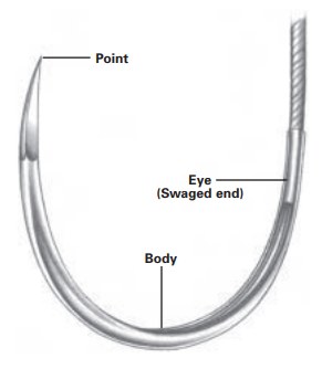
Figure 16.7. Parts of a surgical needle.
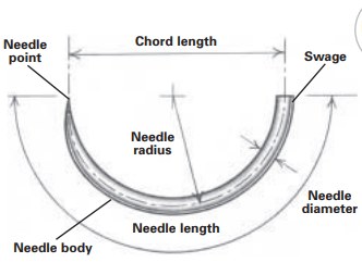
Figure 16.8. Needle dimensions: chord length, needle length, radius, and diameter.
The eye may be closed (like a sewing needle), French (split, with a slit that the suture is pressed into), or swaged (eyeless), in which needle and suture form one continuous unit (Figures 16.9–16.11). Almost all needles today are swaged, which reduces tissue trauma (the strand is no thicker than the needle), prevents premature unthreading, saves threading and sterilizing time, and eliminates fraying from the sharp corners of an eye. The body, the part grasped by the needle holder, should be as close as possible to the diameter of the suture.
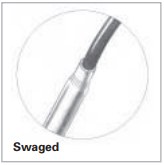
Figure 16.9. Swaged (eyeless) needle.
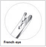
Figure 16.10. French (split) eye.
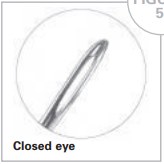
Figure 16.11. Closed eye.
Needles come in a range of curvatures (quarter, 3/8, half, and 5/8 circle, plus straight) and point types (Figure 16.12 and Table 16.8). Select the needle's length, diameter, and curvature for where the suture must go and the space available: deep or confined spaces need tighter curves. Taper points are used for tissues that are easy to penetrate, and cutting needles for tough tissue; if unsure, use a taper point for everything but skin.
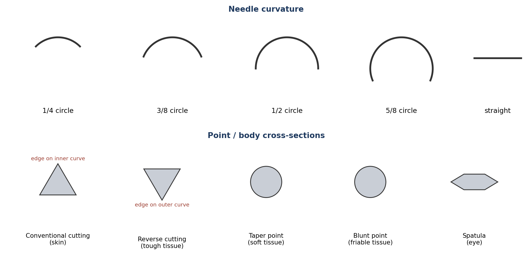
Figure 16.12. Needle curvatures and point (body) cross-sections (schematic).
Table 16.8. Needle point types.
| Type | Design | Use |
|---|---|---|
| Conventional cutting | Triangular body with a cutting edge on the inside (concave) curve | Primarily skin closure |
| Reverse cutting | Triangular body with the cutting edge on the outside (convex) curve; less likely to cut out of tissue | Tough, difficult-to-penetrate tissue |
| Taper point | Round body tapering smoothly to a point; spreads rather than cuts tissue | Soft, easily penetrated tissue (viscera, vessels, fascia) |
| Blunt point | Taper body with a rounded, blunt tip | Friable tissue (such as liver and kidney); blunt dissection; reduces needlestick risk |
| Spatula | Flat on top and bottom, with cutting edges at the sides | Primarily eye surgery |
16.7 Knot Tying
A knot is the weakest point of a suture line, so knot technique matters. General principles:
The completed knot must be firm and tied so that slipping is virtually impossible; the simplest secure knot for the material is best.
Keep knots as small as possible to limit tissue reaction, and cut the ends as short as possible (about 3 mm for most sutures; about 6 mm for gut).
Avoid "sawing" (friction between strands) as throws are cinched down; it weakens the suture.
Avoid crushing or crimping the strand with instruments, except at the free end.
Avoid excessive tension, which breaks suture and cuts tissue; practicing gentle tension allows finer sutures to be used.
Do not tie approximating sutures too tightly, which can strangulate tissue and cause necrosis.
After the first throw, maintain traction on one end to stop the throw loosening, if the tissue is under tension.
Keep the final tension as horizontal as possible and lay throws flat; change your stance if needed to do so.
Extra throws do not add strength to a properly tied, squared knot, only bulk; however, some synthetic materials need additional throws for knot security.
16.7.1 Common Knots
Square knot: the easiest and most reliable two-handed tie for surgical gut, silk, and stainless steel; other materials need additional throws. If the strands are crossed incorrectly, a granny knot results, which tends to slip under stress.
Surgeon's knot: an initial double throw (which resists slipping while the second throw is placed), followed by one or two single throws, is generally sufficient. The exception is nylon monofilament, for which two successive double throws help prevent slippage.
[ PHOTO PLACEHOLDER ] Suggested source: Photograph knots tied in your training lab, or use a published knot-tying manual with permission. |
|---|
Figure 16.13. Square knot, granny knot, and surgeon's knot, showing the direction of each throw.
16.8 Suture Patterns
Suture patterns are either interrupted (each stitch tied separately) or continuous (one strand with a knot at each end). They also differ in how they position the wound edges: appositional patterns bring edges together flush, everting patterns turn them outward, and inverting patterns turn them inward (used to seal hollow organs). Tension patterns spread the load to resist pull-through.
Why it matters
Interrupted patterns are more secure (one failed stitch does not open the whole wound) but slower and use more suture. Continuous patterns are faster, use less suture, and seal better, but the entire line depends on two knots and one strand. Choose based on tension, contamination, and the tissue.
16.8.1 Interrupted Sutures
Ligatures occlude the lumen of a vessel or duct to achieve hemostasis or prevent leakage. In a free tie, the structure is clamped, the strand is passed around it beneath the tip of the clamp, the first throw is placed, the clamp is removed as the knot is tightened, and further throws are added to square and secure the knot.
Figure 16.14. Simple Interrupted |
Simple Interrupted
|
|---|---|
Figure 16.15. Horizontal Mattress |
Horizontal Mattress
|
Figure 16.16. Vertical Mattress
Figure 16.17. Vertical mattress: correct apposition (A) versus excessive eversion (B) |
Vertical Mattress
|
Figure 16.18. Cross Mattress (Cruciate) |
Cross Mattress (Cruciate)
|
Figure 16.19. Gambee |
Gambee
|
Figure 16.20. Quilled |
Quilled
|
Figure 16.21. Far-Far–Near-Near |
Far-Far–Near-Near
|
Figure 16.22. Near-Far–Far-Near |
Near-Far–Far-Near
|
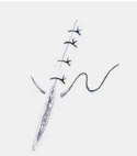

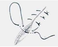
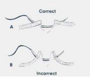
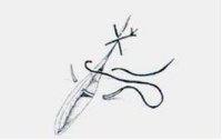
16.8.2 Continuous Sutures
Figure 16.23. Simple Continuous |
Simple Continuous
|
|---|---|
Figure 16.24. Running (Whip) Stitch |
Running (Whip) Stitch
|
Figure 16.25. Ford Interlocking |
Ford Interlocking
|
Figure 16.26. Lembert |
Lembert
|
Figure 16.27. Halsted |
Halsted
|
Figure 16.28. Connell |
Connell
|
Figure 16.29. Cushing |
Cushing
|
Figure 16.30. Parker-Kerr: first (Cushing) layer
Figure 16.31. Parker-Kerr: inversion with the second (Lembert) layer |
Parker-Kerr
|
Figure 16.32. Guard |
Guard
|
Figure 16.33. Continuous Everting Mattress |
Continuous Everting Mattress
|
Figure 16.34. Subcuticular |
Subcuticular
|
Figure 16.35. Subcutaneous |
Subcutaneous
|
Figure 16.36. Bunnell |
Bunnell
|
Figure 16.37. Modified Bunnell |
Modified Bunnell
|
Figure 16.38. Cerclage Wiring |
Cerclage Wiring
|
Figure 16.39. Cerclage (left) and hemicerclage (right), cross-sections (schematic) |
Hemicerclage
|
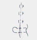
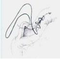
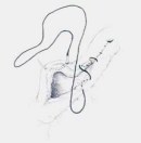
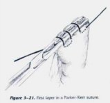
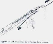
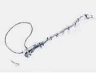
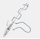
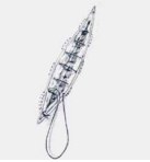
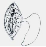
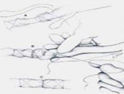
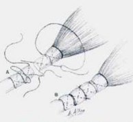
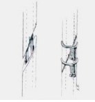
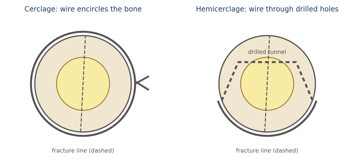
16.8.3 Suture Patterns for Specific Tissues
Table 16.9. Suture patterns commonly used for specific tissues.
| Tissue | Patterns |
|---|---|
| Skin | Simple interrupted, horizontal mattress, vertical mattress, continuous appositional or everting patterns; subcuticular |
| Subcutaneous tissue | Simple continuous |
| Fascia | Simple continuous (primary), simple interrupted, vertical mattress, far-near–near-far |
| Peritoneum | Simple continuous (two layers) and simple interrupted; in the horse it is very thin and fragile, so close the muscle instead |
| Vessels | Simple interrupted and simple continuous |
| Viscera | Inverting patterns such as Cushing, Lembert, and Connell; appositional patterns such as Gambee |
| Muscle | Simple continuous, simple interrupted, horizontal mattress |
| Tendon | Bunnell and modified Bunnell |
| Bone | Cerclage and hemicerclage |
⚠ Check current guidance
Image credits: Figure 16.3 and the suture pattern and needle illustrations in this chapter were supplied from compiled study materials and appear to come from published sources (for example, surgical textbooks and the Ethicon Wound Closure Manual). Before this textbook is printed for distribution, shared, or published, confirm the source of each image, obtain permission where required, and add a credit line. The hemicerclage illustration supplied was identical to the cerclage illustration, so an original diagram is used instead.
Chapter Summary
Good technique minimizes time, trash, and trauma: plan adequate incisions (wounds heal side to side), cut cleanly, handle and retract gently, avoid crushing, keep tissue moist, remove contamination, control bleeding with pressure, patience, and perseverance, and eliminate dead space. Wounds heal through hemostasis, inflammation (neutrophils, then macrophages; strength depends on the suture), proliferation (angiogenesis, granulation with collagen, epithelialization), and remodeling (cross-linking and alignment over months, to about 70–90% of original strength). Healing is by first intention, second intention, or delayed primary closure, and wounds are classed as clean, clean-contaminated, contaminated, or dirty-infected.
Sutures are sized by diameter (more 0s = smaller), may be monofilament (smooth, resists infection, has memory) or multifilament (strong, easy to tie, may wick bacteria), and absorbable (gut by enzymes; synthetics by hydrolysis) or nonabsorbable (silk, steel, nylon, polyester, polypropylene). Needles are swaged, curved to suit the space, and have cutting, reverse cutting, taper, blunt, or spatula points. Knots should be flat, square, small, and secure. Interrupted patterns are secure; continuous patterns are fast and seal well; inverting patterns (Lembert, Cushing, Connell) close hollow viscera; and specialized patterns serve tension, tendon, and bone repair.
Key Terms
Apposition: Bringing tissue edges together in their normal alignment.
Dead space: An open area within closed tissue.
Dehiscence: Breakdown (opening) of a closed incision.
First intention: Healing of a wound closed at the time of surgery.
Granulation tissue: New vascular connective tissue formed during the proliferative phase.
Hydrolysis: Breakdown by water; how synthetic absorbable sutures degrade.
Inverting pattern: A suture pattern that turns tissue edges inward (e.g., Lembert, Cushing).
Ligature: A suture tied around a vessel or duct to occlude it.
Memory: A suture's tendency to return to its packaged shape.
Myofibroblast: A contractile fibroblast that pulls wound edges together.
Second intention: Healing of an open wound by granulation and contraction.
Seroma: A collection of serum in dead space.
Swaged needle: An eyeless needle permanently attached to its suture.
Tensile strength: Load per unit cross-sectional area at the point of rupture.
Third intention: Delayed primary closure after a period of open healing.
Review Questions
1. The three "T's" of tissue handling are:
A. Time, trash, and trauma
B. Tension, tightness, and technique
C. Temperature, tone, and tissue
D. Tape, ties, and thread
2. Which phase of wound healing involves neutrophils peaking at 24–48 hours and macrophages arriving at about 72 hours?
A. Hemostasis
B. Inflammation
C. Proliferation
D. Remodeling
3. During the inflammatory phase, the wound's strength depends on:
A. Collagen cross-links
B. The closure material (suture)
C. Granulation tissue
D. The epidermis
4. A wound left open that heals by granulation and contraction is healing by:
A. First intention
B. Second intention
C. Third intention
D. Primary union
5. A surgical wound in which the gastrointestinal tract is entered under controlled conditions without unusual contamination is classified as:
A. Clean
B. Clean-contaminated
C. Contaminated
D. Dirty-infected
6. Which suture size is the smallest?
A. 2-0
B. 0
C. 5-0
D. 2
7. Why should braided suture not be used for percutaneous (skin) sutures?
A. It is too weak
B. It may harbor microorganisms and wick them into the wound
C. It cannot be knotted
D. It is absorbed too quickly
8. Synthetic absorbable sutures are broken down by:
A. Proteolytic enzymes
B. Hydrolysis
C. Phagocytosis only
D. They are not broken down
9. Which needle has its cutting edge on the outer curve and is used for tough, difficult-to-penetrate tissue?
A. Conventional cutting
B. Reverse cutting
C. Taper point
D. Blunt point
10. Which pattern is a modified Connell in which the needle does not enter the lumen?
A. Lembert
B. Cushing
C. Ford interlocking
D. Gambee
Answer Key
1. A. Keep time to a minimum, remove existing contamination (trash) and prevent new contamination, and keep trauma to a minimum.
2. B. Neutrophils are the first cells to infiltrate the wound; macrophages, the key regulatory cells of the inflammatory phase, arrive at about 72 hours.
3. B. The tissue gains no appreciable tensile strength during the acute inflammatory phase and depends on the suture to hold it together.
4. B. Second-intention healing occurs from the inside out by granulation and contraction. Delayed primary closure is also called third intention.
5. B. Entry into the respiratory, alimentary, genital, or urinary tract under controlled conditions makes a wound clean-contaminated.
6. C. The more 0s, the smaller the diameter: 5-0 is smaller than 2-0, which is smaller than 0. Above 0, larger numbers are larger.
7. B. The interstices of braided suture can harbor bacteria and wick them along the strand into the tissue.
8. B. Synthetic absorbables are hydrolyzed, causing less tissue reaction than natural absorbables (surgical gut), which are digested by enzymes.
9. B. A reverse cutting needle has its cutting edge on the outer (convex) curve. A conventional cutting needle has it on the inner curve; taper points are for soft tissue.
10. B. The Cushing pattern is a modified Connell that does not enter the lumen, giving a better fluid-tight seal.
Chapter 17: Endoscopic Procedures
Learning objectives
After studying this chapter, you should be able to:
Define endoscopy and minimally invasive surgery, and describe their advantages, requirements, and planning.
Describe endoscopes and the video, light, insufflation, electrosurgical, and access equipment used in endoscopic surgery.
Explain how insufflation affects respiratory, acid–base, and cardiovascular physiology.
Describe anesthetic management for laparoscopic and thoracoscopic procedures, including one-lung ventilation.
17.1 General
Endoscopy means "to look inside." It is used to examine the interior of hollow viscera, such as the bronchi and intestinal tract, and the term is also applied to laparoscopy (the abdomen), arthroscopy (joints), and thoracoscopy (the chest). Endoscopic surgery is also known as minimally invasive surgery (MIS): surgical techniques designed to minimize the anatomical approach to the target site, working through small incisions rather than one large one.
Compared with open surgery, MIS generally causes less tissue trauma and pain, smaller wounds, and faster recovery, which benefits animal welfare and can reduce variability in research data. Its disadvantages include the cost of equipment, a two-dimensional view on a monitor, reduced tactile feedback, a long learning curve, and specific complications such as injury to organs or vessels during port placement and problems caused by the insufflation gas.
Endoscopic surgery requires extensive equipment, training, and practice, not only for the operating team but also for preparation and especially anesthesia personnel. Everyone must know their role and be able to perform it without supervision. It also requires careful preparation:
Open surgical packs should be readily available in case the procedure must be converted to an open procedure (for example, because of bleeding or poor visibility).
The room layout must be planned in advance for a smooth workflow without obstructions, with cables and tubing routed safely.
The surgeon(s) and camera operator need an easy, direct view of the monitor, usually placed opposite the surgeon, in line with the target.
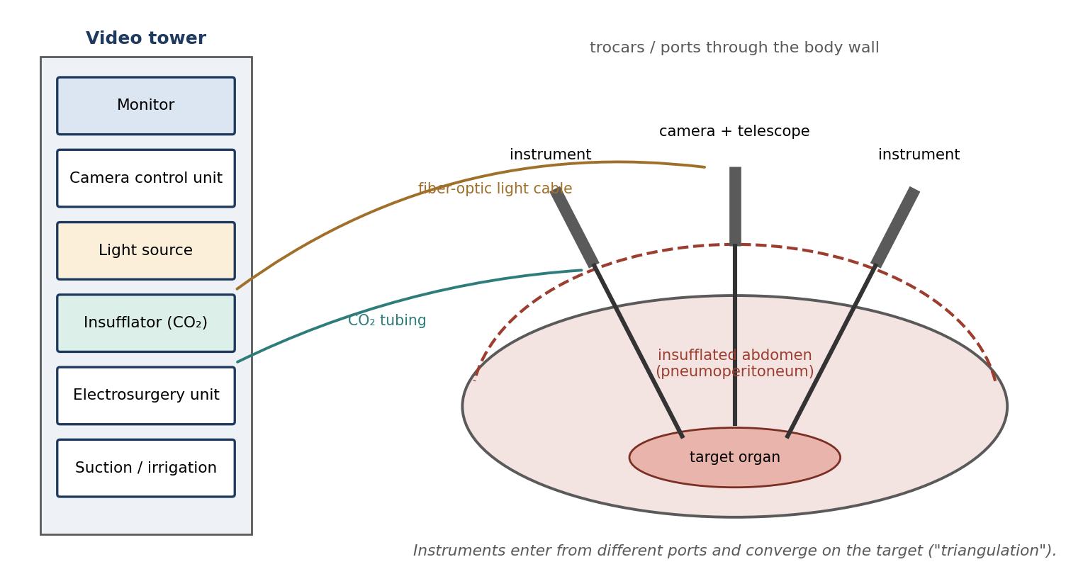
Figure 17.1. Typical laparoscopic setup: video tower, insufflated abdomen, and ports for the telescope and instruments (schematic).
17.2 Endoscopic Equipment
Endoscopes
Endoscopes may be flexible or rigid. Flexible endoscopes are usually used to examine the gastrointestinal and respiratory tracts, where they must follow curves. Rigid endoscopes (telescopes) are more commonly used for surgery because they provide the most light, the largest field of view, and the greatest resolution and clarity. Operating endoscopes are rigid scopes with working channels through which instruments such as biopsy forceps can be passed.
Larger scopes transmit more light and give a better field of view: 10 mm laparoscopes are preferred for most procedures, while scopes smaller than 5 mm are ideal for diagnostic laparoscopy and thoracoscopy, and for small animals. The viewing angle may be straight ahead (0°) or angled (for example, 30° or 70°), which lets the operator see around structures by rotating the scope (Figure 17.2). The field of vision is the area visible through the scope; the closer the tip is to the tissue, the greater the magnification (and the smaller the field). The camera operator may need to adjust the focus as the distance changes.
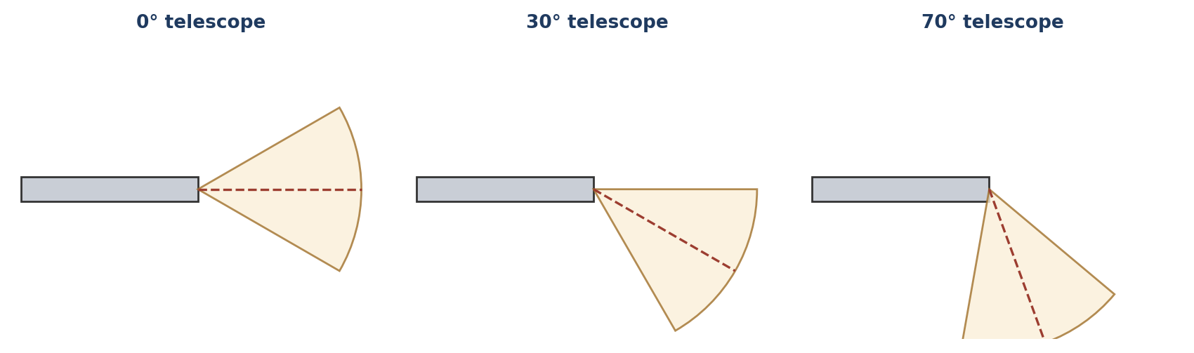
Figure 17.2. Telescope viewing angles. Angled scopes show a field offset from the shaft axis, and rotating the scope sweeps the view around.
Table 17.1. Comparison of flexible and rigid endoscopes.
| Flexible | Rigid (telescope) | |
|---|---|---|
| Typical use | Examination of the GI and respiratory tracts | Laparoscopy, thoracoscopy, arthroscopy; most surgical endoscopy |
| Light, field, and resolution | Less | Most light, largest field, greatest resolution |
| Working channels | Often present | Present in operating endoscopes |
| Sizes | Various | 10 mm for most procedures; < 5 mm for diagnostic work and small patients |
An endoscope must be attached to a camera, camera control unit, and monitor. Some older endoscopes can be looked through directly by eye like a telescope, but this is poorly suited to much more than biopsies. Endoscopic surgical instruments look identical to standard instruments at the working end, but are mounted on a long shaft that passes through a trocar or port.
[ PHOTO PLACEHOLDER ] Suggested source: Wikimedia Commons (commons.wikimedia.org), search "laparoscope" or "laparoscopic tower." Check the image's license and give the attribution it requires. |
|---|
Figure 17.3. A rigid telescope (laparoscope) with its light cable connection, and a laparoscopic video tower.
Video and Light Equipment
Light sources: older systems use tungsten bulbs; newer systems use mercury, xenon, or halogen bulbs and are much brighter. Brightness must be adjustable to avoid washing out the image. Light travels to the scope through a fiber-optic cable, which should be kept clean at both ends and replaced when enough fibers break (commonly about 20%).
Cameras use either one or three chips to convert the image to an electronic signal. A single-chip camera offers about 450 lines of resolution; a three-chip camera uses separate chips for red, green, and blue and offers 600–700 lines. Better systems avoid "whiteout" from glare off pale tissues.
The camera control unit adjusts the image (focus, sharpness, and so on) and transmits it to the monitor.
Monitors offer 400–700 lines of resolution, should be matched to the camera, and must be medical grade and properly grounded. Images may be recorded.
⚠ Check current guidance
Video and lighting technology has advanced considerably: modern systems typically use high-definition (or 4K) digital cameras, LED light sources, and digital recording rather than VHS. The principles (adequate, adjustable light; matched camera and monitor; grounded, medical-grade equipment) still apply.
Insufflation, Electrosurgery, and Irrigation
The abdominal cavity is usually inflated with an inert gas, commonly carbon dioxide (or nitrogen), creating a pneumoperitoneum that lifts the body wall away from the organs and gives room to work. Without it, the cavity collapses and surgery becomes difficult. The insufflator, which may be electronic or mechanical, delivers gas to maintain a constant set pressure in the cavity. CO₂ is widely used because it is non-flammable (so electrosurgery can be used safely) and highly soluble in blood, which reduces the risk from gas embolism, but its absorption raises PaCO₂ (Section 17.3).
Electrosurgery is used for hemostasis and cutting; ultrasonic or laser devices may also be used (Chapter 15). Irrigation and suction, commonly combined in one device, keep the field clean and visible.
Access Equipment
Insufflation needles (such as the Veress needle) create a sealed opening into the abdominal cavity through which gas is delivered. A spring-loaded blunt stylet advances once the sharp needle enters the cavity, protecting the organs. They may be disposable or reusable.
Trocars consist of an obturator (a pointed or blunt inner rod) enclosed in a sleeve (cannula). They are used to create ports for instruments and materials. After placement the obturator is removed, leaving the cannula.
Ports hold an incision open to allow endoscopic instruments to pass into the body cavity. Ports may be placed by an open technique (a small cut-down under direct vision) rather than with a trocar. This is more time-consuming but useful when adhesions may be present or when organs such as the rumen must be avoided.
Reducers act as additional gaskets inside or on top of the trocar so that smaller instruments can pass without gas escaping, maintaining the pneumoperitoneum.
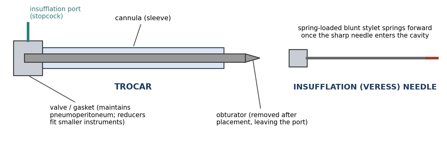
Figure 17.4. A trocar (cannula, obturator, valve, and insufflation port) and an insufflation (Veress) needle (schematic).
Why it matters
Port placement is the most dangerous step of laparoscopy, because the first entry into the abdomen is made blind. Blunt-tipped needles and trocars, open placement, and lifting the body wall all reduce the risk of puncturing a viscus or large vessel. Once the camera is in, every later port can be placed under direct vision.
17.3 Anesthesia for Endoscopy & Endosurgery
Anesthesia for endoscopic surgery differs from standard anesthesia mainly because of the increased cavity pressure caused by insufflation (Figure 17.5). Thoracic procedures usually do not use insufflation, because collapsing one lung and the rigid rib cage already provide an open working space.
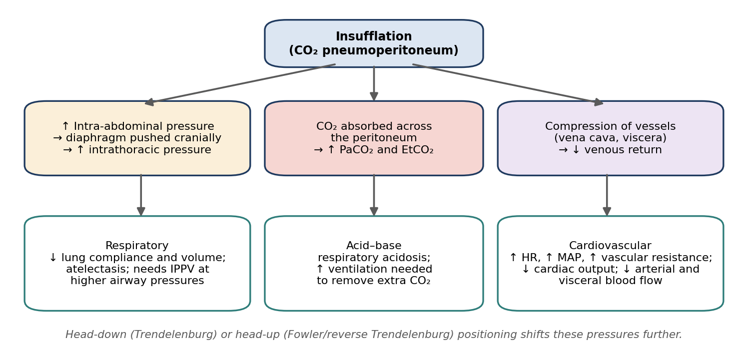
Figure 17.5. Physiological effects of CO₂ pneumoperitoneum.
Effects of Abdominal Insufflation
Respiratory effects. Insufflation raises intra-abdominal pressure (IAP) and pushes the diaphragm into the chest cavity. This increases intrathoracic pressure, reduces lung volume and compliance, and promotes atelectasis, so mechanical ventilation at higher pressures than normal is required.
Carbon dioxide. When the insufflation gas is CO₂, it is absorbed across the peritoneum, so arterial CO₂ (PaCO₂) rises. Ventilation must be increased to remove the extra CO₂ and prevent respiratory acidosis, guided by EtCO₂ and blood gases (Chapter 12). Hypercapnia, together with reduced lung volume, can also be accompanied by a fall in PaO₂, reducing oxygen delivery.
Cardiovascular effects. Raised intra-abdominal pressure and CO₂ absorption increase heart rate, mean arterial pressure (MAP), and vascular resistance, while compression of the vena cava and abdominal vessels decreases venous return, cardiac output, and arterial and visceral blood flow.
Positioning. Placing the animal in Trendelenburg (head-down) or Fowler (head-up, reverse Trendelenburg) position shifts pressures in the abdomen and chest further. Head-down positioning moves the abdominal organs toward the diaphragm, further restricting ventilation; head-up positioning can reduce venous return.
Table 17.2. Summary of the effects of abdominal insufflation.
| System | Effects | Anesthetic response |
|---|---|---|
| Respiratory | ↑ IAP → diaphragm displaced → ↑ intrathoracic pressure; ↓ lung volume and compliance | Intubate; mechanical ventilation at higher pressures |
| Acid–base | CO₂ absorbed → ↑ PaCO₂ and EtCO₂; respiratory acidosis | Increase ventilation; monitor EtCO₂ and blood gases |
| Cardiovascular | ↑ HR, ↑ MAP, ↑ vascular resistance; ↓ venous return, cardiac output, and arterial and visceral blood flow | Monitor BP and ECG; adequate fluid support; use the lowest effective insufflation pressure |
| Positioning | Trendelenburg or Fowler position shifts abdominal and thoracic pressures | Adjust ventilation and monitoring accordingly |
⚠ Check current guidance
Recommended insufflation pressures vary with species, body size, and procedure; using the lowest pressure that gives adequate working space minimizes cardiovascular and respiratory effects. Check current references and your institution's protocols for target pressures.
Thoracoscopy and One-Lung Ventilation
For some procedures, extra working space is needed in the chest, and inert gas is introduced to expand the cavity. This can be dangerous because it limits lung capacity, but only a relatively small pressure increase (about 5 mmHg) is needed to clear space. EtCO₂, MAP, heart rate, and cardiac output will increase.
Thoracoscopy usually requires single-lung (one-lung) ventilation, in which the lung on the operated side is collapsed while the other lung is ventilated. This requires specialized endotracheal tubes (such as double-lumen tubes or bronchial blockers) that isolate the two lungs (Figure 17.6).
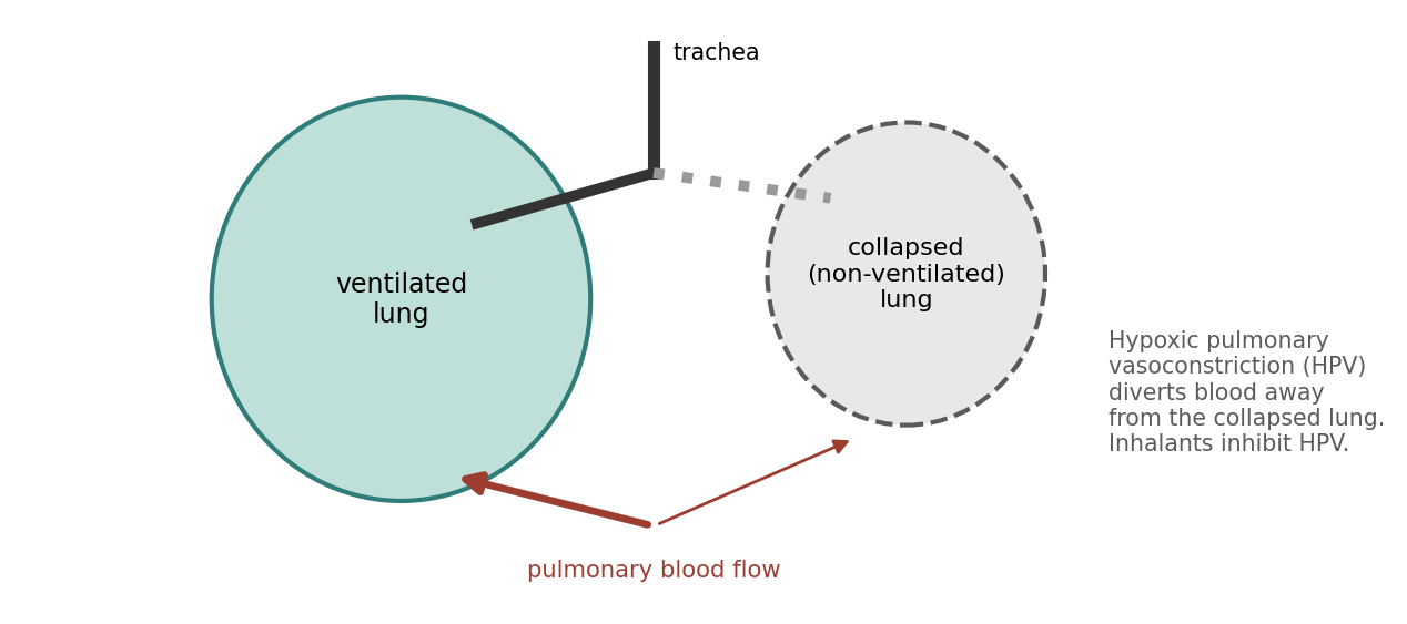
Figure 17.6. One-lung ventilation and hypoxic pulmonary vasoconstriction (schematic).
In lateral recumbency, the lower (dependent) lung receives about 60% of the cardiac output, because gravity favors blood flow to it.
Hypoxic pulmonary vasoconstriction (HPV) is the lung's own protective reflex: blood vessels supplying poorly ventilated (atelectatic) lung constrict, diverting blood flow to the ventilated lung and improving oxygenation.
Inhalation anesthetics inhibit HPV, whereas injectable barbiturates commonly do not. This is one reason oxygenation can be harder to maintain during one-lung ventilation under inhalant anesthesia.
Because insufflation maintains a constant pressure, liquids or inert gases may also be infused between tissues to separate them (hydrodissection or gas dissection), a technique used to open tissue planes in some endoscopic procedures.
Why it matters
During endoscopic procedures, the drapes, darkened room, and focus on the monitor make the animal especially easy to overlook. Capnography, pulse oximetry, blood pressure, and ECG monitoring are essential, because the physiological changes caused by insufflation and one-lung ventilation can develop quickly.
Chapter Summary
Endoscopy ("to look inside") underlies minimally invasive surgery: laparoscopy, thoracoscopy, and arthroscopy. MIS reduces trauma and speeds recovery but requires extensive equipment, trained teams (including anesthesia), planned room layout, a clear view of the monitor, and open packs ready for conversion. Rigid telescopes (most light, largest field, best resolution; 10 mm for most procedures, < 5 mm for diagnostic work) and flexible scopes (GI and airway) connect to a camera, control unit, and medical-grade monitor, lit through a fiber-optic cable from a bright, adjustable source. An insufflator maintains a CO₂ pneumoperitoneum; insufflation needles, trocars, ports, and reducers provide sealed access.
Insufflation raises intra-abdominal and intrathoracic pressure, raises PaCO₂ through absorption, increases heart rate, MAP, and vascular resistance, and decreases cardiac output and visceral blood flow, so animals need intubation, mechanical ventilation at higher pressures, increased ventilation, and careful monitoring. Thoracoscopy relies on one-lung ventilation with specialized tubes; the dependent lung receives about 60% of cardiac output, and hypoxic pulmonary vasoconstriction (inhibited by inhalants) diverts blood from the collapsed lung.
Key Terms
Arthroscopy: Endoscopic examination or surgery of a joint.
Endoscopy: "To look inside"; examination of body interiors with an endoscope.
Hypoxic pulmonary vasoconstriction (HPV): Constriction of pulmonary vessels in poorly ventilated lung, diverting blood to ventilated lung.
Insufflation: Inflation of a body cavity with gas.
Laparoscopy: Endoscopic examination or surgery of the abdomen.
Minimally invasive surgery (MIS): Surgery performed through small incisions using endoscopic techniques.
Obturator: The inner rod of a trocar, removed after placement.
One-lung ventilation: Ventilating one lung while the other is collapsed, for thoracic access.
Pneumoperitoneum: Gas within the peritoneal cavity.
Port: A device holding an incision open for passage of endoscopic instruments.
Reducer: A gasket that lets smaller instruments pass through a larger port without gas leakage.
Thoracoscopy: Endoscopic examination or surgery of the chest.
Trendelenburg position: Head-down tilt; reverse Trendelenburg (Fowler) is head-up.
Trocar: An obturator within a cannula, used to create a port.
Review Questions
1. Endoscopy literally means:
A. To cut inside
B. To look inside
C. To inflate
D. To repair
2. Compared with flexible endoscopes, rigid endoscopes (telescopes):
A. Provide less light and a smaller field
B. Provide the most light, the largest field, and the greatest resolution
C. Are used mainly in the GI tract
D. Cannot be attached to a camera
3. Bringing the tip of the endoscope closer to the tissue:
A. Decreases magnification
B. Increases magnification
C. Has no effect
D. Increases the field of view
4. The gas most commonly used to insufflate the abdomen is:
A. Oxygen
B. Nitrous oxide
C. Carbon dioxide
D. Room air
5. A trocar consists of:
A. A camera and light cable
B. An obturator enclosed in a sleeve (cannula)
C. A needle and syringe
D. A retractor and clamp
6. When might ports be placed by an open technique rather than with trocars?
A. Always, because it is faster
B. When adhesions may be present or organs such as the rumen must be avoided
C. Only in rodents
D. Only for thoracoscopy
7. Increased intra-abdominal pressure from insufflation typically:
A. Decreases intrathoracic pressure
B. Increases intrathoracic pressure and requires higher ventilation pressures
C. Increases cardiac output
D. Has no effect on breathing
8. Why does PaCO₂ rise during CO₂ insufflation?
A. The animal stops breathing
B. CO₂ is absorbed across the peritoneum into the blood
C. Soda lime releases CO₂
D. Oxygen is converted to CO₂
9. During one-lung ventilation, hypoxic pulmonary vasoconstriction (HPV):
A. Diverts blood toward the collapsed lung
B. Diverts blood away from the collapsed lung
C. Is enhanced by inhalant anesthetics
D. Occurs only with barbiturates
10. A fiber-optic light cable should be replaced when approximately what proportion of its fibers are broken?
A. 1%
B. 5%
C. 20%
D. 100%
Answer Key
1. B. Endoscopy means "to look inside"; it is used for hollow viscera and applied to laparoscopy, arthroscopy, and thoracoscopy.
2. B. Rigid telescopes are more commonly used and provide the most light, the largest viewing field, and the greatest resolution and clarity.
3. B. The closer the tip is to the target tissue, the greater the magnification (and the smaller the field of view).
4. C. Inert gas, commonly CO₂ (or nitrogen), is used. CO₂ is non-flammable and highly soluble, which lowers the risk from gas embolism, but it is absorbed and raises PaCO₂.
5. B. A trocar is an obturator within a sleeve; once placed, the obturator is removed, leaving a port for instruments.
6. B. Open placement is more time-consuming but safer when adhesions may be present or when a structure such as the rumen must be avoided.
7. B. Insufflation pushes the diaphragm cranially, raising intrathoracic pressure, so mechanical ventilation at higher pressures is needed.
8. B. Transperitoneal absorption of the insufflation gas raises arterial CO₂, so ventilation must be increased to remove it.
9. B. HPV diverts blood away from the non-ventilated (atelectatic) lung, improving oxygenation. Inhalants inhibit HPV; injectable barbiturates commonly do not.
10. C. The cable should be kept clean at both ends and replaced when enough fibers fail, commonly about 20%.
Chapter 18: Emergency Procedures
Learning objectives
After studying this chapter, you should be able to:
Describe the components of an emergency intervention plan, including crash cart supplies, emergency equipment, and routes of emergency drug administration.
Select appropriate crystalloid, colloid, and dextrose fluids for emergencies, and recognize their complications.
Recognize and describe the treatment of shock and its types, respiratory distress, and cardiac arrest.
Describe cardiopulmonary cerebrovascular resuscitation (CPCR) and the drugs used.
Recognize anesthetic emergencies, acid–base imbalances, allergic reactions, reperfusion injury, and malignant hyperthermia, and describe their treatment.
Emergencies during anesthesia and surgery develop quickly, and the outcome depends on recognizing problems early and responding immediately with the right equipment and drugs. Preparation is everything: an emergency plan, a stocked crash cart, and a team that knows its roles. Many emergencies can be prevented altogether by careful pre-anesthetic evaluation (Chapter 5) and attentive monitoring (Chapter 12).
⚠ Check current guidance
Veterinary resuscitation guidelines have been revised since many references were written. The RECOVER (Reassessment Campaign on Veterinary Resuscitation) guidelines, first published in 2012 and since updated, are the current evidence-based standard for small animal CPR; they emphasize starting chest compressions immediately (often summarized as "CAB": compressions, airway, breathing), compressing at 100–120 per minute in 2-minute cycles, and limiting some drugs. Several points in this chapter are flagged where current guidance differs. Check the current guidelines and your institution's protocols.
18.1 Intervention Plan Considerations
Every emergency response is built on basic life support and resuscitation: Airway, Breathing, and Circulation (ABC):
Airway patency: intubate if the animal is not already intubated.
Breathing: administer oxygen and assist respirations if needed.
Circulation and vascular access: place a percutaneous catheter. Post-operatively, best practice is to leave the IV catheter in place until the animal has recovered (or, in nonhuman primates, until recovery has progressed), so that access is available if an emergency develops.
Therapeutic interventions: fluid therapy and drug therapy.
Crash Cart and Emergency Equipment
A crash cart keeps everything needed for an emergency in one place, checked and restocked on a schedule (Table 18.1). Emergency drug doses are best displayed on a chart listing dose volumes by body weight, so no one has to calculate under pressure (Appendix A).
Table 18.1. Crash cart supplies and emergency equipment.
| Category | Items |
|---|---|
| Airway and breathing | Endotracheal tubes; syringe to inflate the cuff; laryngoscope and blades; Ambu (resuscitation) bag; suction unit with various tips |
| Vascular access | IV catheters; catheter flushing solutions; hair clippers; adhesive tape; gauze; bandaging materials |
| Drugs and fluids | Emergency drugs with a dose chart by body weight; syringes and needles; isotonic crystalloids; synthetic colloids |
| Monitoring and equipment | Stethoscope; defibrillator; ECG; indirect blood pressure monitor; pulse oximeter; capnograph; blood gas analyzer |
[ PHOTO PLACEHOLDER ] Suggested source: Photograph your own facility's crash cart, or search Wikimedia Commons (commons.wikimedia.org) for "crash cart." Check the image's license and give the attribution it requires. |
|---|
Figure 18.1. A stocked crash cart with drawers labeled by category, an Ambu bag, and a posted emergency drug dose chart.
Cardiopulmonary Resuscitation Drugs
Table 18.2. Drugs used in cardiopulmonary resuscitation.
| Drug | Indication |
|---|---|
| Epinephrine | Ventricular fibrillation, asystole, pulseless electrical activity (PEA); early administration is crucial |
| Atropine | Bradycardia, atrioventricular (AV) block, asystole (vagally mediated arrest) |
| Lidocaine | Ventricular arrhythmias |
| Magnesium chloride | Ventricular fibrillation, ventricular tachycardia |
| Dobutamine | Myocardial failure (improves contractility) |
| Dopamine | Low cardiac output |
| Naloxone | Reversal of opioids that may have contributed to arrest (listed for PEA) |
| Sodium bicarbonate | Cardiac arrest with severe metabolic acidosis |
⚠ Check current guidance
Current RECOVER guidance recommends sodium bicarbonate only for prolonged arrest or documented severe metabolic acidosis, and recommends reversal agents (naloxone for opioids, atipamezole for alpha-2 agonists, flumazenil for benzodiazepines) whenever these drugs may have contributed to arrest. Vasopressin is also used as a vasopressor in CPR. Check current guidelines.
Emergency Vascular Access
Emergency drugs, fluids, and blood products must reach the circulation quickly. The main routes are compared in Table 18.3.
Table 18.3. Routes of emergency drug administration.
| Route | Characteristics | Notes |
|---|---|---|
| Intravenous (IV) | Rapid onset, shortest duration; maintains and restores fluid and electrolyte balance | Route of choice; place a catheter early |
| Intraosseous (IO) | Injected into the bone marrow cavity; rapid delivery to the central circulation through intramedullary vessels | Small animals, neonates, or poor venous access: tibia, femur, humerus, iliac wing, or ischium. 15–18 g bone marrow needle in large animals. Requires technical skill and strict asepsis to avoid osteomyelitis |
| Intratracheal (IT) | Rapid absorption from the airway | Atropine, lidocaine, and epinephrine can be given IT; dose usually about 2× the IV dose, diluted and delivered deep in the airway |
| Intracardiac (IC) | Injected through the chest wall into a heart chamber; immediate access | Historically used in CPR; used for euthanasia (only in unconscious or anesthetized animals) |
⚠ Check current guidance
Intracardiac injection is no longer recommended during CPR in current guidelines, because of the risk of lacerating the heart or coronary vessels and of interrupting compressions. Recommended intratracheal doses also vary (some references use higher multiples for epinephrine). A common memory aid for drugs that can be given intratracheally is NAVEL: naloxone, atropine, vasopressin, epinephrine, lidocaine.
[ PHOTO PLACEHOLDER ] Suggested source: Search Wikimedia Commons (commons.wikimedia.org) for "intraosseous needle," or photograph a training model. Check the image's license and give the attribution it requires. |
|---|
Figure 18.2. An intraosseous needle placed in the proximal tibia or femur of a small animal.
18.2 Fluid Therapy
Fluid therapy was introduced in Chapter 5. In emergencies, the choice of fluid depends on whether the goal is rapid volume expansion, replacement of losses, or maintenance.
18.2.1 Crystalloid Solutions
In crystalloids, sodium (Na⁺) is the main osmotically active electrolyte; they may also contain chloride, potassium, calcium, and lactate (a buffer). Used alone, they decrease colloid oncotic pressure by diluting plasma proteins. They are classified by tonicity (Table 18.4).
Table 18.4. Crystalloid solutions in emergencies.
| Type | Examples | Uses | Cautions |
|---|---|---|---|
| Isotonic (osmolality similar to serum, ~300 mOsm/L) | Lactated Ringer's, 0.9% saline, Normosol-R | Replacement fluid for dehydration; rapid volume expansion | Large volumes dilute plasma proteins |
| Hypotonic | 0.45% saline, 2.5% dextrose | Maintenance fluid (supplemented with KCl); congestive heart failure, liver disease, and sodium-retention conditions or models | Contraindicated in shock: water distributes rapidly out of the vessels |
| Hypertonic (high osmolality) | 7% (to 7.5%) saline | Rapid volume expansion with small volumes; hemorrhagic and endotoxic shock; used with colloids | Hypernatremia, hyperosmolality, increased bleeding, thrombosis, tissue irritation or sloughing, electrolyte imbalance; given too fast: bronchoconstriction and bradycardia |
18.2.2 Colloid Solutions
Colloids are used for resuscitation: they expand plasma volume rapidly with smaller volumes than crystalloids and restore oncotic pressure. Their large molecules are restricted to the plasma compartment, and because they are negatively charged they attract and hold water in the vessels, drawing it from the interstitial space. Examples are albumin, plasma, and synthetic colloids such as hetastarch and oxypolygelatin. Colloids can be used for maintenance together with crystalloids, and are a mainstay of therapy for hypovolemic and septic shock. Their complications relate mainly to clotting deficiencies and allergic reactions.
18.2.3 Dextrose Solutions
Dextrose solutions (2.5–50% dextrose in water, 5% dextrose in LRS, and 2.5% dextrose in 0.45% or 0.9% saline) are specialty fluids for replacing insensible losses and for congestive heart failure, and as nutritive therapy with crystalloids. They are contraindicated in shock, because the dextrose is rapidly metabolized to CO₂ and water, giving no lasting intravascular expansion. They must not be given SC, because electrolytes redistribute into the injected tissue, causing tissue necrosis.
⚠ Check current guidance
Synthetic colloids (such as hetastarch) carry safety warnings in human medicine (Chapter 5), and their use in animals is being reassessed. Check current guidance.
18.3 Peri-Operative Emergencies
18.3.1 Shock
Shock is a condition of decreased tissue perfusion and oxygen delivery to vital organs. The imbalance between tissue oxygen demand and delivery leads to tissue injury, organ failure, and death; untreated, it is rapidly fatal. Treatment aims to restore oxygen delivery, treat the underlying cause, and use fluid therapy to prevent circulatory collapse and restore effective vascular volume and blood pressure. Shock doses of crystalloids are given as 90 mL/kg/hr (large animals) or 45–60 mL/kg/hr (small animals).
⚠ Check current guidance
Shock fluid doses are more commonly cited as about 90 mL/kg for dogs and 45–60 mL/kg for cats, given as incremental boluses (for example, a quarter of the dose over 15 minutes, then reassessing) rather than as a fixed hourly rate. Check how your exam references express shock doses.
The body responds to shock in two stages (Figure 18.3):
Hyperdynamic (compensated) shock: impaired perfusion triggers compensatory mechanisms (vasoconstriction, tachycardia, and increased cardiac contractility) that maintain blood pressure and increase cardiac output. Once blood loss exceeds about 40% of blood volume, these mechanisms fail over time and shock becomes irreversible.
Hypodynamic (uncompensated) shock: blood flow is preferentially directed to the brain and heart at the expense of other tissues, which worsens the oxygen deficit and fluid imbalance elsewhere and leads to organ failure. Signs of circulatory failure include hypotension, tachycardia, weak pulse, prolonged CRT, pale mucous membranes, hypothermia, overt weakness, depression, and loss of consciousness.
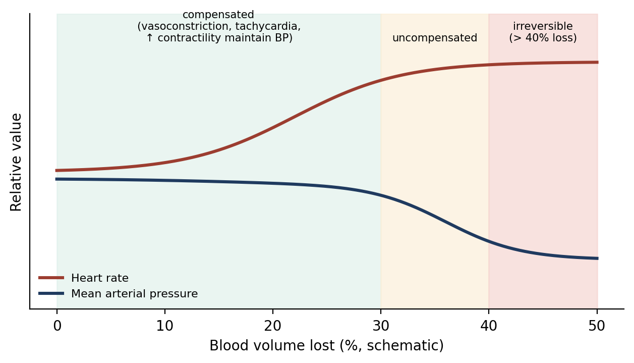
Figure 18.3. Compensated, uncompensated, and irreversible stages of hemorrhagic shock (schematic).
Why it matters
Blood pressure is a late sign of shock. During compensation, heart rate rises and mucous membranes pale while blood pressure stays near normal, so a "normal" blood pressure does not rule out shock. Watching trends in heart rate, CRT, and membrane color catches shock earlier (Chapter 12).
18.3.2 Types of Shock
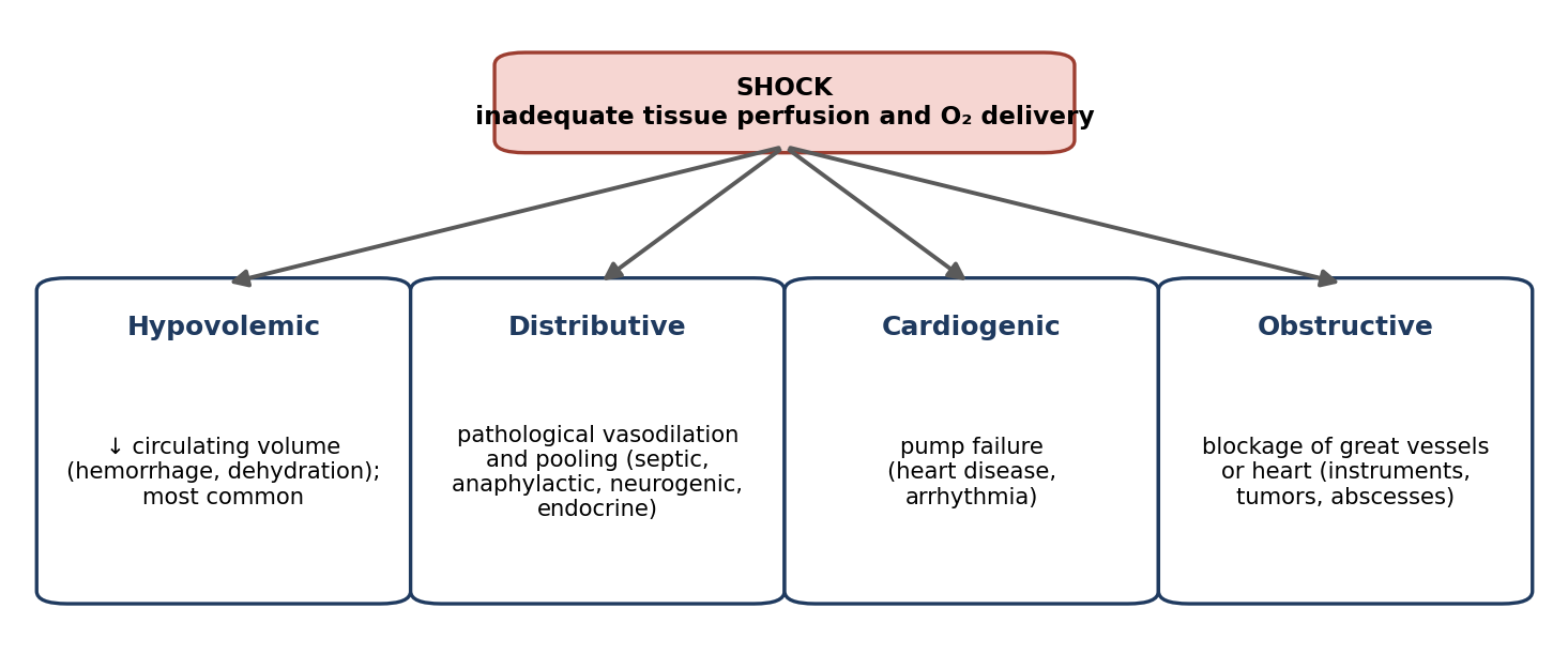
Figure 18.4. The main types of shock.
Table 18.5. Types of shock.
| Type | Mechanism | Signs and causes | Treatment |
|---|---|---|---|
| Hypovolemic (most common) | Decreased circulating blood volume | Pallor, cyanosis, disorientation, tachycardia, cold extremities, dysrhythmias, tachypnea, hypotension, oliguria, DIC, progressive metabolic acidosis | Colloids to expand plasma volume; hypertonic saline early; whole blood or packed RBCs for massive blood loss (to restore O₂-carrying capacity) |
| Distributive | Pathological vasodilation and pooling of blood, decreasing effective volume | Infectious, anaphylactic, endocrine, and neurogenic causes; trauma, heatstroke, anaphylaxis | Treat the cause; fluids; vasopressors |
| Cardiogenic | Primary failure of the ventricles to pump | Heart failure from many primary heart diseases | Oxygen; fluid therapy (cautiously); epinephrine, norepinephrine, dobutamine |
| Obstructive | Physical obstruction of the great vessels or heart | Vessels blocked by instruments during surgery, tumors, abscesses | Relieve the obstruction |
| Septic (distributive) | Overwhelming infection → vasodilation, vascular permeability, poor cardiac function, activation of coagulation | Sepsis; decreased perfusion if not controlled | Aggressive fluids; antibiotics; surgical removal of infected or necrotic tissue; supportive care ± corticosteroids |
| Anaphylactic (distributive) | Immediate hypersensitivity reaction | Brick-red mucous membranes, rapid CRT (1 second or less), labored breathing or wheezing, tachycardia; can progress to respiratory arrest, cardiovascular collapse, and death | Antihistamine; IV fluids; oxygen; corticosteroids; epinephrine in severe cases; life support |
⚠ Check current guidance
Anaphylactic shock is sometimes described with a capillary refill time of "1 second or greater"; the brick-red membranes of early distributive shock are associated with a rapid (short) CRT. Epinephrine is the first-line treatment for anaphylaxis in current guidance, not only in severe cases. Check current references.
18.3.3 Respiratory Distress
Respiratory distress is labored, exaggerated breathing effort with insufficient ventilation and oxygenation, moving only a limited amount of air. Evaluate it by observing the breathing pattern and mucous membrane color and by auscultating the lungs. Signs include dyspnea, tachypnea, orthopnea (difficulty breathing except in certain positions), hyperventilation, hypoventilation, and apnea (Chapter 12).
Supplement oxygen by face mask, flow-by, nasal cannula, oxygen cage, or intubation with manual or mechanical ventilation (Chapter 5). Treatment may include CPR, manual or mechanical ventilation, and respiratory stimulants such as doxapram HCl, which stimulates the respiratory centers in the medulla. Jen Chung (GV26) acupuncture, in which a needle is inserted at the base of the nose where it meets the upper lip, may be used to stimulate breathing when other methods fail or until they are available.
18.3.4 Cardiac Arrest
Cardiac arrest is the cessation of an effective heartbeat. Initially the ECG often shows pulseless electrical activity (PEA), formerly called electromechanical dissociation (EMD): electrical activity continues, but the heart does not pump. Signs of cardiac arrest include:
Non-auscultable or weak heart sounds; a weak, thready, or non-palpable pulse.
Dyspnea or apnea.
Lack of surgical bleeding and absent or severely low blood pressure.
Cyanosis, no muscle tone, and (later) dilated pupils.
Common causes during surgery include anesthetic overdose, hypovolemia, acute cardiogenic shock, severe acidosis, and hypoxemia.
18.3.5 Other Cardiac Emergencies
Asystole: no cardiac electrical activity, and no cardiac output or blood flow.
Ventricular fibrillation (VF): uncoordinated contraction of the ventricular muscle (Chapter 12). Successful treatment requires early diagnosis and intervention, because the brain is the organ most susceptible to hypoxia and ischemia, with serious injury after 4–5 minutes. Effective blood flow and a heartbeat must be re-established.
Cardiopulmonary Cerebrovascular Resuscitation (CPCR)
CPCR is commonly referred to as CPR; the extra "C" emphasizes the importance of maintaining perfusion and oxygen delivery to the brain during and after an arrest. The most important goal is early restoration of brain perfusion. The classic plan is "ABCDE" (Figure 18.5):
A = Airway: ensure the airway is patent; intubate if possible.
B = Breathing: supply oxygen and ventilate.
C = Cardiac massage: external (closed-chest) or internal (open-chest) compressions.
D = Drugs (Section 18.4).
E = Electrical: ECG diagnosis and defibrillation.
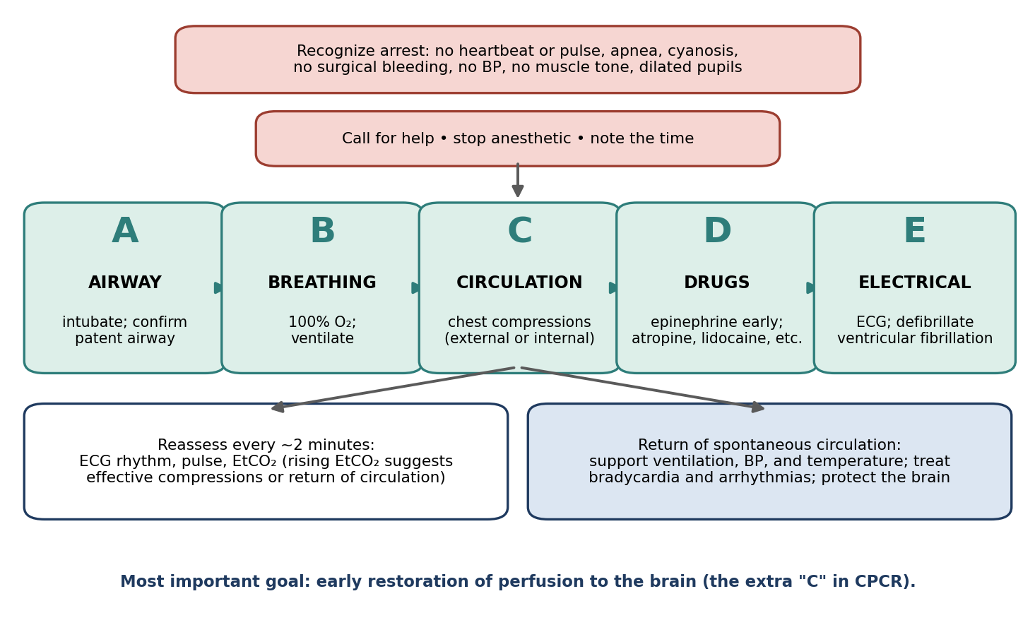
Figure 18.5. Cardiopulmonary cerebrovascular resuscitation (CPCR), following the ABCDE plan.
External chest compressions produce cardiac output by one or both of two mechanisms:
The thoracic pump theory: compressing the chest raises pressure throughout the thoracic cavity, pushing blood out of the chest during the compression phase. This predominates in larger, broad-chested animals.
The cardiac pump theory: the chest wall directly compresses the heart. This explains blood flow in smaller animals and those with a narrow (side-to-side) chest.
Abdominal compressions may be interposed between chest compressions to increase blood return from the lower half of the body. Positioning depends on the animal's size and chest shape: animals under 7 kg (15 lb) are placed in lateral recumbency; animals over 7 kg in lateral or dorsal recumbency; and deep-chested breeds in dorsal recumbency. Compressions are given at 120 per minute for animals under 7 kg and 80–100 per minute for animals over 7 kg.
Open-chest (internal) CPR is more effective at perfusing the heart and brain during the critical first minutes, producing higher blood pressure and cardiac output, and may be practical when the chest or abdomen is already open during surgery.
Defibrillation delivers an electrical shock of 3–5 joules (watt-seconds) to depolarize the heart and allow a normal rhythm to resume. Ventricular fibrillation is the least common form of cardiac arrest in animals; asystole and PEA are more common, and these are not treated with defibrillation.
⚠ Check current guidance
Current RECOVER guidelines recommend chest compressions at 100–120 per minute for dogs and cats of all sizes, in uninterrupted 2-minute cycles, with compression to one-third to one-half of chest width. Defibrillation energy is usually calculated per kilogram (for example, about 2–4 J/kg for external monophasic defibrillation and much lower for internal paddles), so "3–5 joules" likely refers to joules per kilogram. Check current guidelines.
18.4 Drug Therapy
Epinephrine (adrenaline): early administration is crucial.
Vasopressors (listed as isoproterenol and phenylephrine): augment coronary blood flow by constricting vessels, increasing systemic vascular resistance and diastolic perfusion pressure.
Lidocaine: used after resuscitation if ventricular dysrhythmias are compromising cardiac output.
Atropine or glycopyrrolate: reflex (vagal) bradycardia may have contributed to the arrest, and bradycardia often occurs after the heartbeat is re-established.
⚠ Check current guidance
Isoproterenol is a beta-adrenergic agonist that increases heart rate and causes vasodilation; it is not a vasopressor. Phenylephrine, norepinephrine, and vasopressin are vasopressors. Check current references.
18.5 Anesthetic Emergencies
Anesthetic emergencies have many general causes: adverse drug reactions, equipment malfunction, anesthetic overdose, surgical complications, pre-existing medical problems, and human error. The first interventions are the same for almost all of them:
Reduce or turn off the anesthetic.
Provide oxygen.
Verify that the equipment is working properly, and replace it if needed (Chapter 9).
Further treatment may require manual or mechanical ventilation, IV fluid therapy, thermotherapy (warming), reversal agents, and other drugs.
Respiratory Arrest
Cardiopulmonary arrest most often follows uncorrected excessive anesthetic depth, which can happen at any time during anesthesia, and requires prompt CPR. Respiratory arrest (apnea) may be caused by excessive anesthetic depth, induction with respiratory-depressant drugs, or a fall in blood CO₂ below the apneic threshold (for example, after over-ventilation), which removes the drive to breathe. Treatment is to adjust the anesthetic and bag the animal 2–10 times per minute until normal breathing resumes; use the low end of the range in apnea, deliberately hypoventilating so that CO₂ can rise back to normal and restart spontaneous breathing.
Hypotension
Causes of hypotension include blood loss, shock, cardiac arrhythmias, excessive anesthetic depth, and adverse drug effects. It is evaluated directly (Doppler, oscillometric, or direct monitoring) and indirectly (pale membranes, prolonged CRT, weak pulses; Chapter 12). Treatment is to decrease the anesthetic, give IV fluids and oxygen, provide warmth, and give drugs such as sympathomimetics (dopamine or dobutamine).
Hypoxia
Hypoxia is a deficiency of oxygen at the tissue level, caused by reduced perfusion or reduced oxygen content of the blood. It can be present without cyanosis: about 5 g/dL of deoxygenated hemoglobin must be present before cyanosis can be seen, so anemic animals may be severely hypoxic and never look blue. In most species, oxygen saturation may fall below 50% before cyanosis is detected; saturation below 90% indicates hypoxia, and below 80% can lead to organ failure. Causes include lung disease, decreased cardiac output, and severe anemia. Prolonged hypoxia leads to vascular paralysis, systemic vasodilation, and cardiovascular collapse, which are irreversible and rapidly fatal. Treatment depends on the cause and includes oxygen therapy, mechanical ventilation, and respiratory stimulants.
Table 18.6. Common anesthetic emergencies.
| Emergency | Main causes | Treatment |
|---|---|---|
| Respiratory arrest | Excessive depth; respiratory-depressant induction drugs; hypocapnia below the apneic threshold | Adjust anesthetic; ventilate 2–10 breaths/min (low end in apnea) |
| Cardiopulmonary arrest | Uncorrected excessive depth; hypovolemia; cardiogenic shock; severe acidosis; hypoxemia | Immediate CPCR |
| Hypotension | Blood loss; shock; arrhythmias; excessive depth; drug effects | Decrease anesthetic; IV fluids; oxygen; warming; dopamine or dobutamine |
| Hypoxia | Lung disease; decreased cardiac output; severe anemia | Treat cause; oxygen; ventilation; respiratory stimulants |
18.6 Acid–Base Imbalances
Acid–base status is defined by pH, the result of processes in the body tending toward acidosis or alkalosis. Normal values are given as pH 7.36–7.41, with acidosis below 7.35 and alkalosis above 7.42.
⚠ Check current guidance
Most references give the normal blood pH range as 7.35–7.45. Check which values your exam references use.
18.6.1 Mechanisms That Regulate pH
pH is maintained by three systems that work over different time scales:
Chemical buffers act within seconds: the bicarbonate–carbonic acid system, phosphate (in red blood cells and kidneys), and hemoglobin.
The respiratory system acts within minutes: by changing ventilation, the lungs regulate CO₂ and therefore the concentration of carbonic acid.
The kidneys act over hours to days, eliminating excess acid or base and regulating bicarbonate.
These systems link through the carbonic acid–CO₂ equilibrium: CO₂ + H₂O ⇌ H₂CO₃ ⇌ H⁺ + HCO₃⁻. More CO₂ pushes the reaction to the right, producing more H⁺ (acidosis); less CO₂ pulls it to the left (alkalosis). Figure 18.6 shows how to interpret a blood gas.
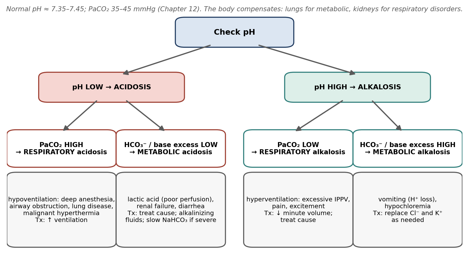
Figure 18.6. Interpreting acid–base disorders from pH, PaCO₂, and bicarbonate or base excess.
18.6.2 Respiratory Acidosis
Respiratory acidosis occurs when CO₂ production exceeds CO₂ excretion. The blood gas shows increased CO₂, which adds acid and lowers pH. Causes include anything that impairs CO₂ excretion (deep anesthesia, pulmonary disease, respiratory obstruction) or increases CO₂ production (malignant hyperthermia). Other signs include increased cardiac output (hypertension), vasodilation, and ventricular arrhythmias. The kidneys compensate over time. Treatment is to lower CO₂ by ventilating with a higher minute volume (increased tidal volume and/or rate) than before.
18.6.3 Respiratory Alkalosis
Respiratory alkalosis occurs when CO₂ excretion exceeds production. The blood gas shows decreased CO₂, with loss of H⁺ and relative gain in base. Causes include anything that drives hyperventilation: excessive controlled ventilation, pain, and excitement. Signs include tachycardia and ECG changes. The kidneys compensate over time. Treatment is to decrease minute volume if the animal is mechanically ventilated, or, if it is hyperventilating spontaneously, to find and treat the cause (for example, light anesthesia).
18.6.4 Metabolic Acidosis
In metabolic acidosis, the blood gas shows a low adjusted base excess (ABE) or low bicarbonate (HCO₃⁻); loss of bicarbonate is equivalent to a gain in H⁺. Causes include lactic acid accumulation (commonly from decreased tissue perfusion), renal failure, and loss of bicarbonate-rich secretions (such as diarrhea). The respiratory system compensates rapidly by hyperventilating. Treatment of mild imbalance uses alkalinizing IV solutions (containing lactate, gluconate, or acetate); more severe imbalance may require sodium bicarbonate, given slowly IV over 15–30 minutes, because deaths have occurred with fast administration in dehydrated animals.
18.6.5 Metabolic Alkalosis
In metabolic alkalosis, the blood gas shows a high adjusted base excess or high bicarbonate. Causes include vomiting (loss of H⁺ in stomach acid) and hypochloremia (which increases renal reabsorption of bicarbonate). The respiratory system compensates by hypoventilating, causing a mild respiratory acidosis. Treatment is to replace what is lacking: potassium if hypokalemic, and chloride after vomiting.
Table 18.7. Summary of acid–base disorders.
| Disorder | pH | Primary change | Common causes | Compensation | Treatment |
|---|---|---|---|---|---|
| Respiratory acidosis | ↓ | ↑ PaCO₂ | Deep anesthesia, lung disease, obstruction, MH | Kidneys (slow) | ↑ ventilation |
| Respiratory alkalosis | ↑ | ↓ PaCO₂ | Over-ventilation, pain, excitement | Kidneys (slow) | ↓ minute volume; treat cause |
| Metabolic acidosis | ↓ | ↓ HCO₃⁻ / base excess | Lactic acidosis, renal failure, diarrhea | Hyperventilation (fast) | Alkalinizing fluids; slow IV NaHCO₃ |
| Metabolic alkalosis | ↑ | ↑ HCO₃⁻ / base excess | Vomiting, hypochloremia | Hypoventilation | Replace Cl⁻, K⁺ |
18.7 Allergic Reactions
Allergic (anaphylactic) reactions are mediated by the immune system. Reactions to anesthetics are uncommon and usually follow prior sensitization to the drug. They are more likely with repeated exposure to an allergen; for example, an allergy to eggs could lead to a reaction to the egg lecithin in propofol (Chapter 8). Anaphylactic reactions to thiopental have been reported, as have allergic reactions in dogs to IV injection of the radiographic contrast agent diatrizoic acid. Signs include tachypnea, bronchoconstriction, and mucoid diarrhea, and may progress to anaphylactic shock (Section 18.3.2). Treatment is IV fluids, antihistamines, corticosteroids, and epinephrine.
18.8 Miscellaneous Emergencies
18.8.1 Reperfusion Injury
Reperfusion injury is cell injury that develops when blood flow returns to a tissue that was previously deprived of it. When oxygenation is restored, altered enzyme systems generate harmful oxygen free radicals, and white blood cells release inflammatory mediators in response to damaged cell membranes, causing inflammation, vessel injury, thrombosis, and edema. Reperfusion injury can cause systemic disorders such as disseminated intravascular coagulation (DIC), systemic inflammatory response syndrome (SIRS), and multi-organ dysfunction. All vital organs can be affected after resuscitation from shock or cardiopulmonary arrest, and after release of a vascular clamp or tourniquet (Chapter 11, Bier block).
18.8.2 Malignant Hyperthermia (MH)
Malignant hyperthermia is a life-threatening pharmacogenetic myopathy: in genetically predisposed animals, triggering drugs cause uncontrolled release of calcium within skeletal muscle, producing sustained contraction and a massive increase in metabolism. Swine (notably Yorkshire and other susceptible lines) are particularly susceptible. Halothane is the most potent triggering agent among volatile anesthetics, but others (and succinylcholine) can also trigger it. MH causes a rapid increase in body temperature, and death can result if it is not treated quickly.
Early signs typically include a rapidly rising EtCO₂ (often the first sign), tachycardia, tachypnea, and muscle rigidity, followed by hyperthermia, metabolic and respiratory acidosis, and hyperkalemia.
Treatment and prevention:
Prophylactic dantrolene before anesthesia in susceptible animals. Dantrolene depresses excitation–contraction coupling in skeletal muscle.
If MH occurs: stop the triggering agent, ventilate with 100% oxygen at a high minute volume, give dantrolene IV, actively cool the animal, and correct acid–base and electrolyte abnormalities.
Replace potassium lost due to muscle tremors, as indicated by blood electrolyte measurements.
⚠ Check current guidance
MH typically causes hyperkalemia (potassium released from damaged muscle) rather than potassium loss, and treatment may require lowering potassium. Potassium replacement should be guided by measured electrolytes. Check current references.
Why it matters
MH is a reason to know the genetic background of research swine, to avoid triggering agents in susceptible lines, and to monitor EtCO₂ and temperature continuously in every pig. A rising EtCO₂ that does not respond to increased ventilation is an early warning that should never be ignored.
Chapter Summary
Emergency response starts with ABC (airway, breathing, circulation), vascular access (IV, IO, IT; IC historically), a stocked crash cart with a weight-based dose chart, and monitoring equipment. Isotonic crystalloids replace losses; hypertonic saline and colloids expand volume rapidly; hypotonic and dextrose fluids are contraindicated in shock, and dextrose must not be given SC. Shock (hypovolemic, distributive [septic, anaphylactic], cardiogenic, obstructive) progresses from compensated to uncompensated and becomes irreversible beyond about 40% blood loss. Respiratory distress is treated with oxygen, ventilation, and stimulants.
Cardiac arrest (often PEA initially) requires immediate CPCR, with early epinephrine and the goal of restoring brain perfusion. Anesthetic emergencies (respiratory arrest, hypotension, hypoxia) are first managed by reducing anesthetic, giving oxygen, and checking equipment. Acid–base disorders are respiratory (PaCO₂) or metabolic (HCO₃⁻/base excess), each with characteristic causes and treatments. Allergic reactions, reperfusion injury, and malignant hyperthermia (prevented and treated with dantrolene) complete the emergencies a surgical research professional must be ready to recognize.
Key Terms
Anaphylaxis: An immediate, immune-mediated hypersensitivity reaction.
Asystole: Absence of cardiac electrical activity.
Base excess: A blood gas measure of metabolic acid–base status.
CPCR: Cardiopulmonary cerebrovascular resuscitation; CPR with emphasis on brain perfusion.
Crash cart: A cart stocked with emergency supplies, drugs, and equipment.
Dantrolene: A muscle relaxant used to prevent and treat malignant hyperthermia.
Defibrillation: Delivery of an electrical shock to terminate ventricular fibrillation.
Intraosseous (IO): Into the bone marrow cavity.
Malignant hyperthermia (MH): A genetic, drug-triggered hypermetabolic crisis of skeletal muscle.
Oncotic pressure: Osmotic pressure exerted by large molecules (colloids) in plasma.
Pulseless electrical activity (PEA): Electrical activity on the ECG without effective cardiac contraction.
Reperfusion injury: Tissue injury that occurs when blood flow returns after ischemia.
Shock: Decreased tissue perfusion and oxygen delivery to vital organs.
Review Questions
1. The basic order of emergency life support is:
A. Drugs, fluids, oxygen
B. Airway, breathing, circulation
C. Circulation, drugs, electrical
D. Oxygen, sedation, recovery
2. Which emergency drug can be given intratracheally?
A. Sodium bicarbonate
B. Epinephrine
C. Dobutamine
D. Dextrose 50%
3. Which fluid is contraindicated in shock because water distributes rapidly out of the vessels?
A. Lactated Ringer's
B. Hypertonic saline
C. Hypotonic crystalloids (e.g., 0.45% saline, 2.5% dextrose)
D. Hetastarch
4. Shock becomes irreversible when blood loss exceeds approximately:
A. 5%
B. 15%
C. 40%
D. 80%
5. The most common type of shock is:
A. Cardiogenic
B. Obstructive
C. Hypovolemic
D. Anaphylactic
6. Brick-red mucous membranes, rapid capillary refill, wheezing, and tachycardia after a drug injection suggest:
A. Hypovolemic shock
B. Anaphylactic shock
C. Cardiogenic shock
D. Respiratory alkalosis
7. Which drug is the cornerstone of CPR, and should be given early?
A. Lidocaine
B. Epinephrine
C. Naloxone
D. Doxapram
8. During anesthesia, the most common cause of cardiopulmonary arrest is:
A. Allergic reaction
B. Uncorrected excessive anesthetic depth
C. Malignant hyperthermia
D. Hypothermia
9. A blood gas shows low pH and high PaCO₂. This is:
A. Metabolic acidosis
B. Respiratory acidosis
C. Respiratory alkalosis
D. Metabolic alkalosis
10. Which drug is used to prevent and treat malignant hyperthermia?
A. Dantrolene
B. Halothane
C. Succinylcholine
D. Acepromazine
Answer Key
1. B. Basic life support follows ABC: airway, breathing, circulation. CPCR extends this to ABCDE: drugs and electrical defibrillation.
2. B. Atropine, lidocaine, and epinephrine can be given intratracheally (at a higher dose than IV).
3. C. Hypotonic fluids distribute rapidly into all body water and do little to expand plasma volume, so they are contraindicated in shock.
4. C. Compensatory mechanisms maintain blood pressure early, but once loss exceeds about 40% of blood volume they fail and shock becomes irreversible.
5. C. Hypovolemic shock, from decreased circulating volume, is the most common type.
6. B. These are signs of anaphylaxis, an immediate hypersensitivity reaction that can progress to respiratory arrest and cardiovascular collapse.
7. B. Early administration of epinephrine is crucial during cardiopulmonary resuscitation.
8. B. Cardiopulmonary arrest most often follows uncorrected excessive anesthetic depth, which can happen at any time during anesthesia.
9. B. Low pH is acidosis; high PaCO₂ shows a respiratory cause (CO₂ production exceeds excretion). Treatment is increased ventilation.
10. A. Dantrolene depresses excitation–contraction coupling in skeletal muscle. Halothane and succinylcholine are triggers.
Appendix A: Dose Calculations
Learning objectives
After studying this chapter, you should be able to:
Convert between common units of weight, volume, and concentration, including percentage and ratio solutions.
Calculate a drug dose and the volume to administer from a dose rate, body weight, and concentration.
Calculate fluid rates and drip rates, and constant rate infusions.
Prepare dilutions using the "amount needed" approach and C₁V₁ = C₂V₂.
Recognize when a calculated volume is too small to measure accurately, and correct it by dilution.
Dose calculations make up an estimated 5–15% of the certification exams (Chapter 1), and they are a daily part of work in surgical research. Exam questions are standard word problems that provide all the information you need; simple problems take a single step, while harder ones require unit conversion or several steps. The method is always the same: convert the units, calculate the amount of drug, convert that amount to a volume, and check that the answer makes sense.
⚠ Check current guidance
The doses used in this appendix are examples for practicing arithmetic only. They are not dosing recommendations. Always use doses prescribed by a veterinarian or specified in the approved protocol.
A.1 Units and Conversions
Table A.1. Common conversions.
| Quantity | Conversion |
|---|---|
| Weight | 1 kg = 1,000 g; 1 g = 1,000 mg; 1 mg = 1,000 µg (mcg) |
| Pounds to kilograms | 1 kg = 2.2 lb, so kg = lb ÷ 2.2 (e.g., 22 lb = 10 kg) |
| Grams to kilograms | kg = g ÷ 1,000 (e.g., 350 g = 0.35 kg; 25 g = 0.025 kg) |
| Volume | 1 L = 1,000 mL; 1 mL = 1 cc |
| Percentage solution (w/v) | % = grams per 100 mL, so 1% = 1 g/100 mL = 10 mg/mL (e.g., 2% lidocaine = 20 mg/mL) |
| Ratio solution | 1:1,000 = 1 g in 1,000 mL = 1 mg/mL; 1:10,000 = 0.1 mg/mL |
| Units (U, IU) | Some drugs (such as heparin and insulin) are measured in units of activity rather than weight; calculate with units exactly as with mg |
Why it matters
To convert a percentage to mg/mL, multiply by 10. A 5% solution contains 5 g per 100 mL, which is 5,000 mg ÷ 100 mL = 50 mg/mL.
A.2 Calculating a Dose and Volume
The standard calculation has two steps (Figure A.1):
Dose (mg) = dose rate (mg/kg) × body weight (kg)
Volume (mL) = dose (mg) ÷ concentration (mg/mL)
Combined: Volume (mL) = (dose rate × body weight) ÷ concentration. Units must match: if the dose rate is in mg/kg, the weight must be in kg and the concentration in mg/mL. Writing the units at every step shows when a conversion is needed, because units that do not cancel indicate an error.
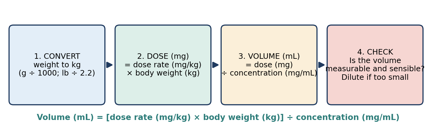
Figure A.1. The steps of a dose calculation.
Example 1. A 3.5 kg nonhuman primate is to receive buprenorphine at 0.03 mg/kg. The concentration is 0.3 mg/mL. What volume is given?
Dose = 0.03 mg/kg × 3.5 kg = 0.105 mg
Volume = 0.105 mg ÷ 0.3 mg/mL = 0.35 mL
Example 2. A 350 g rat is to receive buprenorphine at 0.01 mg/kg. The concentration is 0.3 mg/mL. What volume is given?
Convert: 350 g ÷ 1,000 = 0.35 kg
Dose = 0.01 mg/kg × 0.35 kg = 0.0035 mg
Volume = 0.0035 mg ÷ 0.3 mg/mL = 0.0117 mL (about 0.01 mL)
Check: about 0.01 mL is far too small to measure accurately with a standard syringe. In practice, the drug would be diluted first (Section A.5). Diluted 1:10 to 0.03 mg/mL, the volume becomes 0.0035 ÷ 0.03 = 0.12 mL, which can be measured accurately.
Example 3 (percentage solution). A 5 kg dog is prescribed lidocaine at 2 mg/kg. The solution is 2% lidocaine. What volume is given?
Convert the concentration: 2% × 10 = 20 mg/mL
Dose = 2 mg/kg × 5 kg = 10 mg
Volume = 10 mg ÷ 20 mg/mL = 0.5 mL
Example 4 (pounds). A dog weighs 44 lb and is prescribed a drug at 2 mg/kg; the drug is supplied at 20 mg/mL. What volume is given?
Convert: 44 lb ÷ 2.2 = 20 kg
Dose = 2 mg/kg × 20 kg = 40 mg
Volume = 40 mg ÷ 20 mg/mL = 2 mL
A.3 Fluid Rates and Drip Rates
Fluid rate (mL/h) = rate (mL/kg/h) × body weight (kg). When fluids are given by gravity through a drip set, the rate is set by counting drops. Each drip set delivers a fixed number of drops per mL (the drip factor): commonly 60 drops/mL for a microdrip set, and 10, 15, or 20 drops/mL for macrodrip sets.
Drip rate (drops/min) = fluid rate (mL/h) × drip factor (drops/mL) ÷ 60 (min/h)
Example 5. A 10 kg beagle is to receive fluids at 5 mL/kg/h. What is the flow rate?
Rate = 5 mL/kg/h × 10 kg = 50 mL/h
Example 6. The drip set delivers 60 drops/mL. How many drops per minute give 50 mL/h?
Drops per hour = 50 mL/h × 60 drops/mL = 3,000 drops/h
Drops per minute = 3,000 ÷ 60 = 50 drops/min
Note: with a 60 drops/mL microdrip set, drops per minute always equals mL per hour.
Example 7. Using a 15 drops/mL set for the same 50 mL/h, what is the drip rate?
Drip rate = 50 × 15 ÷ 60 = 12.5 drops/min (about one drop every 4.8 seconds, since 60 ÷ 12.5 = 4.8)
Constant Rate Infusions
For a drug given as a continuous infusion, the rate is calculated the same way, with a time unit added. Infusion rate (mL/h) = dose rate per hour × body weight ÷ concentration. If the dose is given per minute, multiply by 60 to convert to per hour.
Example 8. A drug is ordered at 2 µg/kg/min for a 20 kg dog. The infusion solution contains 100 µg/mL. What is the infusion rate in mL/h?
Dose per minute = 2 µg/kg/min × 20 kg = 40 µg/min
Dose per hour = 40 µg/min × 60 = 2,400 µg/h
Rate = 2,400 µg/h ÷ 100 µg/mL = 24 mL/h
A.4 Dilutions
For dilutions, think: "How much drug do I need in my final solution?" Calculate the total amount of drug required (final concentration × final volume), then divide by the stock concentration to find the volume of stock to add. This is the same as the formula C₁V₁ = C₂V₂, where C₁ and V₁ are the stock concentration and the volume of stock used, and C₂ and V₂ are the final concentration and final volume.
Example 9. Make 500 mL of heparinized saline at 100 U/mL from heparin stock containing 10,000 U/mL. How much heparin is needed?
Amount needed = 100 U/mL × 500 mL = 50,000 U
Volume of stock = 50,000 U ÷ 10,000 U/mL = 5 mL
Note: adding 5 mL to a full 500 mL bag gives 505 mL (99 U/mL), which is usually close enough; for an exact final volume, add the heparin to 495 mL of saline.
Example 10. Make 10 mL of buprenorphine at 0.03 mg/mL from a 0.3 mg/mL stock.
C₁V₁ = C₂V₂: 0.3 mg/mL × V₁ = 0.03 mg/mL × 10 mL, so V₁ = 0.3 ÷ 0.3 = 1 mL
Add 1 mL of stock to 9 mL of diluent (a 1:10 dilution).
A.5 Multi-Step Problems and Small Volumes
Small animals often need volumes too small to measure. A good rule of thumb is that a volume below about 0.05–0.1 mL in a 1 mL syringe is difficult to measure accurately; diluting the drug produces a larger, more accurate volume. The diluent must be compatible with the drug (commonly sterile saline or sterile water), and diluted solutions must be labeled with the drug, concentration, date, and expiration according to institutional policy.
Example 11. A 25 g mouse is to receive a drug at 5 mg/kg. The stock is 50 mg/mL. What volume would be given, and how could it be made measurable?
Convert: 25 g = 0.025 kg
Dose = 5 mg/kg × 0.025 kg = 0.125 mg
Volume of stock = 0.125 mg ÷ 50 mg/mL = 0.0025 mL, which cannot be measured accurately
Dilute 1:50 to 1 mg/mL: 50 mg/mL × V₁ = 1 mg/mL × 10 mL, so V₁ = 0.2 mL of stock made up to 10 mL (0.2 mL stock + 9.8 mL diluent)
Volume of diluted drug = 0.125 mg ÷ 1 mg/mL = 0.125 mL
Tips for Accurate Calculations
Write every unit and make sure they cancel correctly.
Convert first: weights to kg, percentages and ratios to mg/mL, and minutes to hours if needed.
Estimate the answer before calculating, and check that the result is sensible. A volume of 15 mL for a mouse, or 0.001 mL for a dog, signals an error, often a misplaced decimal point or a missed g-to-kg conversion.
Watch the decimal point. A tenfold error is the most common and most dangerous calculation mistake; always write a leading zero (0.5 mL, not .5 mL) and never a trailing zero (5 mL, not 5.0 mL).
Double-check high-risk calculations with a colleague.
A.6 Practice Problems
A 4 kg cat is prescribed a drug at 0.2 mg/kg. The concentration is 10 mg/mL. What volume is given?
A 300 g rat is to receive ketamine at 50 mg/kg. Ketamine is supplied at 100 mg/mL. What volume is given?
A 25 kg pig is to receive fluids at 10 mL/kg/h through a 15 drops/mL drip set. What is the fluid rate in mL/h, and the drip rate in drops/min?
A 1 L bag of fluids is running at 100 mL/h. How long will it last?
How many grams of dextrose are in 500 mL of 5% dextrose?
Epinephrine is supplied as a 1:1,000 solution. What is its concentration in mg/mL? What volume provides 0.01 mg/kg for a 15 kg dog?
How would you make 20 mL of a 2 mg/mL solution from a 10 mg/mL stock?
A 30 g mouse is to receive buprenorphine at 0.1 mg/kg from a 0.3 mg/mL stock. What volume would be needed? What volume would be needed after diluting the stock to 0.03 mg/mL?
How much heparin (1,000 U/mL) is needed to make 250 mL of heparinized saline at 10 U/mL?
A 10 kg dog is to receive a drug by infusion at 1 mg/kg/h. The infusion solution contains 2 mg/mL. What is the infusion rate?
Answers to Practice Problems
Table A.2. Worked answers.
| # | Working | Answer |
|---|---|---|
| 1 | 0.2 mg/kg × 4 kg = 0.8 mg; 0.8 mg ÷ 10 mg/mL | 0.08 mL |
| 2 | 0.3 kg × 50 mg/kg = 15 mg; 15 mg ÷ 100 mg/mL | 0.15 mL |
| 3 | 10 mL/kg/h × 25 kg = 250 mL/h; 250 × 15 ÷ 60 = 62.5 | 250 mL/h; about 63 drops/min |
| 4 | 1,000 mL ÷ 100 mL/h | 10 hours |
| 5 | 5% = 5 g/100 mL; 500 mL = 5 × 5 g | 25 g |
| 6 | 1:1,000 = 1 g/1,000 mL = 1 mg/mL; 0.01 mg/kg × 15 kg = 0.15 mg; 0.15 mg ÷ 1 mg/mL | 1 mg/mL; 0.15 mL |
| 7 | C₁V₁ = C₂V₂: 10 × V₁ = 2 × 20, V₁ = 4 mL | 4 mL stock + 16 mL diluent |
| 8 | 0.03 kg × 0.1 mg/kg = 0.003 mg; 0.003 ÷ 0.3 = 0.01 mL; 0.003 ÷ 0.03 = 0.1 mL | 0.01 mL undiluted (too small to measure); 0.1 mL diluted |
| 9 | 10 U/mL × 250 mL = 2,500 U; 2,500 U ÷ 1,000 U/mL | 2.5 mL |
| 10 | 1 mg/kg/h × 10 kg = 10 mg/h; 10 mg/h ÷ 2 mg/mL | 5 mL/h |
Key Terms
Concentration: The amount of drug per unit volume of solution (e.g., mg/mL).
Constant rate infusion (CRI): Continuous administration of a drug at a steady rate.
Diluent: The fluid used to dilute a drug.
Dose: The amount of drug to be given (e.g., mg).
Dose rate: The amount of drug per unit of body weight (e.g., mg/kg).
Drip factor: The number of drops per mL delivered by an administration set.
Percentage solution (w/v): Grams of drug per 100 mL of solution.
References
The references below were listed as the basis for the ASR Certification Study Guide, on which this textbook is built. Many have since been revised; entries more than about ten years old are marked so that you can check for the current edition. The current ASR Recommended References list, available from the ASR, takes precedence over this list (Chapter 1).
AVMA Guidelines for the Euthanasia of Animals: 2013 Edition. [A newer edition (2020) has been published; use the current edition.]
Welsh E. Anaesthesia for Veterinary Nurses. Blackwell Science, 2003. [Check for a newer edition.]
Popesko P, Rajtová V, Horák J. Colour Atlas of Anatomy of Small Laboratory Animals, Vol. 2. 2002.
Kohn D. Anesthesia and Analgesia in Laboratory Animals. San Diego: Academic Press, 1997. [Later editions have been published; check for the current edition.]
Bassert J, Thomas J. McCurnin's Clinical Textbook for Veterinary Technicians, 8th ed. Elsevier Saunders, 2014. [Later editions have been published.]
Code of Federal Regulations, Title 9, Chapter 1, Subchapter A: Animal Welfare (Parts 1–12). [Use the current electronic CFR (eCFR).]
Bojrab MJ, Waldron DR, Toombs JP. Current Techniques in Small Animal Surgery, 5th ed. Teton NewMedia, 2014. [Check for a newer edition.]
Dunn DL. Ethicon Wound Closure Manual. Johnson & Johnson. [Product information changes; check current Ethicon materials and package inserts.]
Waynforth HB, Flecknell PA. Experimental and Surgical Technique in the Rat, 2nd ed. Academic Press, 1992.
Knecht CD. Fundamental Techniques in Veterinary Surgery, 3rd ed. Elsevier Health Sciences, 1987.
National Research Council. Guide for the Care and Use of Laboratory Animals, 8th ed. National Academies Press, 2011. [Check whether a newer edition has been published and adopted.]
Orpet H, Welsh P. Handbook of Veterinary Nursing, 2nd ed. Wiley-Blackwell, 2010. [Check for a newer edition.]
Kirk and Bistner's Handbook of Veterinary Procedures and Emergency Treatment, 9th ed. Saunders, 2011. [Check for a newer edition.]
Chiasson R. Laboratory Anatomy of the White Rat. McGraw-Hill Education, 1988.
Flecknell P. Laboratory Animal Anaesthesia, 4th ed. Academic Press, 2015. [Check for a newer edition.]
van Dongen JJ, Remie R, Rensema JW, van Wunnik GHJ. Manual of Microsurgery on the Laboratory Rat. Elsevier, 1990.
Taylor R, McGehee R. Manual of Small Animal Postoperative Care. Williams & Wilkins, 1995.
Evans H, de Lahunta A. Miller's Anatomy of the Dog, 4th ed. Saunders, 2012. [Check for a newer edition.]
Loughlin KR, Brooks DC. Principles of Endosurgery. Blackwell Science, 1996.
Lavin L. Radiography in Veterinary Technology, 4th ed. Elsevier Health Sciences, 2006. [Check for a newer edition.]
Research Animal Anesthesia, Analgesia, and Surgery: proceedings of a conference sponsored by SCAW, Atlanta, Georgia, May 12–13, 1994.
Warren RG. Small Animal Anesthesia. Mosby, 1983.
Wingfield W. Small Animal Surgery: An Atlas of Operative Techniques. W.B. Saunders, 1979.
Fossum TW. Small Animal Surgery, 3rd ed. Mosby, 2007. [Later editions have been published.]
Tracy DL. Small Animal Surgical Nursing, 3rd ed. Mosby, 2000.
Turner AS, McIlwraith CW. Techniques in Large Animal Surgery, 2nd ed. Wiley, 1991. [Check for a newer edition.]
Lumb and Jones' Veterinary Anesthesia and Analgesia. Wiley, 2007. [Later editions have been published.]
Hackett TB, Mazzaferro EM. Veterinary Emergency & Critical Care Procedures, 2nd ed. Wiley-Blackwell, 2012. [Check for a newer edition.]
Freeman LJ. Veterinary Endosurgery. Elsevier Health Sciences, 1998.
McCarthy TC. Veterinary Endoscopy for the Small Animal Practitioner, 1st ed. Saunders, 2004. [Check for a newer edition.]
Ettinger SJ, Feldman EC. Textbook of Veterinary Internal Medicine, 7th ed. Saunders, 2009. [Later editions have been published.]
Additional Sources Referred to in This Textbook
Academy of Surgical Research. Certification Study Guide, 1st ed. (Flegal M, and the ASR Certification Committee) and 2nd ed. (ASR Certification Committee, 2012–2016). Updated 19 October 2016.
Academy of Surgical Research. Guidelines for Training in Surgical Research with Animals. J Invest Surg 1989; 2(2): 263–268, and 2009; 22: 218–225.
Academy of Surgical Research. Guidelines for Rodent Survival Surgery. J Invest Surg 2009; 22: 445–451.
Office of Laboratory Animal Welfare. Public Health Service Policy on Humane Care and Use of Laboratory Animals (current edition).
RECOVER (Reassessment Campaign on Veterinary Resuscitation) guidelines for small animal cardiopulmonary resuscitation (published 2012; since updated).
Supplementary study materials: ASRT Study Materials (Overview; Anatomy & Physiology; Anesthesia Definitions; Instruments; Suture; Wound Healing).
Glossary
This glossary combines the key terms from every chapter (263 terms). The chapter numbers after each definition show where the term is introduced or discussed.
#
3 Rs: Replacement, reduction, and refinement; the guiding principles for the humane use of animals in research. (Ch. 2)
A
AAALAC International: Association for the Assessment and Accreditation of Laboratory Animal Care; offers voluntary accreditation, formed in 1965. (Ch. 2)
Abduction / adduction: Movement of an extremity away from / toward the center of the body. (Ch. 14)
Academy of Surgical Research (ASR): The professional organization that administers the SRT, SRS, and SRA certification exams and publishes guidelines for surgical research with animals. (Ch. 1)
A-delta fiber: A myelinated, fast-conducting nociceptor fiber carrying sharp first pain. (Ch. 13)
Agonist-antagonist: An opioid that activates some opioid receptors while blocking others (e.g., butorphanol). (Ch. 6)
Allodynia: Pain from a stimulus that does not normally provoke pain. (Ch. 13)
Amide-linked local anesthetic: A stable local anesthetic metabolized by the liver (e.g., lidocaine, bupivacaine). (Ch. 11)
Analgesia: Freedom from, or absence of, pain. (Ch. 7)
Anaphylaxis: An immediate, immune-mediated hypersensitivity reaction. (Ch. 18)
Anatomical dead space: The part of each breath that fills the airways but does not reach the alveoli. (Ch. 12)
Animal Welfare Act (AWA): Federal law passed in 1966, administered by USDA APHIS, regulating the treatment of covered animals in research. (Ch. 2)
Animal Welfare Regulations: The regulations implementing the AWA, published in Title 9 of the Code of Federal Regulations. (Ch. 2)
Anticholinergic: A drug that blocks muscarinic acetylcholine receptors (e.g., atropine, glycopyrrolate). (Ch. 6)
Anticholinesterase: A drug that inhibits acetylcholinesterase, increasing acetylcholine (e.g., neostigmine). (Ch. 6)
Antiseptic: A substance that inhibits or destroys microorganisms on or in living tissue. (Ch. 3)
APL (pop-off) valve: Adjustable pressure-limiting valve that releases excess gas from the breathing system to the scavenger. (Ch. 9)
Apneic threshold: The PaCO₂ below which spontaneous breathing stops. (Ch. 12)
Apposition: Bringing tissue edges together in their normal alignment. (Ch. 16)
Arthroscopy: Endoscopic examination or surgery of a joint. (Ch. 17)
Asepsis: A state of freedom from disease-causing contaminants. (Ch. 3)
Aseptic technique: Procedures that prevent contamination of the surgical site by microorganisms. (Ch. 2, 3)
Asystole: Absence of cardiac electrical activity. (Ch. 18)
Atelectasis: Collapse of part of the lung, often in the dependent (lower) lung of a recumbent animal. (Ch. 5)
Atrioventricular (AV) node: Conduction tissue that delays the impulse between atria and ventricles. (Ch. 14)
Autoclave: A chamber that sterilizes using saturated steam under pressure. (Ch. 3)
B
Bactericidal: Kills bacteria. (Ch. 4)
Bacteriostatic: Preventing the growth of bacteria without necessarily killing them. (Ch. 3, 4)
Balanced anesthesia: Anesthesia using multiple drugs, each targeting a component of the anesthetic state. (Ch. 7)
Barbiturate slough: Tissue injury and sloughing caused by perivascular injection of a barbiturate. (Ch. 8)
Basal metabolic rate (BMR): The rate of energy use by the body at rest. (Ch. 7)
Base excess: A blood gas measure of metabolic acid–base status. (Ch. 18)
Bier block: IV regional anesthesia of a limb isolated by a tourniquet. (Ch. 11)
Blood-brain barrier: The barrier between blood and brain tissue that limits passage of non-lipophilic, ionized, or protein-bound drugs. (Ch. 7)
Blood/gas partition coefficient: A measure of an inhalant's solubility in blood; lower values mean faster induction and recovery. (Ch. 9)
Box lock: The hinge joint of a ringed instrument. (Ch. 15)
C
Capillary refill time (CRT): Time for a blanched mucous membrane to regain color; normally 1–2 seconds. (Ch. 12)
Capnograph: A monitor that measures and displays CO₂ in respired gas. (Ch. 12)
Cardiac output: Heart rate × stroke volume. (Ch. 14)
Cardioplegic solution: A solution used to stop the heart in a relaxed state during cardiac surgery. (Ch. 10)
Cardiopulmonary bypass: Circulation of blood outside the body through a machine that oxygenates it and, if needed, cools or warms it. (Ch. 10)
Cataleptoid state: A trance-like state with open eyes, increased muscle tone, and movements unrelated to stimulation, produced by dissociatives. (Ch. 8)
Ceiling effect: The point beyond which increasing the dose produces little additional effect. (Ch. 6)
Central venous pressure (CVP): Pressure in the intrathoracic vena cava. (Ch. 12)
C fiber: An unmyelinated, slow-conducting nociceptor fiber carrying dull, burning pain. (Ch. 13)
Closed gloving: Gloving in which the hands remain inside the gown sleeves; the recommended method. (Ch. 4)
Cold sterilant: A liquid chemical sterilant used at room temperature by full immersion. (Ch. 3)
Cole tube: An uncuffed endotracheal tube with a shoulder that seats against the arytenoid cartilages. (Ch. 5)
Colloid: A fluid containing large molecules that remain in the vascular compartment. (Ch. 5)
Colloid osmotic pressure: Osmotic pressure created by large molecules (such as proteins) that holds water in the vessels. (Ch. 5)
Concentration: The amount of drug per unit volume of solution (e.g., mg/mL). (Ch. App. A)
Constant rate infusion (CRI): Continuous administration of a drug at a steady rate. (Ch. App. A)
CPCR: Cardiopulmonary cerebrovascular resuscitation; CPR with emphasis on brain perfusion. (Ch. 18)
Crash cart: A cart stocked with emergency supplies, drugs, and equipment. (Ch. 18)
Crystalloid: A solution of crystalline solids dissolved in water that distributes throughout the extracellular fluid. (Ch. 5)
Cyanosis: Blue or purple discoloration of mucous membranes due to poorly oxygenated blood. (Ch. 12)
Cyclooxygenase (COX): The enzyme family inhibited by NSAIDs to reduce prostaglandin production. (Ch. 13)
Cycloplegia: Paralysis of the eye's focusing (ciliary) muscle. (Ch. 6)
D
Dantrolene: A muscle relaxant used to prevent and treat malignant hyperthermia. (Ch. 18)
Dead space: An open area within closed tissue. (Ch. 16)
Defibrillation: Delivery of an electrical shock to terminate ventricular fibrillation. (Ch. 18)
Dehiscence: Breakdown (opening) of a closed incision. (Ch. 16)
Depolarizing block: Neuromuscular block caused by sustained stimulation of acetylcholine receptors (e.g., succinylcholine). (Ch. 6)
Diaphysis / epiphysis: The shaft / end of a long bone. (Ch. 14)
Diffusion hypoxia: Hypoxia caused by nitrous oxide flooding into the alveoli when it is discontinued. (Ch. 9)
Diluent: The fluid used to dilute a drug. (Ch. App. A)
Disinfection: Inactivation of most pathogenic organisms on inanimate objects, including vegetative bacteria but not spores. (Ch. 3)
Dissociative anesthesia: Anesthesia in which drugs such as ketamine dissociate the thalamocortical and limbic systems, producing a cataleptoid state with open eyes, intact swallowing, and muscle hypertonus. (Ch. 7)
Dissociative anesthetic: An anesthetic that interrupts transmission between unconscious and conscious parts of the brain (e.g., ketamine). (Ch. 8)
Distress: The outward expression of suffering that an observer can see. (Ch. 13)
Dorsal recumbency: Lying on the back. (Ch. 4)
Dose: The amount of drug to be given (e.g., mg). (Ch. App. A)
Dose rate: The amount of drug per unit of body weight (e.g., mg/kg). (Ch. App. A)
Drip factor: The number of drops per mL delivered by an administration set. (Ch. App. A)
E
Electronarcosis: Deep narcosis produced by passing electrical current through the cerebrum. (Ch. 10)
Endomysium, perimysium, epimysium: Connective tissue around a muscle fiber, a fascicle, and a whole muscle. (Ch. 14)
Endoscopy: "To look inside"; examination of body interiors with an endoscope. (Ch. 17)
Endotoxin: A toxic component of certain bacteria that damages host cells. (Ch. 4)
End-tidal CO₂ (EtCO₂): CO₂ concentration at the end of exhalation; approximates PaCO₂. (Ch. 12)
Epidural space: The space outside the dura mater surrounding the spinal cord and nerve roots. (Ch. 11)
Erythema: Redness of the skin, a sign of inflammation or infection. (Ch. 4)
Esmarch bandage: An elastic bandage used to exsanguinate a limb. (Ch. 11)
Ester-linked local anesthetic: A local anesthetic with a short half-life that may cause allergic reactions (e.g., procaine). (Ch. 11)
Exsanguinate: To drive blood out of a limb or region. (Ch. 11)
Extracorporeal: Outside the body. (Ch. 10)
F
Fasciculation: Brief, uncoordinated muscle twitching. (Ch. 6)
Fenestrated: Having an opening (window), as in a ring-tipped forceps. (Ch. 15)
Fenestrated drape: A drape with an opening (fenestration) that exposes the surgical site. (Ch. 4)
Field block: A wall of local anesthetic infiltrated around, rather than into, an area. (Ch. 11)
First intention: Healing of a wound closed at the time of surgery. (Ch. 16)
First-order kinetics: Elimination of a constant fraction of drug per unit time. (Ch. 7)
G
GABA: Gamma-aminobutyric acid, the main inhibitory neurotransmitter of the brain. (Ch. 8)
Gas: A substance in gaseous form at room temperature and sea-level pressure (e.g., N₂O). (Ch. 9)
General anesthesia: Drug-induced, reversible unconsciousness with analgesia, in which the patient is not aroused by noxious stimuli. (Ch. 7)
Glucose effect: Re-anesthetization caused by giving glucose during recovery from barbiturates in susceptible species. (Ch. 8)
Good Laboratory Practices (GLPs): FDA and EPA regulations governing the conduct of studies submitted to those agencies, emphasizing training, SOPs, quality assurance, and records. (Ch. 2)
Granulation tissue: New vascular connective tissue formed during the proliferative phase. (Ch. 16)
Grimace scale: A pain assessment tool that scores changes in facial expression. (Ch. 13)
H
Hanging drop: A drop of saline in the needle hub that is drawn in when the needle enters the epidural space. (Ch. 11)
Heart block: Impaired electrical conduction through the AV node. (Ch. 12)
Heat exchanger: A device that cools or warms blood as it passes through. (Ch. 10)
Heat-up time: The time for an autoclave to reach temperature and saturate the pack with steam. (Ch. 3)
Hematocrit: The percentage of blood volume made up of red blood cells. (Ch. 5)
Hemostat: A ratcheted forceps used to clamp blood vessels. (Ch. 15)
Hofmann elimination: Spontaneous, pH- and temperature-dependent breakdown of atracurium in the body. (Ch. 6)
Hydrolysis: Breakdown by water; how synthetic absorbable sutures degrade. (Ch. 16)
Hyperalgesia: An increased response to a stimulus that is normally painful. (Ch. 13)
Hypercapnia / hypocapnia: Elevated / lowered PaCO₂. (Ch. 12)
Hyperkalemia: Elevated blood potassium. (Ch. 7)
Hypnosis: Artificially induced sleep from which the patient is readily aroused. (Ch. 7)
Hypoproteinemia: Abnormally low protein levels in the blood. (Ch. 5)
Hypoxemia: Insufficient oxygenation of the blood (PaO₂ below 80 mmHg). (Ch. 12)
Hypoxic pulmonary vasoconstriction (HPV): Constriction of pulmonary vessels in poorly ventilated lung, diverting blood to ventilated lung. (Ch. 17)
I
IACUC: Institutional Animal Care and Use Committee; interprets, implements, oversees, and evaluates an institution's animal care and use program. (Ch. 2)
Incise drape: A sterile adhesive plastic drape applied directly to the skin over the incision site. (Ch. 4)
Induced hypothermia: Deliberate lowering of body temperature to support anesthesia or protect organs. (Ch. 10)
Infiltration: Injection of local anesthetic directly into the tissue to be anesthetized. (Ch. 11)
Insertion / origin: The more movable / more fixed attachment of a muscle. (Ch. 14)
Insufflation: Inflation of a body cavity with gas. (Ch. 17)
Intercalated disc: Junction between cardiac muscle cells that allows rapid impulse spread. (Ch. 14)
Intercostal nerve block: Local anesthetic placed at the caudal border of ribs to anesthetize the chest wall. (Ch. 11)
Intra-osseous (IO): Administered into the marrow cavity of a bone. (Ch. 5)
Intraosseous (IO): Into the bone marrow cavity. (Ch. 18)
Inverting pattern: A suture pattern that turns tissue edges inward (e.g., Lembert, Cushing). (Ch. 16)
Iodophor: Iodine complexed with a surfactant or polymer that releases free iodine slowly (e.g., povidone-iodine). (Ch. 3)
Isotonic: Having the same solute concentration as body fluids, so no fluid shifts occur across cell membranes. (Ch. 5)
K
Kick bucket: A wheeled waste container used to collect used sponges and disposables. (Ch. 4)
L
Laparoscopy: Endoscopic examination or surgery of the abdomen. (Ch. 17)
Laryngeal mask airway (LMA): A device whose mask seals over the laryngeal opening without entering the trachea. (Ch. 5)
Laryngospasm: Involuntary closure of the larynx, which obstructs the airway. (Ch. 5)
Latent heat of vaporization: Calories required to change 1 g of liquid to vapor. (Ch. 9)
Lateral recumbency: Lying on the side (named for the side in contact with the table). (Ch. 4)
Ligament: Fibrous tissue connecting bone to bone. (Ch. 14)
Ligature: A suture tied around a vessel or duct to occlude it. (Ch. 16)
M
MAC: Minimum (median) alveolar concentration: the end-tidal inhalant concentration that prevents movement to a noxious stimulus in 50% of subjects. (Ch. 7)
Major surgical procedure: A procedure that penetrates and exposes a body cavity or produces substantial impairment of physical or physiologic function. (Ch. 2)
Malignant hyperthermia (MH): A genetic, drug-triggered hypermetabolic crisis of skeletal muscle. (Ch. 18)
Mapleson system: A non-rebreathing breathing system that relies on high fresh gas flow to remove CO₂. (Ch. 9)
Mean arterial pressure (MAP): The average arterial pressure, which drives organ perfusion. (Ch. 12)
Mediastinum: The partition between the two sides of the thoracic cavity. (Ch. 14)
Memory: A suture's tendency to return to its packaged shape. (Ch. 16)
Meninges: The dura mater, arachnoid membrane, and pia mater covering the brain and spinal cord. (Ch. 14)
Methemoglobinemia: A condition in which oxidized hemoglobin cannot carry oxygen. (Ch. 11)
Minimally invasive surgery (MIS): Surgery performed through small incisions using endoscopic techniques. (Ch. 17)
Minor surgical procedure: A procedure that does not expose a body cavity and causes little or no physical impairment (Guide definition; not defined by the AWA). (Ch. 2)
Miosis: Constriction of the pupil. (Ch. 6)
Multimodal analgesia: Using analgesics that act on more than one part of the nociceptive process. (Ch. 13)
Multiple major survival surgery: More than one major surgery from which the same animal is allowed to recover; restricted by all major oversight documents. (Ch. 2)
Murphy eye: A side hole near the tip of a Murphy tube that allows gas flow if the end hole is blocked. (Ch. 5)
Mydriasis: Dilation of the pupil. (Ch. 6)
Myofibroblast: A contractile fibroblast that pulls wound edges together. (Ch. 16)
N
Narcosis: Drug-induced deep sleep from which the patient is not easily aroused; may or may not include analgesia. (Ch. 7)
Needle holder: A ratcheted instrument that grips a suture needle. (Ch. 15)
Neuroleptanalgesia: Sedation and analgesia produced by combining a neuroleptic and a potent opioid. (Ch. 6)
Neurosteroid: A steroid anesthetic that enhances GABA-mediated inhibition (e.g., alphaxalone). (Ch. 8)
NMDA receptor: A glutamate receptor in the spinal cord implicated in wind-up. (Ch. 13)
Nociception: Nervous system processing of signals generated by noxious stimuli. (Ch. 13)
Nociceptor: A receptor preferentially sensitive to noxious stimuli. (Ch. 13)
Non-depolarizing block: Neuromuscular block caused by competitive blockade of acetylcholine receptors. (Ch. 6)
Normothermia: Normal body temperature. (Ch. 5)
O
Obturator: The inner rod of a trocar, removed after placement. (Ch. 17)
OLAW: Office of Laboratory Animal Welfare; implements the PHS Policy. (Ch. 2)
Oncotic pressure: Osmotic pressure exerted by large molecules (colloids) in plasma. (Ch. 18)
One-lung ventilation: Ventilating one lung while the other is collapsed, for thoracic access. (Ch. 17)
Open gloving: Gloving with the hands extended from the sleeves; greater risk of contamination. (Ch. 4)
Oscillometry: Automatic indirect blood pressure measurement based on cuff pressure oscillations. (Ch. 12)
Oxybarbiturate: A barbiturate with an oxygen atom at the key position (e.g., pentobarbital, methohexital). (Ch. 8)
P
Pain: An unpleasant sensory and emotional experience associated with actual or potential tissue damage. (Ch. 13)
Pain tolerance: The greatest level of pain a subject will tolerate. (Ch. 13)
Parasympathetic: The craniosacral division of the ANS; slows the heart and dilates vessels. (Ch. 14)
Partial agonist: A drug that only partially activates its receptor (e.g., buprenorphine). (Ch. 6)
Partial pressure gradient: A difference in concentration that drives passive movement of molecules from higher to lower concentration. (Ch. 7)
Partial (side-biting) clamping: Clamping part of a vessel wall while blood continues to flow past. (Ch. 15)
Partition coefficient: The ratio of an agent's concentration in two media at equilibrium. (Ch. 9)
Patent: Open and unobstructed. (Ch. 5)
PEEP: Positive end-expiratory pressure. (Ch. 9)
Percentage solution (w/v): Grams of drug per 100 mL of solution. (Ch. App. A)
Periosteum: Connective tissue covering bone. (Ch. 14)
Perivascular: Outside a blood vessel (an injection that misses the vein). (Ch. 8)
Phagocyte: An immune cell that engulfs and consumes foreign material. (Ch. 4)
Pharmacokinetics: The absorption, distribution, metabolism, and elimination of drugs. (Ch. 7)
Phase II block: A non-depolarizing-like block that can develop after large or prolonged doses of a depolarizing agent. (Ch. 6)
PHS Policy: Public Health Service Policy on Humane Care and Use of Laboratory Animals; mandated by the Health Research Extension Act of 1985. (Ch. 2)
Physical status: The patient's medical condition and the function of its organ systems, assessed before anesthesia. (Ch. 5)
Pin-index safety system: Gas-specific pin arrangement on small cylinders that prevents connection to the wrong yoke. (Ch. 9)
pKa: The pH at which a drug is half ionized; affects local anesthetic onset. (Ch. 11)
Pneumoperitoneum: Gas within the peritoneal cavity. (Ch. 17)
Port: A device holding an incision open for passage of endoscopic instruments. (Ch. 17)
Portal vein: Vein carrying blood from the GI tract, spleen, and pancreas to the liver. (Ch. 14)
Potency: The amount of drug needed to produce a given effect. (Ch. 6)
Precision vaporizer: An agent-specific, calibrated vaporizer that delivers a set concentration regardless of flow and temperature. (Ch. 9)
Pre-emptive analgesia: Analgesia given before a painful stimulus to prevent sensitization of the nervous system. (Ch. 6, 13)
Premature ventricular contraction (PVC): An early beat originating in the ventricles. (Ch. 12)
Prophylactic: Preventive; given before infection develops. (Ch. 4)
Pseudocholinesterase: A plasma enzyme that rapidly breaks down succinylcholine. (Ch. 6)
Pulseless electrical activity (PEA): Electrical activity on the ECG without effective cardiac contraction. (Ch. 18)
Pulse oximetry (SpO₂): Non-invasive measurement of hemoglobin oxygen saturation. (Ch. 12)
R
Ratchet: The interlocking teeth that lock a ringed instrument closed. (Ch. 15)
Recommended References: The reading list posted by the ASR on its website; the source from which certification exam questions are drawn. (Ch. 1)
Redistribution: Movement of a drug from vessel-rich tissues (including the brain) to muscle and fat, ending its effect. (Ch. 7)
Reducer: A gasket that lets smaller instruments pass through a larger port without gas leakage. (Ch. 17)
Reperfusion injury: Tissue injury that occurs when blood flow returns after ischemia. (Ch. 18)
Residual activity: Continued antimicrobial action of an agent after it has been applied. (Ch. 3)
Retractor: An instrument that holds tissue aside to expose the surgical field. (Ch. 15)
Retrograde intubation: Intubation over a catheter passed from the trachea up through the larynx into the mouth. (Ch. 5)
Ring block: Local anesthetic injected around a limb's circumference to block the distal limb. (Ch. 11)
Rongeur: A heavy, cupped-jaw instrument for removing bone in small pieces. (Ch. 15)
Rumen tympany: Bloat; distension of the rumen with gas. (Ch. 5)
S
Sanitization: Removal of organic and inorganic material and infectious debris to reduce pathogen numbers. (Ch. 3)
Saturated vapor pressure: The maximum vapor pressure of a liquid at a given temperature. (Ch. 9)
Scavenging system: Equipment that collects and removes waste anesthetic gases. (Ch. 9)
Second intention: Healing of an open wound by granulation and contraction. (Ch. 16)
Sedation: Central depression with drowsiness; the patient is unaware of surroundings but responds to pain. (Ch. 7)
Self-retaining retractor: A retractor that holds itself open with a ratchet, rack, or screw. (Ch. 15)
Seroma: A collection of serum in dead space. (Ch. 16)
Serrations: Ridges on the jaws of an instrument that improve grip. (Ch. 15)
Shock: Decreased tissue perfusion and oxygen delivery to vital organs. (Ch. 18)
Sinoatrial (SA) node: The heart's pacemaker. (Ch. 14)
Soda lime: A CO₂ absorbent used in rebreathing circuits. (Ch. 9)
Splash block: Local anesthetic applied directly to exposed tissues before closure. (Ch. 13)
SRA: Surgical Research Anesthetist certification. (Ch. 1)
SRS: Surgical Research Specialist certification. (Ch. 1)
SRT: Surgical Research Technician certification. (Ch. 1)
Standard operating procedure (SOP): A written, approved procedure describing exactly how a task is performed. (Ch. 2)
Stereotaxic frame: A device that holds the head rigidly, used for procedures requiring skull stability. (Ch. 4)
Sterilant: A product designed to kill all microorganisms; must be labeled as a sterilant. (Ch. 3)
Sterile field: The area created by sterile drapes in which surgery is performed. (Ch. 4)
Sterilization: The physical or chemical destruction of all microbial life, including transmissible agents. (Ch. 3)
Sterilization indicator: A device that changes appearance when exposed to sterilizing conditions, used to verify processing. (Ch. 3)
Sternal (ventral) recumbency: Lying on the sternum. (Ch. 4)
Subarachnoid: Within the space beneath the arachnoid membrane, containing cerebrospinal fluid. (Ch. 11)
Suffering: An unpleasant emotional state that is internalized and not outwardly expressed. (Ch. 13)
Surgical asepsis: The set of best practices that prevent or minimize contamination of the surgical site. (Ch. 4)
Survival surgery: Surgery from which the animal is allowed to recover from anesthesia. (Ch. 2)
Swaged needle: An eyeless needle permanently attached to its suture. (Ch. 16)
Sympathetic: The thoracolumbar division of the ANS; accelerates the heart and constricts vessels. (Ch. 14)
Synovial joint: A freely movable joint with a fluid-filled cavity. (Ch. 14)
T
Tendon: Fibrous tissue connecting muscle to bone. (Ch. 14)
Tensile strength: Load per unit cross-sectional area at the point of rupture. (Ch. 16)
The Guide: The NRC's Guide for the Care and Use of Laboratory Animals (8th ed., 2011). (Ch. 2)
Therapeutic: Used to treat an established condition. (Ch. 4)
Therapeutic index: The ratio between a drug's toxic dose and its effective dose; a high index means a wide margin of safety. (Ch. 8)
Thermodilution: Measurement of cardiac output from the temperature change after injecting cold saline. (Ch. 12)
Thiobarbiturate: A barbiturate with a sulfur atom at the key position (e.g., thiopental, thiamylal). (Ch. 8)
Third intention: Delayed primary closure after a period of open healing. (Ch. 16)
Thoracoscopy: Endoscopic examination or surgery of the chest. (Ch. 17)
Thumb forceps: Spring-tensioned, tweezer-like forceps without a lock. (Ch. 15)
To effect: Dosing in increments until the desired response is reached, rather than giving a fixed calculated dose. (Ch. 8)
Tranquilization: Relief of anxiety; the patient is relaxed but aware. (Ch. 7)
Trendelenburg position: Head-down tilt; reverse Trendelenburg (Fowler) is head-up. (Ch. 17)
Trephine: A cylindrical cutting instrument for removing a disc or core of bone. (Ch. 15)
Trocar: An obturator within a cannula, used to create a port. (Ch. 17)
Tunica intima, media, adventitia: The inner, middle, and outer layers of a blood vessel wall. (Ch. 14)
V
Vapor: The gaseous state of a substance that is liquid at room temperature and pressure. (Ch. 9)
Vegetative bacteria: Bacteria in their actively growing (non-spore) form. (Ch. 3)
Venous admixture: Blood reaching the arterial circulation without being properly oxygenated. (Ch. 12)
Ventilation: Movement of gas into and out of the alveoli. (Ch. 12)
Ventricular fibrillation: Chaotic, ineffective electrical activity of the ventricles, in which the heart does not pump blood. (Ch. 10)
Ventricular fibrillation (VF): Chaotic ventricular electrical activity with no effective pumping. (Ch. 12)
VIC / VOC: Vaporizer in circuit / vaporizer out of circuit. (Ch. 9)
W
Wicking: Movement of microorganisms through wet packaging, compromising sterility. (Ch. 3)
Wind-up: Central sensitization of dorsal horn neurons caused by ongoing nociceptor stimulation. (Ch. 13)
Z
Zero-order kinetics: Elimination of a constant amount of drug per unit time, occurring when metabolism is saturated. (Ch. 7)
Image Credits and Permissions
All diagrams, charts, and schematic drawings not listed below are original to this textbook. The images listed below were supplied from compiled study materials. Their original sources must be confirmed, permission obtained where required, and a credit line added to each caption before this textbook is printed for distribution, shared, or published. If permission cannot be obtained, replace the image with an original photograph or drawing.
Table IC.1. Supplied images requiring source confirmation and permission.
| Chapter | Figures | Supplied from / apparent source |
|---|---|---|
| Chapter 14 | 17 images: Figures 14.2–14.26 (photographs and illustrations as captioned; original diagrams in this range are not included) | ASRT Study Materials: Anatomy & Physiology (several labeled Merriam-Webster or Encyclopaedia Britannica; most depict human anatomy) |
| Chapter 15 | 126 images: Figures 15.3–15.130 (photographs and illustrations as captioned; original diagrams in this range are not included) | ASRT Study Materials: Instruments (several appear to be manufacturer catalog images, e.g., GerMedUSA, Sontec, Scatter Instruments) |
| Chapter 16 | 31 images: Figures 16.3–16.38 (photographs and illustrations as captioned; original diagrams in this range are not included) | ASRT Study Materials: Suture and Wound Healing (illustrations appear to come from surgical textbooks and the Ethicon Wound Closure Manual) |
Chapter 15 contains 126 supplied instrument photographs, one or more per instrument, in Figures 15.3 through 15.130; Figures 15.1, 15.2, and the jaw and scissor diagrams in that chapter are original.
Photo Placeholders to Complete
The following 18 figures are placeholders, marked "[ PHOTO PLACEHOLDER ]" in the text, with suggested sources for a suitable image.
Table IC.2. Photo placeholders.
| Chapter | Figure | Image needed | Suggested source |
|---|---|---|---|
| Chapter 3 | Figure 3.3 | A hot bead (glass bead) sterilizer used for re-sterilizing instrument tips between rodent surgeries. | Wikimedia Commons (commons.wikimedia.org), search "glass bead sterilizer." Check the image's license and give the attribution it requires. |
| Chapter 3 | Figure 3.6 | Chemical sterilization indicators (autoclave tape or indicator strips) before and after a steam cycle, showing the color change. | Wikimedia Commons (commons.wikimedia.org), search "autoclave tape" or "sterilization indicator." Check the image's license and give the attribution it requires. |
| Chapter 4 | Figure 4.4 | A rodent stereotaxic frame, showing the ear bars, nose clamp, and manipulator arm. | Wikimedia Commons (commons.wikimedia.org), search "stereotaxic frame" or "stereotactic apparatus rat." Check the image's license and give the attribution it requires. |
| Chapter 4 | Figure 4.5 | The closed gloving sequence: glove placed on the sleeve cuff, glove cuff pulled over the gown cuff, and fingers advanced into the glove. | Wikimedia Commons (commons.wikimedia.org), search "closed gloving" or "surgical gloving technique." Alternatively, photograph your own facility's staff. Check any image's license and give the attribution it requires. |
| Chapter 5 | Figure 5.4 | Straight (Miller) and curved (Macintosh) laryngoscope blades. | Wikimedia Commons (commons.wikimedia.org), search "Miller blade" and "Macintosh blade." Check the image's license and give the attribution it requires. |
| Chapter 6 | Figure 6.4 | A peripheral nerve stimulator used to assess neuromuscular block (for example, by train-of-four stimulation). | Wikimedia Commons (commons.wikimedia.org), search "peripheral nerve stimulator" or "train of four." Check the image's license and give the attribution it requires. |
| Chapter 8 | Figure 8.4 | Propofol, showing its characteristic milky white emulsion. | Wikimedia Commons (commons.wikimedia.org), search "propofol." Check the image's license and give the attribution it requires. |
| Chapter 9 | Figure 9.4 | An agent-specific precision vaporizer (for example, an isoflurane vaporizer) mounted on an anesthesia machine. | Wikimedia Commons (commons.wikimedia.org), search "isoflurane vaporizer" or "anaesthetic vaporizer." Check the image's license and give the attribution it requires. |
| Chapter 9 | Figure 9.7 | An E-size cylinder valve showing the pin-index safety holes, and a cylinder secured to a cart. | Wikimedia Commons (commons.wikimedia.org), search "pin index safety system" or "E cylinder." Check the image's license and give the attribution it requires. |
| Chapter 10 | Figure 10.3 | A heart-lung (cardiopulmonary bypass) machine with its heat exchanger, as used for extracorporeal cooling. | Wikimedia Commons (commons.wikimedia.org), search "heart-lung machine" or "cardiopulmonary bypass machine." Check the image's license and give the attribution it requires. |
| Chapter 11 | Figure 11.5 | A spinal needle with stylet, as used for lumbosacral epidural injection. | Wikimedia Commons (commons.wikimedia.org), search "spinal needle" or "Tuohy needle." Check the image's license and give the attribution it requires. |
| Chapter 12 | Figure 12.7 | Doppler blood pressure measurement: a cuff on the limb with the Doppler probe placed over the artery distal to it. | Wikimedia Commons (commons.wikimedia.org), search "Doppler blood pressure" or "veterinary blood pressure." Check the image's license and give the attribution it requires. |
| Chapter 13 | Figure 13.5 | A grimace scale for a laboratory species (for example, the Mouse Grimace Scale), showing facial action units scored 0, 1, and 2. | The NC3Rs (nc3rs.org.uk) publishes grimace scale posters for several species; check its terms of use and request permission if needed. Alternatively, cite the original published scale and photograph your own animals. |
| Chapter 14 | Figure 14.27 | A labeled dissection or radiograph of a common research species (for example, a rat), showing the major thoracic and abdominal organs. | Search Wikimedia Commons (commons.wikimedia.org) for labeled rat or mouse anatomy, or use images from your institution's training materials. Check the image's license and give the attribution it requires. |
| Chapter 16 | Figure 16.13 | Square knot, granny knot, and surgeon's knot, showing the direction of each throw. | Photograph knots tied in your training lab, or use a published knot-tying manual with permission. |
| Chapter 17 | Figure 17.3 | A rigid telescope (laparoscope) with its light cable connection, and a laparoscopic video tower. | Wikimedia Commons (commons.wikimedia.org), search "laparoscope" or "laparoscopic tower." Check the image's license and give the attribution it requires. |
| Chapter 18 | Figure 18.1 | A stocked crash cart with drawers labeled by category, an Ambu bag, and a posted emergency drug dose chart. | Photograph your own facility's crash cart, or search Wikimedia Commons (commons.wikimedia.org) for "crash cart." Check the image's license and give the attribution it requires. |
| Chapter 18 | Figure 18.2 | An intraosseous needle placed in the proximal tibia or femur of a small animal. | Search Wikimedia Commons (commons.wikimedia.org) for "intraosseous needle," or photograph a training model. Check the image's license and give the attribution it requires. |